Radiological Case Study: Head CT Scan Evaluation Post Trauma
VerifiedAdded on 2023/04/17
|4
|1415
|414
Case Study
AI Summary
This case study presents a radiological assessment of a patient who suffered head trauma from being kicked by a racehorse, focusing on the findings from a Head CT scan. The report details the suitability of CT scans for evaluating skull base fractures and intracranial bleeding, outlining the methodology, including the use of axial CT scans and specific window settings. Key findings include a depressed skull fracture, comminuted frontal bone marrow fracture, contusion hemorrhage, and extradural hematoma. The study emphasizes the limitations of the CT scan, particularly in assessing the extent of blood hemorrhage, and compares the findings with normal CT scans. The primary diagnosis points to a deep skull injury with minimal internal brain damage, while the secondary diagnosis highlights the risk of intracranial hemorrhage. The case study concludes with relevant references, underscoring the importance of CT scans in diagnosing and managing head trauma.

Study Request
Patient Name: UNKNOWN MR#: 00490224
Gender: Female DOB: 28/02/1969
Exam Date: 14/03/2019 Exam Time: 08:12 am
Patient
Location: ED
Requesting
Physician:
Type of
Examination
:
CT Scan exmaination
Clinical Indications for Exam (Mandatory): Kicked and trampled by a racehorse. Significant
bleeding and soft tissue swelling to the patient's face.
Body of Report
What radiological test (and modality) was conducted? Discuss the suitability given
clinical indications
Head CT Scan –
Different protocols can be employed in the assessment and evaluation of the skull base. CT and
MRI techniques complement each other and are essential in diagnosing the skull base area.
Multi-slice scanner with the acquisition of 0.5-0.6 mm thin section base of the skull can be
performed through multiplanar reconstruction. The images can be viewed under the bone
window and soft tissue protocols. Computerized tomography is essential in getting a detailed
overview of the patient’s brain. It visualizes the fractures and uncovering evidence of the
bleeding in the brain the patient is facing.
CT offers modality of choice in the defining modalities of the skull base are assessed ad depict
cortical margins of the skull neurovascular foramina. CT scan offers a gold assessment standard
in evaluating skull fractures as observed from the patient case study (Bou-Assaly, S Srinivasan A,
Mukherji, 2012).
CT scan assessment offers information for the patient concerning the anatomy, bone destruction,
periosteal reaction, ossification, and calcification.
Skull base information in CT scan is an essential modality technique which is key in detecting
bone malfunctioned and deformation. The erosion and bone formation alteration can be viewed
by the CT. MR offers detection of marrow invasion, extension of intracranial extension (Aghadasi
et al., 2015).
The patient assessment involves physical and neurological examination of the scalp due to
trauma on the skull with a bone deformation of the skull scalp. The CT scan for the patient
requires a continuous volume of contiguous slices. This is essential for bigger anatomical view to
be undertaken during single breath hold.
How was the radiological test conducted?
(e.g., views, contrast/non-contrast, kVp and mAs, slice thickness) Search discussion
board
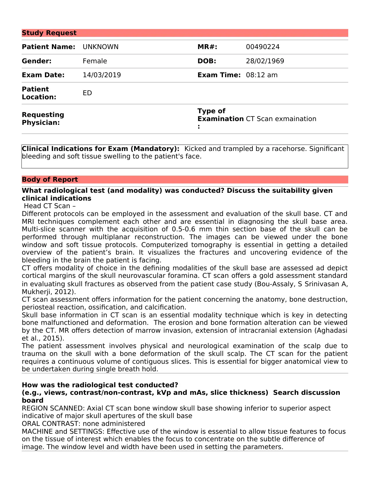
REGION SCANNED: Axial CT scan bone window skull base showing inferior to superior aspect
indicative of major skull apertures of the skull base
ORAL CONTRAST: none administered
MACHINE and SETTINGS: Effective use of the window is essential to allow tissue features to focus
on the tissue of interest which enables the focus to concentrate on the subtle difference of
image. The window level and width have been used in setting the parameters.
Paraphrase This Document
Need a fresh take? Get an instant paraphrase of this document with our AI Paraphraser

What radiological test (and modality) was conducted? Discuss the suitability given
clinical indications
Head CT Scan –
Different protocols can be employed in the assessment and evaluation of the skull base. CT and
MRI techniques complement each other and are essential in diagnosing the skull base area.
Multi-slice scanner with the acquisition of 0.5-0.6 mm thin section base of the skull can be
performed through multiplanar reconstruction. The images can be viewed under the bone
window and soft tissue protocols. Computerized tomography is essential in getting a detailed
overview of the patient’s brain. It visualizes the fractures and uncovering evidence of the
bleeding in the brain the patient is facing.
CT offers modality of choice in the defining modalities of the skull base are assessed ad depict
cortical margins of the skull neurovascular foramina. CT scan offers a gold assessment standard
in evaluating skull fractures as observed from the patient case study (Bou-Assaly, S Srinivasan A,
Mukherji, 2012).
CT scan assessment offers information for the patient concerning the anatomy, bone destruction,
periosteal reaction, ossification, and calcification.
Skull base information in CT scan is an essential modality technique which is key in detecting
bone malfunctioned and deformation. The erosion and bone formation alteration can be viewed
by the CT. MR offers detection of marrow invasion, extension of intracranial extension (Aghadasi
et al., 2015).
The patient assessment involves physical and neurological examination of the scalp due to
trauma on the skull with a bone deformation of the skull scalp. The CT scan for the patient
requires a continuous volume of contiguous slices. This is essential for bigger anatomical view to
be undertaken during single breath hold.
The Head CT image was reviewed using center level and window with settings of CL=30HU AND
WW=80HU, aimed at reviewing the influence of the lesion diagnostic accuracy.
Main Radiological findings:
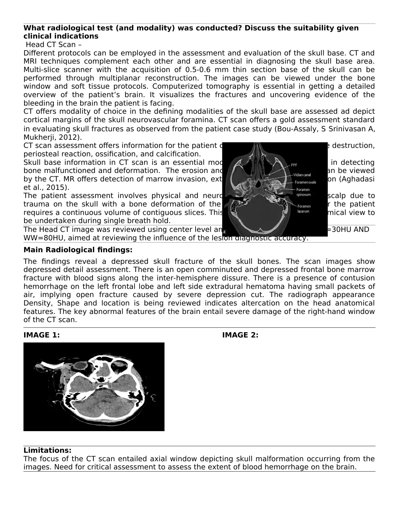
The findings reveal a depressed skull fracture of the skull bones. The scan images show
depressed detail assessment. There is an open comminuted and depressed frontal bone marrow
fracture with blood signs along the inter-hemisphere dissure. There is a presence of contusion
hemorrhage on the left frontal lobe and left side extradural hematoma having small packets of
air, implying open fracture caused by severe depression cut. The radiograph appearance
Density, Shape and location is being reviewed indicates altercation on the head anatomical
features. The key abnormal features of the brain entail severe damage of the right-hand window
of the CT scan.
IMAGE 1: IMAGE 2:
Limitations:
The focus of the CT scan entailed axial window depicting skull malformation occurring from the
images. Need for critical assessment to assess the extent of blood hemorrhage on the brain.

What radiological test (and modality) was conducted? Discuss the suitability given
clinical indications
Head CT Scan –
Different protocols can be employed in the assessment and evaluation of the skull base. CT and
MRI techniques complement each other and are essential in diagnosing the skull base area.
Multi-slice scanner with the acquisition of 0.5-0.6 mm thin section base of the skull can be
performed through multiplanar reconstruction. The images can be viewed under the bone
window and soft tissue protocols. Computerized tomography is essential in getting a detailed
overview of the patient’s brain. It visualizes the fractures and uncovering evidence of the
bleeding in the brain the patient is facing.
CT offers modality of choice in the defining modalities of the skull base are assessed ad depict
cortical margins of the skull neurovascular foramina. CT scan offers a gold assessment standard
in evaluating skull fractures as observed from the patient case study (Bou-Assaly, S Srinivasan A,
Mukherji, 2012).
CT scan assessment offers information for the patient concerning the anatomy, bone destruction,
periosteal reaction, ossification, and calcification.
Skull base information in CT scan is an essential modality technique which is key in detecting
bone malfunctioned and deformation. The erosion and bone formation alteration can be viewed
by the CT. MR offers detection of marrow invasion, extension of intracranial extension (Aghadasi
et al., 2015).
The patient assessment involves physical and neurological examination of the scalp due to
trauma on the skull with a bone deformation of the skull scalp. The CT scan for the patient
requires a continuous volume of contiguous slices. This is essential for bigger anatomical view to
be undertaken during single breath hold.
Comparative data:
Normal CT scan with and without contrasts used as a comparative measure tool.
Impression:
Primary diagnosis
The CT scan image reflect impression related to deep skull injury linked to skull depression on
the left upper region. The anatomical show impairment with a minimal invasive internal injury to
the brain parts. Noncontrast images can further reveal dissidence mass at the anterior horn
occurring on the left lateral with possible hemorrhage (Yi et al., 2016).
Secondary diagnosis
Risk of intracranial hemorrhage hence the needs to manage bleeding due to risks of leading to
internal bleeding as a result of exposed canal opening on the brain skull. This depicts in focal
brain injury occurring in a localized spot rather damage over wider area surface. Thus suspected
hemorrhage can be assessed and checked with contrast application being undertaken (Kodama,
2016).
References:
Aghdasi, N., Li, Y., Berens, A., Moe, K. S., Bly, R. A., & Hannaford, B. (2015). Atlas and feature based 3D
pathway visualization enhancement for skull base pre-operative fast planning from head CT. In Medical
Imaging 2015: Image-Guided Procedures, Robotic Interventions, and Modeling (Vol. 9415, p. 941519).
International Society for Optics and Photonics.
Bou-Assaly, W., Srinivasan, A., & Mukherji, S. K. (2012). Head and neck high-field imaging: Oncology
applications. Neuroimaging Clinics, 22(2), 285-296.
Kodama, S. (2016). Surgical Simulation of Endoscopic Endonasal Anterior Skull Base Surgery using 3D Head
Model and Cone Beam CT. Journal of Neurological Surgery Part B: Skull Base, 77(S 01), P130.
⊘ This is a preview!⊘
Do you want full access?
Subscribe today to unlock all pages.

Trusted by 1+ million students worldwide

Yi, W., Liu, Z. G., Li, X., Tang, J., Jiang, C. B., Hu, J. Y., ... & Xia, Y. F. (2016). CT-diagnosed severe skull
base bone destruction predicts distant bone metastasis in early N-stage nasopharyngeal carcinoma.
OncoTargets and therapy, 9, 7011.
base bone destruction predicts distant bone metastasis in early N-stage nasopharyngeal carcinoma.
OncoTargets and therapy, 9, 7011.
1 out of 4
Your All-in-One AI-Powered Toolkit for Academic Success.
+13062052269
info@desklib.com
Available 24*7 on WhatsApp / Email
![[object Object]](/_next/static/media/star-bottom.7253800d.svg)
Unlock your academic potential
Copyright © 2020–2026 A2Z Services. All Rights Reserved. Developed and managed by ZUCOL.

