Analysis of CT Scan Technology: Composition, Functions, and Biophysics

Verified

Added on  2023/05/29

|8
|1660
|281
Report
AI Summary
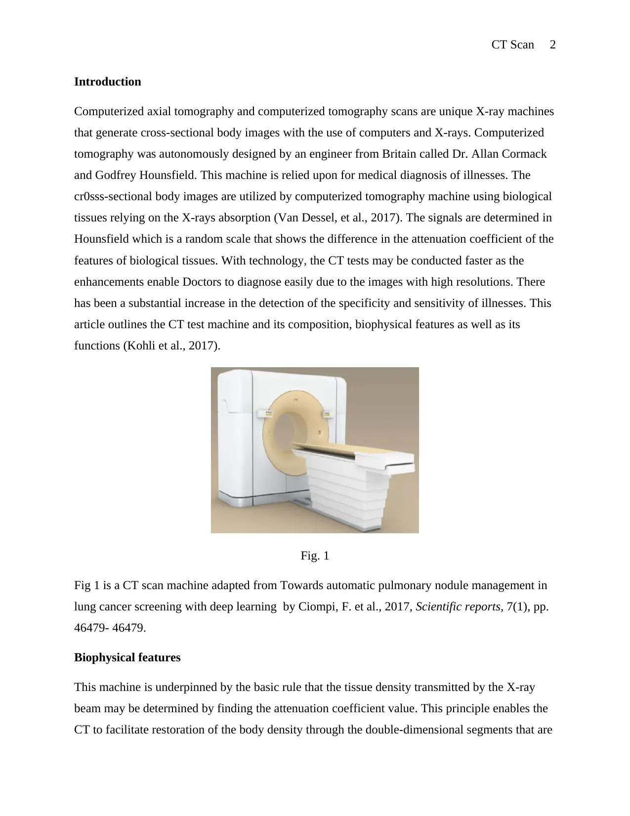
This report provides a detailed overview of CT scan technology, including its composition, functions, and biophysical features. It highlights the importance of CT scans in detecting various diseases and conditions, such as heart diseases, cancer, and internal bleeding. The report discusses the two major components of a CT scan machine: the laser system and the CT scanner gantry, explaining the function of each. Furthermore, it delves into the biophysical principles, emphasizing how the attenuation coefficient is used to restore body density. The report concludes that CT scan technology is significant in medical diagnostics, enabling the detection of internal injuries and various illnesses through the application of X-ray technology and advanced image reconstruction techniques. Desklib provides students access to solved assignments and resources.
Document Page
CT Scan 1
CT SCAN
By (Name of Student)
The Name of the Class (course)
Professor (Tutor)
The Name of the School (University)
The City and State where it is located
Date
tabler-icon-diamond-filled.svg

Paraphrase This Document

Need a fresh take? Get an instant paraphrase of this document with our AI Paraphraser
Document Page
CT Scan 2
Introduction
Computerized axial tomography and computerized tomography scans are unique X-ray machines
that generate cross-sectional body images with the use of computers and X-rays. Computerized
tomography was autonomously designed by an engineer from Britain called Dr. Allan Cormack
and Godfrey Hounsfield. This machine is relied upon for medical diagnosis of illnesses. The
cr0sss-sectional body images are utilized by computerized tomography machine using biological
tissues relying on the X-rays absorption (Van Dessel, et al., 2017). The signals are determined in
Hounsfield which is a random scale that shows the difference in the attenuation coefficient of the
features of biological tissues. With technology, the CT tests may be conducted faster as the
enhancements enable Doctors to diagnose easily due to the images with high resolutions. There
has been a substantial increase in the detection of the specificity and sensitivity of illnesses. This
article outlines the CT test machine and its composition, biophysical features as well as its
functions (Kohli et al., 2017).
Fig. 1
Fig 1 is a CT scan machine adapted from Towards automatic pulmonary nodule management in
lung cancer screening with deep learning by Ciompi, F. et al., 2017, Scientific reports, 7(1), pp.
46479- 46479.
Biophysical features
This machine is underpinned by the basic rule that the tissue density transmitted by the X-ray
beam may be determined by finding the attenuation coefficient value. This principle enables the
CT to facilitate restoration of the body density through the double-dimensional segments that are
Document Page
CT Scan 3
perpendicular to the acquisition system's axis (Rashid et al., 2017). The X-ray tube released N
photons which attenuate as they go through the biological function layers and the information
used to develop scanned object (Van Dessel et al., 2017). Generally, there are two absorption
processes which include Compton effect and the photoelectric effect. This feature is
characterized by one coefficient, mju.
When using the CT machine, the X-rays emitters are rotated all over the patient while the
detector is put in opposite sides of the diameters which collects up the body part images, in this
case, the detector and the beam moving in a synchronized manner. The detectors of the scanner
do not generate images. They determine the X-rays thin beams that are transmitted via a
complete body scan. The images of particular parts are taken at various angles which enables for
the retrieval of data inside the third dimension. The CT detector produces measurements that are
equivalent to the attenuation coefficient value (Oh, et al., 2017). The computer applies unique
mathematical algorithms to reconstruct images to achieve the patient's tomographic images
relying on raw scan information (Oh, et al., 2017).
One can find the proportionate attenuation coefficient to the amount of irradiated tissue if the X-
ray is converted in to quasi-monochromatic or monochromatic with the most suitable filters
using the basic absorption formula of X-rays in practice (Rashid, et al., 2017). The location
determines the emitted intensity I (x) of the photons beams obtained. Whereby, I(x) is less while
the body is greatly radio-opaque. The sectional image of the Irradiated X-ray object is restored
from a huge amount of attenuation coefficient values. It collects all the information received
from the material’s elementary volumes via the detectors (Tu et al., 2018).
The CT image scanner involves the element’s square matrix (pixel) every single of it
representing a voxel of the patient’s tissue. The measurement of the attenuated coefficient for
every proportionate voxel to the pixels must be estimated while processing the image. Every
point of the image is bordered by a star with halo-shape which degrades the difference and blurs
the object's boundary. The filter procedure works in a way that the obtained negative value
amounts to the projection filtered while it is eliminated when projected backward thus producing
the image (Moifo et al., 2017). Before information is displayed on the screen, the CT values are
generated from the conventional rescaling presented in Hounsfield Units. Therefore, the PC
Document Page
CT Scan 4
processes the signal dispatched by the detector the transmitted signal using the detector in digital
data form (Tu et al., 2018).
CT Scan composition
The imaging function of the CT consists of two major constituents found in the simulator which
includes the Laser system and the CT Scanner Gantry (Kohli, et al., 2017).
a. The Laser System
The CT scan process involves three kinds of lasers which are an overhead saggital laser, internal
laser and wall-mounted laser (Tootooni et al., 2017).
i. Internal Laser; It is used to locate the patient during the processes of treatment and
simulation.
ii. Wall-mounted Laser is located at the left and right of the room as a reference of the
image information which coordinates the beam isocenter.
b. Overhead Saggital Laser; they function similarly as the wall-mounted laser.
c. Scanner Gantry
The gantry consists of the detector array, a high-voltage generator, patient support couch, X-ray
and their mechanical support. The scanner receives the image information to be restored by the
simulated virtual application to form a 3D virtual representation of the patient (Ciompi, et al.,
2017).
Functions of the CT Scan
The CT test may be proposed by the Doctor to assist in (Ciompi, et al., 2017):
Detect internal bleeding and injuries
To locate infections, tumors, and blood clots
In the diagnosis of bone and muscle deformities which includes fractures and tumors
tabler-icon-diamond-filled.svg

Paraphrase This Document

Need a fresh take? Get an instant paraphrase of this document with our AI Paraphraser
Document Page
CT Scan 5
Fig. 2
Fig. 3
Fig. 2 and 3 are lasers adapted from Accuracy and reliability of different cone beam computed
tomography (CBCT) devices for structural analysis of alveolar bone in comparison with
multislice CT and micro-CT by Van Dessel, J. et al., 2017, European journal of oral
implantology, , 10(1), pp. 95-105.
Conclusion
Document Page
CT Scan 6
The CT machine is significant in the detection of various diseases and conditions such as heart
diseases, cancer, and liver masses. It may also be used to detect internal bleedings and injuries
which are very important in the diagnosis of illnesses and treatment of such conditions. This
paper has analyzed the composition, functions, and properties of this test machine. The CT
consists of two critical constituents such as the Laser system and the CT Scanner Gantry. The
principle of the tissue density transmitted by X-ray beams being able to be estimated by
obtaining the attenuation coefficient facilitates for the restoration of the body density. The
attenuation X-ray beam estimations are documented while the information is applied in obtaining
a 3D reflection of the object scanned (Tu et al., 2018).
Document Page
CT Scan 7
References
Ciompi, F. et al. (2017) Towards automatic pulmonary nodule management in lung cancer
screening with deep learning. Scientific reports,7(1), pp. 46479- 46479.
Kohli, M., Prevedello, L., Filice, R. and Geis, J. (2017) Implementing machine learning in
radiology practice and research. American Journal of Roentgenology, 208(4), pp. 754-760.
Moifo, B. et al. (2017) Diagnostic Reference Levels of Adults CT-Scan Imaging in Cameroon: A
Pilot Study of Four Commonest CT-Protocols in Five Radiology Departments. Departments.
Open Journal of Medical Imaging, , 7(01), pp. 1- 1.
Oh, E. et al. (2017) Prediction of pathologic femoral fractures in patients with lung cancer using
machine learning algorithms: Comparison of computed tomography-based radiological features
with clinical features versus without clinical features.. ournal of Orthopaedic Surgery, 25(2), pp.
2309499017716243 - 2309499017716243.
Rashid, R. et al. (2017) Effect of scan strategy on density and metallurgical properties of 17-4PH
parts printed by Selective Laser Melting. Journal of Materials Processing Technology, 24(1), pp.
502-511.
Tootooni, M. et al. (2017) Classifying the Dimensional Variation in Additive Manufactured Parts
From Laser-Scanned Three-Dimensional Point Cloud Data Using Machine Learning
Approaches. ournal of Manufacturing Science and Engineering 139, no. 9 (2017): , 139(9), pp.
091005- 091005.
Tu, S. et al. (2018) Localized thin-section CT with radiomics feature extraction and machine
learning to classify early-detected pulmonary nodules from lung cancer screening. Physics in
Medicine & Biology, 63(6), pp. 065005- 065005.
tabler-icon-diamond-filled.svg

Paraphrase This Document

Need a fresh take? Get an instant paraphrase of this document with our AI Paraphraser
Document Page
CT Scan 8
Van Dessel, J. et al. (2017) Accuracy and reliability of different cone beam computed
tomography (CBCT) devices for structural analysis of alveolar bone in comparison with
multislice CT and micro-CT. European journal of oral implantology, , 10(1), pp. 95-105.
chevron_up_icon
1 out of 8
circle_padding
hide_on_mobile
zoom_out_icon
[object Object]