Effectiveness of Ultrasound in Detecting Breast Cancer & Anomalies
VerifiedAdded on 2023/04/24
|13
|3458
|409
Report
AI Summary
This report critically evaluates the effectiveness of the CT ultrasound technique in detecting congenital anomalies in developing babies and its established use in breast cancer detection. The study aims to determine if CT ultrasound can effectively detect birth defects and genetic defects within the developing fetus, and if it can ensure a safer pregnancy. The methodology involves a systematic review of electronic databases such as Google Scholar, MedLine, and PubMed, using specific keywords and inclusion/exclusion criteria to retrieve relevant research papers. The retrieved studies will be analyzed using thematic data analysis to determine the success rate of the technique in detecting congenital anomalies and its potential for preventing associated risks. The report also addresses the limitations of traditional ultrasound techniques and the potential benefits of early detection in reducing infant mortality and healthcare costs.

Running head: MEDICAL IMAGING
MEDICAL IMAGING
Name of the Student:
Name of the University:
Author Note:
MEDICAL IMAGING
Name of the Student:
Name of the University:
Author Note:
Paraphrase This Document
Need a fresh take? Get an instant paraphrase of this document with our AI Paraphraser

1MEDICAL IMAGING
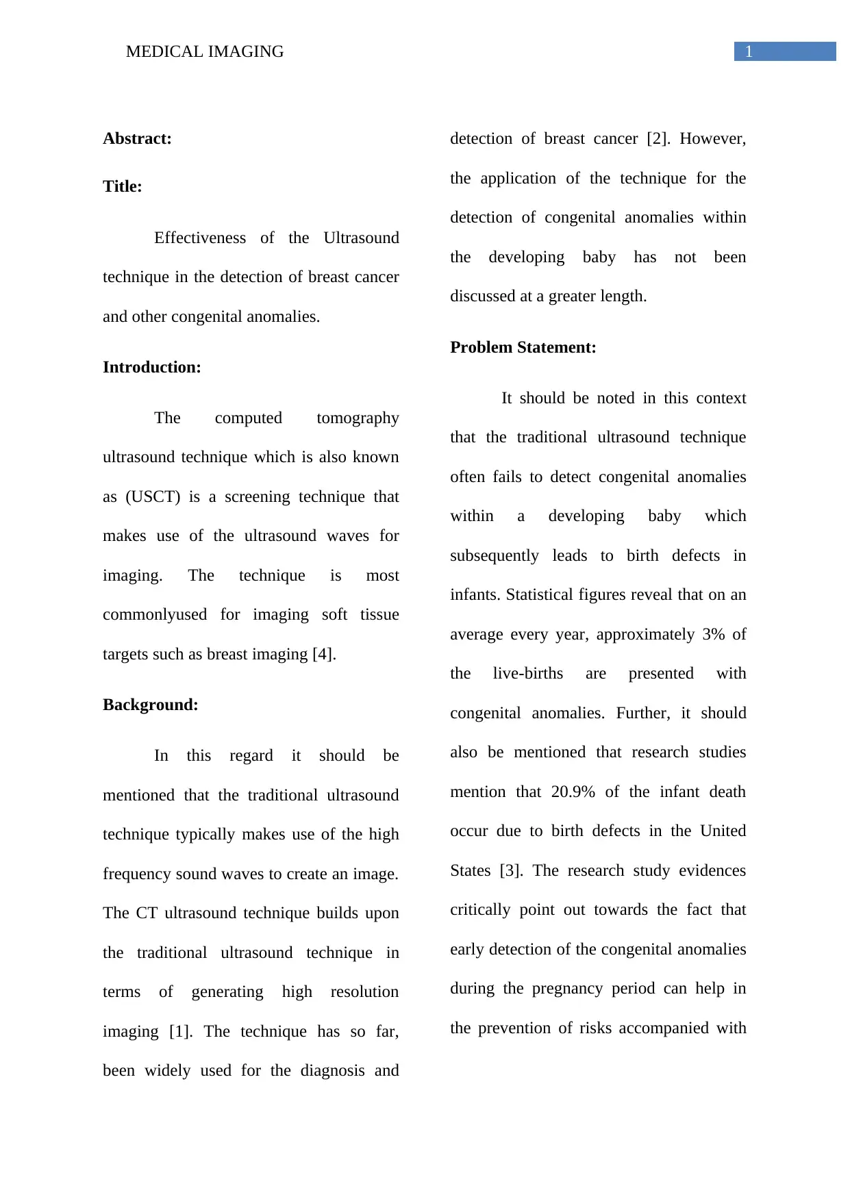
Abstract:
Title:
Effectiveness of the Ultrasound
technique in the detection of breast cancer
and other congenital anomalies.
Introduction:
The computed tomography
ultrasound technique which is also known
as (USCT) is a screening technique that
makes use of the ultrasound waves for
imaging. The technique is most
commonlyused for imaging soft tissue
targets such as breast imaging [4].
Background:
In this regard it should be
mentioned that the traditional ultrasound
technique typically makes use of the high
frequency sound waves to create an image.
The CT ultrasound technique builds upon
the traditional ultrasound technique in
terms of generating high resolution
imaging [1]. The technique has so far,
been widely used for the diagnosis and
detection of breast cancer [2]. However,
the application of the technique for the
detection of congenital anomalies within
the developing baby has not been
discussed at a greater length.
Problem Statement:
It should be noted in this context
that the traditional ultrasound technique
often fails to detect congenital anomalies
within a developing baby which
subsequently leads to birth defects in
infants. Statistical figures reveal that on an
average every year, approximately 3% of
the live-births are presented with
congenital anomalies. Further, it should
also be mentioned that research studies
mention that 20.9% of the infant death
occur due to birth defects in the United
States [3]. The research study evidences
critically point out towards the fact that
early detection of the congenital anomalies
during the pregnancy period can help in
the prevention of risks accompanied with

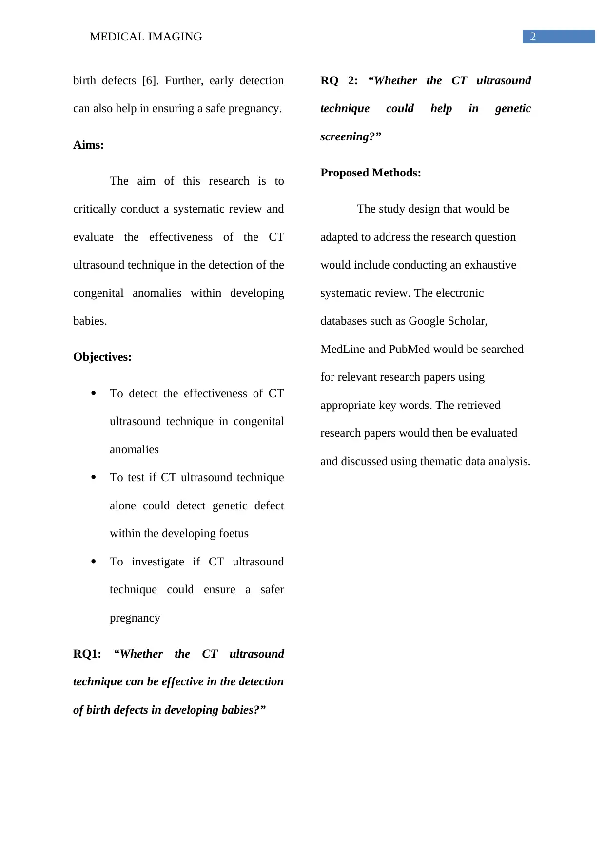
2MEDICAL IMAGING
birth defects [6]. Further, early detection
can also help in ensuring a safe pregnancy.
Aims:
The aim of this research is to
critically conduct a systematic review and
evaluate the effectiveness of the CT
ultrasound technique in the detection of the
congenital anomalies within developing
babies.
Objectives:
To detect the effectiveness of CT
ultrasound technique in congenital
anomalies
To test if CT ultrasound technique
alone could detect genetic defect
within the developing foetus
To investigate if CT ultrasound
technique could ensure a safer
pregnancy
RQ1: “Whether the CT ultrasound
technique can be effective in the detection
of birth defects in developing babies?”
RQ 2: “Whether the CT ultrasound
technique could help in genetic
screening?”
Proposed Methods:
The study design that would be
adapted to address the research question
would include conducting an exhaustive
systematic review. The electronic
databases such as Google Scholar,
MedLine and PubMed would be searched
for relevant research papers using
appropriate key words. The retrieved
research papers would then be evaluated
and discussed using thematic data analysis.
⊘ This is a preview!⊘
Do you want full access?
Subscribe today to unlock all pages.

Trusted by 1+ million students worldwide

3MEDICAL IMAGING
Expected Outcome:
On the basis of the background
information about the application of the
technique, it can be mentioned that the
technique is extremely useful in the
detection of breast cancer [5]. However,
research papers discussing the application
of the technique to determine birth defects
in the developing babies are limited. On
conducting a systematic review the
findings of the retrieved studies would
help in determining the success rate of the
technique in the detection of congenital
anomalies. Thus, it can be said that the CT
ultrasound technique is an extremely
proficient technique used in breast cancer
screening. The findings of the systematic
review would help in determining the
effectiveness of the technique in detecting
congenital anomalies in developing babies
and preventing associated risks.
Paraphrase This Document
Need a fresh take? Get an instant paraphrase of this document with our AI Paraphraser

4MEDICAL IMAGING
Introduction:
The CT ultrasound technique can
be defined as a form of medical imaging
technique that uses the ultrasound waves
for the purpose of imaging. The method
comprises of a series of steps with the first
step comprising of generating an
ultrasound wave. This is done with the
help of Piezoelectric ultrasound
transducers [6] [8]. The waves are
generated in the similar direction of the
object that is to be measured and the waves
are received at the other end by the similar
or a different set of transducers [8]. The
mechanism operates in such a way that
once the generated waves interacts with
the object, the waves are modified and
automatically carry the information about
the structure of the object [8]. The waves
are then recorded and the information
about the modulated waves is used for the
creation of the object that was being
measures in the subsequent step [8].
Background:
The first kind of Ultrasound
computed tomography scan can be
retrieved to the twentieth century when the
diagnosis field was largely based upon
compounding setups [9]. The first digital
technology of ultrasound computed
tomography was used only in the 1970s
[8]. The use of a computer in the technique
clearly indicates the advancement in the
field of technology with respect to digital
signal processing, image recreation and
image processing clear algorithms [8] [9]
[10]. In this regard, it should be mentioned
that the technique can be considered as an
evolution in the field of medical imaging
as it provides multiple information about
the object properties unlike the traditional
methods such as X-ray that only provides
information about the physical properties
of an object [9]. The object’s attenuation
coefficient can be detected with the help of
the attenuation of the sound wave’s
experience [10]. Also, the time of flight

5MEDICAL IMAGING
provides information about the speed of
sound and other important components
such as the surface morphology, refractive
index is easily measure with the scattering
property of the wave. It should further be
noted that the technique is different from
the convention ultrasound technique which
uses the technology of phased array for the
formation of beam [8]. The CT ultrasound
technique typically uses unfocused
spherical waves for the purpose of
imaging. The advantages of the technique
can be attributed to the production of a 3-D
image by stacking 2D images with the help
of 3-D aperture setups [9] [10]. Also, the
technique helps in retrieving quantitative
imaging rather than qualitative imaging.
Therefore, computing power along with
the availability of the data bandwidth can
be attributed as the integral factors that has
led to the development of this technique
[8] [9].
Analysis of Problems:
The CT ultrasound technique is
widely used for soft tissue imaging.
Studies reveal that the technique is
extensively used for the detection of breast
cancer [7] [9]. However, studies also
confirm that the method uses low sound
pressures and is a risk-free imaging
procedure which can be deemed
appropriate for periodic screenings [8]. In
addition to this, research studies also prove
that the USCT setups are either fixed or
motor moved and can reproduce images
without the direct contact with the soft
tissue or organs [10] [11]. Further, the
procedure has been studied to be economic
and extremely feasible. However, the
usability of the technique in the detection
of congenital anomalies in the developing
foetus remains questionable. On an
average, approximately 17% to 42% of
infant mortality is attributed because of a
congenital anomaly during the period of
gestation [4]. The use of the new born
screening (NBS) methods to detect the
⊘ This is a preview!⊘
Do you want full access?
Subscribe today to unlock all pages.

Trusted by 1+ million students worldwide

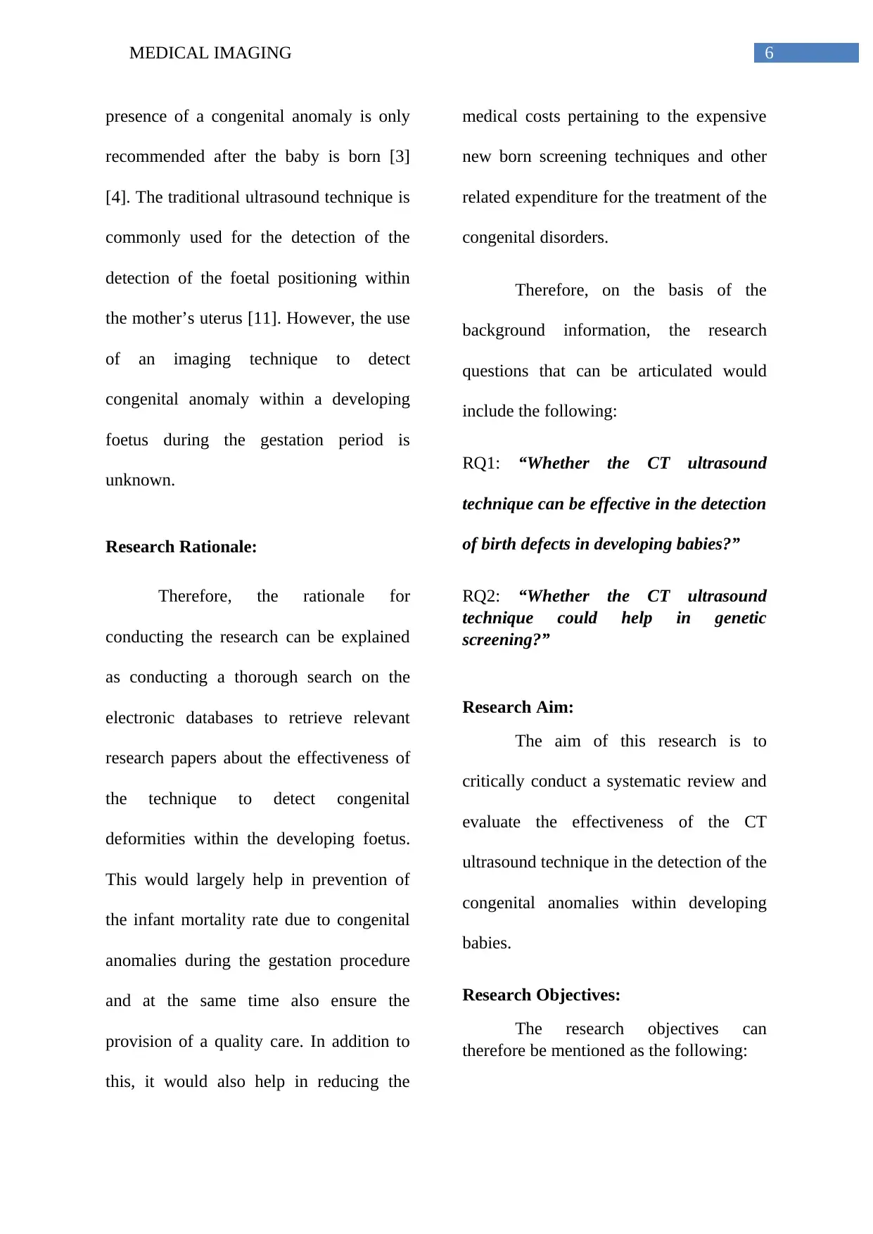
6MEDICAL IMAGING
presence of a congenital anomaly is only
recommended after the baby is born [3]
[4]. The traditional ultrasound technique is
commonly used for the detection of the
detection of the foetal positioning within
the mother’s uterus [11]. However, the use
of an imaging technique to detect
congenital anomaly within a developing
foetus during the gestation period is
unknown.
Research Rationale:
Therefore, the rationale for
conducting the research can be explained
as conducting a thorough search on the
electronic databases to retrieve relevant
research papers about the effectiveness of
the technique to detect congenital
deformities within the developing foetus.
This would largely help in prevention of
the infant mortality rate due to congenital
anomalies during the gestation procedure
and at the same time also ensure the
provision of a quality care. In addition to
this, it would also help in reducing the
medical costs pertaining to the expensive
new born screening techniques and other
related expenditure for the treatment of the
congenital disorders.
Therefore, on the basis of the
background information, the research
questions that can be articulated would
include the following:
RQ1: “Whether the CT ultrasound
technique can be effective in the detection
of birth defects in developing babies?”
RQ2: “Whether the CT ultrasound
technique could help in genetic
screening?”
Research Aim:
The aim of this research is to
critically conduct a systematic review and
evaluate the effectiveness of the CT
ultrasound technique in the detection of the
congenital anomalies within developing
babies.
Research Objectives:
The research objectives can
therefore be mentioned as the following:
Paraphrase This Document
Need a fresh take? Get an instant paraphrase of this document with our AI Paraphraser

7MEDICAL IMAGING
To detect the effectiveness of CT
ultrasound technique in congenital
anomalies
To test if CT ultrasound technique
alone could detect genetic defect
within the developing foetus
To investigate if CT ultrasound
technique could ensure a safer
pregnancy
Research Methodology:
According to Neuman (2013),
systematic review can be defined as kind
of literature review that applies systematic
methods in order to scan electronic
databases and collect secondary data to
critically appraise the studies and
synthesize the findings [12]. This method
helps in identifying studies and
synthesizing their findings that directly
relate to the developed research question.
The method helps in collecting data from
relevant research studies and conducts an
exhaustive summary of their findings in
order to formulate new theories or develop
a better understanding about the existing
theories [13] [14]. In addition to this, this
method is also considered as an
inexpensive and feasible research study
design to embark upon a new study [14].
The study design thus chosen to address
the research topic is systematic review.
The rationale for the choice of the
research study design can be attributed to
the gamut of the research papers available
on the concerned topic.
Literature Search:
The first step would comprise of
scanning the electronic databases in order
to retrieve relevant research papers aligned
to the research questions. The electronic
databases that would be scanned to retrieve
relevant research papers would include
Google Scholar, CINAHL and Cochrane
Library. The scan would be conducted
after applying filters which would include
the specific inclusion and exclusion
criteria. In addition to this, specific key
terms would be used to conduct the search.

8MEDICAL IMAGING
Key terms:
Key terms are specific terms or
phrases that relate with the major concept
of the research topic and help in retrieving
relevant results. In addition to this, the
BOOLEAN operators, OR/AND would
also be used in combination with the key
terms to retrieve relevant research studies
[14]. The key terms that would be used
would thus include the following:
Computed tomography ultrasound, CT
ultrasound, congenital anomalies, breast
cancer, gestation, safe pregnancy, new
born screening, 3D ultrasound
Exclusion Criteria:
The application of certain specific
parameters would help in retrieving better
results. These parameters are basically the
inclusion as well as the exclusion criteria
[15]. The exclusion criteria basically
comprises of the exclusive characteristics
based on which the research papers are
eliminated and not considered in the
systematic review [13] [15]. These
characteristics would comprise of the
following:
Research papers published before
2013
Research papers published in
foreign languages other than
English
Research papers that include
animal trials
Research papers where the full text
in not accessible
Research papers that are systematic
reviews
Inclusion Criteria:
The inclusion criteria include the
inclusive characteristics that would be
used to include the studies in the
systematic review [15]. These
characteristics would comprise of the
following:
Research studies published in
English
⊘ This is a preview!⊘
Do you want full access?
Subscribe today to unlock all pages.

Trusted by 1+ million students worldwide

9MEDICAL IMAGING
Research studies that include
randomized control trials and are
qualitative research studies
Research studies published
between the timeframe (2013-
2017)
Research studies with accessibility
to full-text
Data Collection and Data Analysis:
The abstract of the research studies
shortlisted would first be read to decide the
compatibility of the paper with the
research topic. The complete research
papers would then be read and re-read with
special attention to the methodology
section and the result section. Accordingly,
the findings from the research studies
would then be noted down and the
research papers would then be appraised in
order to determine the strength and
weakness of each paper. Finally, the
findings retrieved from the research study
would then be analyzed and discussed with
the help of thematic-analysis. Therefore,
the research approach adapted for the data
collection method would be qualitative as
the review would characteristically include
the qualitative research studies. Also, the
research philosophy used would be
transformative in this case [15].
The thematic-data analysis method
is considered as the best data analysis
method that helps in analysing qualitative
data collected from research studies [16].
Thematic analysis helps in examining and
recording the patterns or themes present
within a data set [16] [17]. This form
o
f analysis can be considered as a canopy
that is best fitted to investigate a variety of
research approaches 14]. It should
however, be noted in this context that this
form of analysis is influenced by different
philosophical assumptions. Therefore, the
best data analysis method to analyze the
data collected from the research studies
Paraphrase This Document
Need a fresh take? Get an instant paraphrase of this document with our AI Paraphraser

10MEDICAL IMAGING
would be the thematic analysis method.
The inductive approach would be used to
analyze the collected qualitative data. The
inductive approach is best suited to
analyze secondary data collected from
research studies in order to derive themes
of concepts [18].
Research Philosophy:
The research would be based on the
interpretivism research philosophy. The
rationale for the choice of the philosophy
can be stated as the efficacy of the research
philosophy to conduct in-depth
investigations and also competently handle
a small sample size [18].
Conclusion:
Therefore, to conclude, it can be
said that the systematic review would help
in retrieving a number of research studies
that have effectively tested the efficacy of
the technique in the detection of soft tissue
abnormalities. On the basis of the
background information, it can be
commented that the technique is an
inexpensive and convenient imaging
procedure that is widely used for the
detection of breast cancer. However, the
usability of the technique to detect
congenital anomalies within the
developing foetus has not yet been
investigated and is a mystery. Thus, it can
be hoped that the systematic review would
help in critically analyzing the available
literatures that talk about the effectiveness
of the technique in the detection of
congenital deformities within the
developing foetus. This would help in the
prevention of infant mortality due to
congenital deformities and also help in
improving the quality of care.
Timeline:
1ST
week
of 1st
mont
h
2nd week
of 1st
month
2nd and
3rd
Month
4th
and
5th
Mont
h
6th
month
Select
ion of
resear
ch
topic
Formula
tion of
research
question
and
selection
of study
design
Collect
ion of
data
Analy
sis of
data
Final
submiss
ion

11MEDICAL IMAGING
References:
[1] Stotzka R, Wuerfel J, Mueller TO,
Gemmeke H. Medical imaging by
ultrasound computer tomography.
InMedical Imaging 2002: Ultrasonic
Imaging and Signal Processing 2002 Apr
11 (Vol. 4687, pp. 110-120). International
Society for Optics and Photonics.
[2] Medina-Valdés L, Pérez-Liva M,
Camacho J, Udías JM, Herraiz JL,
González-Salido N. Multi-modal
ultrasound imaging for breast cancer
detection. Physics Procedia. 2015 Jan
1;63:134-40.
[3] Liu L, Oza S, Hogan D, Perin J, Rudan
I, Lawn JE, Cousens S, Mathers C, Black
RE. Global, regional, and national causes
of child mortality in 2000–13, with
projections to inform post-2015 priorities:
an updated systematic analysis. The
Lancet. 2015 Jan 31;385(9966):430-40.
[4] Jalalian A, Mashohor SB, Mahmud
HR, Saripan MI, Ramli AR, Karasfi B.
Computer-aided detection/diagnosis of
breast cancer in mammography and
ultrasound: a review. Clinical imaging.
2013 May 1;37(3):420-6.
[5] Drukteinis JS, Mooney BP, Flowers
CI, Gatenby RA. Beyond mammography:
new frontiers in breast cancer screening.
The American journal of medicine. 2013
Jun 1;126(6):472-9.
[6] Svensson E, Ehrenstein V, Nørgaard
M, Bakketeig LS, Rothman KJ, Sørensen
HT, Pedersen L. Estimating the proportion
of all observed birth defects occurring in
pregnancies terminated by a second-
trimester abortion. Epidemiology. 2014
Nov 1;25(6):866-71.
[7] Werner H, Rolo LC, Júnior EA, Dos
Santos JR. Manufacturing models of fetal
malformations built from 3-dimensional
ultrasound, magnetic resonance imaging,
and computed tomography scan data.
Ultrasound quarterly. 2014 Mar
1;30(1):69-75.
⊘ This is a preview!⊘
Do you want full access?
Subscribe today to unlock all pages.

Trusted by 1+ million students worldwide
1 out of 13
Related Documents
Your All-in-One AI-Powered Toolkit for Academic Success.
+13062052269
info@desklib.com
Available 24*7 on WhatsApp / Email
![[object Object]](/_next/static/media/star-bottom.7253800d.svg)
Unlock your academic potential
Copyright © 2020–2026 A2Z Services. All Rights Reserved. Developed and managed by ZUCOL.


