Detailed Therapeutic Case Study on Cushing's Syndrome Aetiology
VerifiedAdded on 2020/03/04
|12
|4547
|42
Case Study
AI Summary
This case study delves into Cushing's Syndrome, a disorder characterized by elevated cortisol levels. The aetiology is primarily linked to adrenal gland dysfunction or pituitary gland tumors, leading to excessive cortisol or ACTH production. The physiology involves the hypothalamus-pituitary-adrenal axis and its impact on metabolic processes. Pathophysiologically, hypercortisolism results in glucose intolerance, dyslipidemia, and cardiovascular complications. The molecular pathology highlights genetic mutations and abnormalities in the adrenal and pituitary glands. Diagnosis involves laboratory tests like the Dexamethasone suppression test and ACTH measurement. The study explores various aspects of this complex disorder, providing a comprehensive overview of its causes, mechanisms, and diagnostic approaches.
Therapeutic Case Study
1
1
Paraphrase This Document
Need a fresh take? Get an instant paraphrase of this document with our AI Paraphraser
Aetiology:
Elevated levels of cortisol hormone are mainly responsible for the occurrence of Cushing
syndrome. Cortisol is mainly produced by adrenal glands in the body. Cortisol performs
important functions in the body like regulation of blood pressure and normal functioning of
cardiovascular system. Cortisol is also helpful in responding to stress and metabolizing
carbohydrates, proteins and fats in the usable energy. However, elevated levels of cortisol,
disrupt these physiological processes and lead to development of Cushing syndrome. Cortisol
levels in MV might increase due to high levels of stress, malnutrition, depression and high
levels of emotional stress. Cortisol levels in a person can also increase due to athletic
training, alcoholism and panic disorders. High doses of anti-inflammatory medications like
corticosteroids might increase cortisol levels. Pituitary gland releases more
adrenocorticotropic (ACTH) hormone in pituitary gland tumor. This increase in the
adrenocorticotroic hormone is known as Cushing’s disease. Adrenal gland abnormality or
tumor might cuase Cushing’s syndrome. Even though70 – 80 % cases of the Cushing's
syndrome occur due to excessive secretion of ACTH from the pituitary adenomas, it is
evident that only 10 – 15 % pituitary adenomas secret excessive ACTH. Even though
approximately 10 % cases of the Cushing's syndrome occur due to excessive secretion of
cortisol from the adrenal adenomas, it is evident that only 6 % adrenal adenomas secret
excessive cortisol. However, exact aetiology of excessive secretion of ACTH from pituitary
adenomas and cortisol secretion from adrenal adenomas has not been well established.
Genetic studies established that mutation in the ubiquitin-specific protease 8 is responsible
for Cushing's disease 1,2.
Difference between Cushing’s syndrome and Cushing’s disease:
Tumor in the adrenal gland produces more amount of cortisol. This leads to the development
of Cushing’s syndrome. Cushing’s syndrome can also be developed due to tumor in the
pituitary gland. Cushing’s syndrome is the group of symptoms in which cortisol levels
remains elevated for the longer duration. Tumor in the pituitary gland produces more amount
of ACTH. This ACTH stimulates more secretion of cortisol from the adrenal gland. This type
of Cushing’s syndrome is called as Cushing’s disease. Tumors produced by ACTH can be
produced anywhere in the body and these are termed as ectopic tumors. Long term steroid
administration is one of the causes of Cushing’s syndrome. Cushing’s disease may occur due
to the endogenous factors like self-overproduction of cortisol by the body. Endogenous over-
2
production of cortisol may occur due to pituitary tumor (Cushing’s disease), adrenal tumor
and ectopic tumors3.
Physiology:
Hypothalamus is the part of brain below which pituitary gland is present. Corticotropin-
releasing hormone (CRH) is secreted by paraventricular nucleus (PVN) of the hypothalamus.
CRH stimulates pituitary gland to secret ACTH. ACTH gets transported through blood and as
result adrenal gland gets stimulated to secret cortisol. Zona fasciculata is the region of cortex
of adrenal gland which is responsible for the secretion of cortisol. Corticosteroid hormones
are under the control of hypothalamus-pituitary-adrenal axis. Corticosteroids exhibit most
prominent metabolic activity during the postprandial period. During this period, these
hormones behave as contra-insular hormones. Hence, it provides substrates for oxidative
metabolism and stimulate lipolysis and proteolysis. This results in the release of fatty acids
and amino acids which induce glucose production by stimulating gluconeogenesis and
inhibiting glycogen synthesis. Elevated levels of cortisol are responsible for the negative
feedback which is responsible for the reduced secretion of ACTH4,5.
Pathophysiology:
In all types of etiologies of Cushing syndrome, there are raised levels of cortisol. Cortisol
secreting adenoma in the cortex of adrenal gland occurs in Cushing’s syndrome. This
adenoma of cortex of adrenal gland might be of primary hypercortisolism or hypercorticism
types. Cushing’s syndrome due to elevated levels of ACTH is known as ectopic Cushing
syndrome. Patients with severe hypercortisolism are associated with the phenotypes like
glucose intolerance, dyslipidaemia, metabolic bone disease, and abnormal weight gain. On
the other hand patients with mild or moderate hypercortisolism are rarely associated with
glucose intolerance, dyslipidaemia, metabolic bone disease, and abnormal weight gain.
Hence, it would be difficult to differentiate these patients from other patients with metabolic
syndrome. There is presentation of striae, supraclavicular fat pads, and proximal muscle
weakness in patients with increased hypercortisolism6. Elevated levels of corticosteroids
results in the augmented production of glucose at the level of liver and skeletal muscle. In
liver corticosteroids increase levels of glucose by stimulating gluconeogenesis and inhibiting
insulin sensitivity. Corticosteroids stimulate gluconeogenesis by acting on different genes
responsible for glucose metabolism. Corticosteroids reduce insulin sensitivity by exerting
3
⊘ This is a preview!⊘
Do you want full access?
Subscribe today to unlock all pages.

Trusted by 1+ million students worldwide
antagonistic effect on metabolic action of insulin which is most important hormone required
for suppression of glucose production7,8.
Corticosteroids decrease expression and phosphorylation of insulin receptor substrate (IRS)-
1, phosphatidylinositol-3kinase (PI3K) and protein kinase B (PKB/AKT). As a result, there is
decrease in migration of glucose transporter GLUT4 to the cell surface which results in the
reduction of glucose uptake. It also leads to decrease of glycogen synthase kinase (GSK)-3
phosphorylation and results in the decrease of glycogen synthesis. In skeletal muscles,
corticosteroids increase insulin resistance which results in the decreased glucose uptake and
prevention of glycogen synthesis. Corticosteroids exhibit effect on insulin resistance by
directly acting on the insulin receptor or alteration in the insulin function by altering lipid and
protein metabolism9,10.
Corticosteroids also play prominent role in the regulating adipose tissue differentiation,
distribution and metabolism. Corticosteroids can stimulate differentiation of pre-adipocytes in
adipocytes. Increased adipocytes are associated with raised body fat mass. Corticosteroid
specifically act on visceral fat and not on the peripheral fat. Hence, corticosteroids are
associated with central obesity in Cushing’s syndrome. Corticosteroids also play role in the
release of hormone like adipokine which is responsible for the development of insulin
resistance. Co-occurrence of insulin resistance and increased visceral fat can lead to the
development of metabolic syndrome which is associated with impairment in the glucose
metabolism. Metabolic syndrome comprises of visceral obesity, diabetes mellitus, and
dyslipidemia. Effect of corticosteroids on the pancreatic beta cells result in the decreased
insulin secretion. Corticosteroids exhibit effect on insulin secretion in dose dependent and
time dependent manner. Corticosteroid exhibit effect on different steps of insulin secretion
like glucose uptake, glucose utilization and calcium fluxes and exocytosis of granules
containing insulin. Glucose transporter GLUT2 and glucokinase expression gets reduced due
to corticosteroids11,12.
Corticosteroids can change AMP-activated protein kinase (AMPK) activity in tissue specific
manner. Suppression of AMPK is proportional to hypercortisolism. Due to tissue specific
activities, AMPK increases appetite, visceral obesity and dyslipidemia. Upon activation,
AMPK plays key role in glycolysis and fatty acid synthesis which produces ATP. Due to
hormone-sensitive lipase (HSL), there is increased level of lipids form adipose tissue which
lead to development of adipocytes. 11 β- hydroxysteroid dehydrogenase type 1 (11 βH SD1)
4
Paraphrase This Document
Need a fresh take? Get an instant paraphrase of this document with our AI Paraphraser
is responsible for the activation of cortisol from the inactive cortisone. 11 βH SD1
overexpressed in mature adipocytes in conditions like hepatic steatosis and there is increased
levels of cholesterol and triglycerides. 11 βH SD1 is also associated with insulin
resistance13,14.
Cardiovascular complications associated with corticosteroids include systemic arterial
hypertension, atherosclerosis, and thromboembolism. It has been established that elevated
levels of corticosteroid can lead to the increase in renin-angiotensin system, stimulated
sympathetic nervous system and endothelin system. It also leads to reduced nitric oxide (NO)
synthesis and kallikrein-kinin system. Patients of Cushing’s syndrome are associated with
irreversible arterial stiffness. Endothelin, homocysteine, vascular endothelial factor,
adrenomedulin, and cell adhesion molecules are the markers of endothelial dysfunction and
are responsible for vascular endothelial and smooth muscle proliferation and fibrosis around
vessels and consequently development of atherosclerosis plaques. In subclinical Cushing’s
syndrome both systolic and diastolic hypertension can occur. Blood pressure can elevate in
patients with Cushing’s syndrome due to ACTH stimulated cortisol level15,16. Corticosteroids
also produces immunosuppression and neuropsychiatric complications. Ectopic ACTH
production lead to Cushing’s syndrome and neurocognitive impairment. Neuropsychiatric
disorders associated with Cushing’s syndrome are major depression, mania, anxiety, and
cognitive impairment17. Bone complications associated with corticosteroids include
osteoporosis and osteoarthritis. Osteoporosis may occur in patients with Cushing’s syndrome
due to increased bone reabsorption, decreased bone formation, reduced intestinal calcium
absorption and increased urinary calcium excretion18.
Molecular Pathology:
Adrenocortical carcinomas are associated with clinical hypercortisolism and androgen
overproduction. Elevated levels of urinary metabolites of androgens or glucocorticoid
precursors are evident in the adrenocortical carcinomas. It reflects reduced activity of
steroidogenic enzymes. Bilateral macronodular adrenal hyperplasia (BMAH) is seldom
connected with MEN1, familial adenomatous polyposis, and fumarate hydratase gene
mutations. Bilateral adrenal hyperplasias are presented as in the form of nodule diameter
greater than 1 cm (macronodular) or less than 1 cm (micronodular). Patients with Cushing’s
syndrome possesses ARMC5 gene mutations and associated with subclinical BMAH. In
addition to the ARMC5 gene mutation, there is occurrence of mutations in histone H3 lysine
5
79 methyltransferase gene (DOTL1), in a histone deacetylase gene (HDAC9), or in a prune
homologue 2 gene (PRUNE2) in BMAH19,20.
Molecular genetic abnormalities are helpful in the identifying causes of the Cushing’s
syndrome. Glucocorticoid receptor gene mutation, dysfunction of proteins related to
glucocorticoid-receptor function (Brg1 and HDAC2), and TP53-inactivating defects are
responsible for the occurrence of Cushing’s syndrome. Overexpression of testicular orphan
receptor 4 (TR4),30 a nuclear receptor with unknown ligand, the pituitary transforming gene
(PTTG),31 and epidermal growth factor receptor are responsible for occurrence of Cushing’s
syndrome. Overexpression of these genes are useful in identifying causal relationship and
establishing therapeutic roles. Somatic mutations in the ubiquitin specific peptidase 8 gene
(USP8) established role of epidermal growth factor receptor in Cushing’s syndrome. USP8
inactivation produces augmented epidermal growth factor receptor deubiquitination which
leads to impairment in its down regulation and increased growth factor signalling and pro-
opiomelanocortin (POMC) and ACTH synthesis. Mutations in the cAMP-dependent or β-
catenin signalling pathways are expresses in the adrenal adenomas. Mutations of GNAS
(encoding Gsα, which activates adenylate cyclase) are reported in cortisol secreting
adenomas and β-catenin (CTNNB1) mutations in cortisol-secreting adenomas. Protein kinase
A (PRKACA) mutations are reported in adenomas of Cushing’s syndrome.
Phosphodiesterase 11A (PDE11A) or phosphodiesterase 8B (PDE8B) genetic variants are
responsible for adrenal adenomas21.
Diagnosis:
Most of the clinical features of Cushing’s syndrome like body weight gain and blood pressure
are common. Hence, it would be difficult to make exact diagnosis of Cushing’s syndrome.
Hence, laboratory tests need to be performed for the diagnosis of Cushing’s syndrome. These
tests would also be helpful in identifying Cushing disease which mainly occurs due to tumor
of pituitary gland. Dexamethasone suppression test and 24-hour urinary and salivary
measurement for cortisol are useful for the confirmation of Cushing’s syndrome.
Dexamethasone suppression test involves frequent administration of dexamethasone and
continuous monitoring of cortisol and ACTH levels. In this test, if body is monitoring
cortisol, there would be decrease in the cortisol level. However, in cases of Cushing’s
syndrome, there would be increase in the cortisol level. This test is not always useful because
other illness or condition may produce high level of cortisol. This condition is called as
6
⊘ This is a preview!⊘
Do you want full access?
Subscribe today to unlock all pages.

Trusted by 1+ million students worldwide
pseudo-Cushing’s states. Doctors need to treat pseudo-Cushing’s states like depression. Post
treatment, if there is no reduction in cortisol along with other physical worsening, Cushing’s
syndrome would be confirmed22.
ACTH measurement in the blood can be useful in differentiating Cushing’s syndrome and
Cushing’s disease. Blood ACTH levels would be less in cases of Cushing’s syndrome,
however it would be normal or higher in cases of Cushing’s disease. This is because, blood
ACTH levels would be less in patients with adrenal origin Cushing’s syndrome as compared
to the other origin of Cushing’s syndrome. ACTH producing tumor from the pituitary origin
can be differentiated from the other parts of origin by using inferior petrosal sinus sampling,
or IPSS. In this test, small plastic tube need to be inserted in the right and left veins in the
groin and attach them to the veins near the pituitary gland. Blood should be collected form
these veins and other locations far from the pituitary. During this procedure, medications
which increase ACTH levels from the pituitary need to be injected. ACTH levels produced
near pituitary and far from the pituitary need to be measured to assess tumors produced by
pituitary origin. Inferior petrosal sinus sampling (IPSS) is more reliable as compared to the
dexamethasone suppression test. Magnetic resonance imaging (MRI) can also be used as a
diagnostic tool for Cushing’s syndrome. In this test, contrast is useful for showing up tumor
in the MRI scan. Identification of the definitive tumor along with positive dexamethasone
suppression test would be useful in diagnosis of Cushing’s disease. In such cases, there is no
requirement of IPSS test. In few healthy people, there may be abnormal area on the pituitary,
which might resemble tumor in MRI, hence abnormality doesn’t reflect occurrence of
Cushing’s disease. In few cases, tumor may be too small to detect using MRI. Hence, absence
of tumor should not exclude Cushing’s syndrome. Sodium and potassium levels can be
increased in patients with Cushing’s syndrome, hence assessment of electrolyte balance can
be used as a diagnostic tool23.
Treatment and management:
Main aim of the treatment of Cushing’s syndrome should be reduction in the level of cortisol.
Treatment of Cushing’s syndrome may vary based on the cause of the disease. Corticosteroid
medication use should be reduced. Dose of the corticosteroid should be reduced. However,
reduced dose of corticosteroid should be useful in the management of chronic inflammatory
disease. Non-corticosteroids drugs can be administered along with corticosteroids to reduce
dose of corticosteroids. Use of corticosteroids should not be stopped abruptly because it may
7
Paraphrase This Document
Need a fresh take? Get an instant paraphrase of this document with our AI Paraphraser
lead to decrease in cortisol levels. In patients with existence of tumor, surgery should be
performed to remove tumor. Pituitary tumors can be removed by performing surgery through
nose and neurosurgeon can perform this surgery. Adrenal gland tumors can be removed by
standard surgical procedure and using minimally invasive surgical technique. After the
completion of the surgery, cortisol replacement medications should be provided to the
patients to maintain normal levels of cortisol. Cortisol replacement medications usually takes
long duration to maintain normal level of cortisol. Radiation therapy should be provided to
the patients, in whom surgeon can’t remove tumor completely. Hence, in such cases radiation
therapy should be used as an adjuvant therapy along with surgery. Radiation therapy should
be used in patients in whom surgery can not be performed. Radiation should be given in little
doses for a duration of 6 weeks. Stereotactic radiosurgery, which is also called as Gamma
Knife surgery can also be used, in which large single dose of radiation should be given to
tumor part with minimal exposure to the surrounding area. Medications to control cortisol
levels should be administered to patients, whom are very sick and surgery can not be
performed in such patients24.
Ketoconazole, mitotane and metyrapone can be used to reduce excessive production of
cortisol from the adrenal gland. It is evident that, Ketoconazole can reduce plasma cortisone,
however it can not reduce plasma ACTH. Metyrapone exhibits its action by inhibiting 11α-
hydroxylase. Due to use of metyrapone, there may be increase in ACTH levels relative to the
decrease in the cortisol levels. This may result in the stimulation of the adrenal androgen and
increased mineralocorticoid production. Mifepristone is useful in the Cushing’s syndrome
patients with diabetes and glucose intolerance. Mitotane exhibits its action by inhibiting 11α-
hydroxylase, 18-hydroxylase, 3α-hydroxy-lase, hydroxysteroid dehydrogenase, and several
cholesterol side chain cleavage enzymes. Mitotane exhibits adrenolytic action by binding its
metabolite to the macromolecules in adrenal cortical cell mitochondria. This results in the cell
destruction and cellular necrosis. Mifepristone does not reduce cortisol level; however it
blocks action of cortisol on tissues. Pasireotide is useful in the Cushing’s disease because it
reduces the ACTH secretion from pituitary tumor. This medication should be administered
twice daily, if surgery can not be performed or in cases of unsuccessful surgery. If none of
the treatment works, bilateral adrenalectomy should be performed. In bilateral
adrenalectomy, there would be surgical removal of adrenal gland. This surgical intervention
is useful in reducing excess levels of cortisol, however such patients require life-long cortisol
replacement medications25.
8
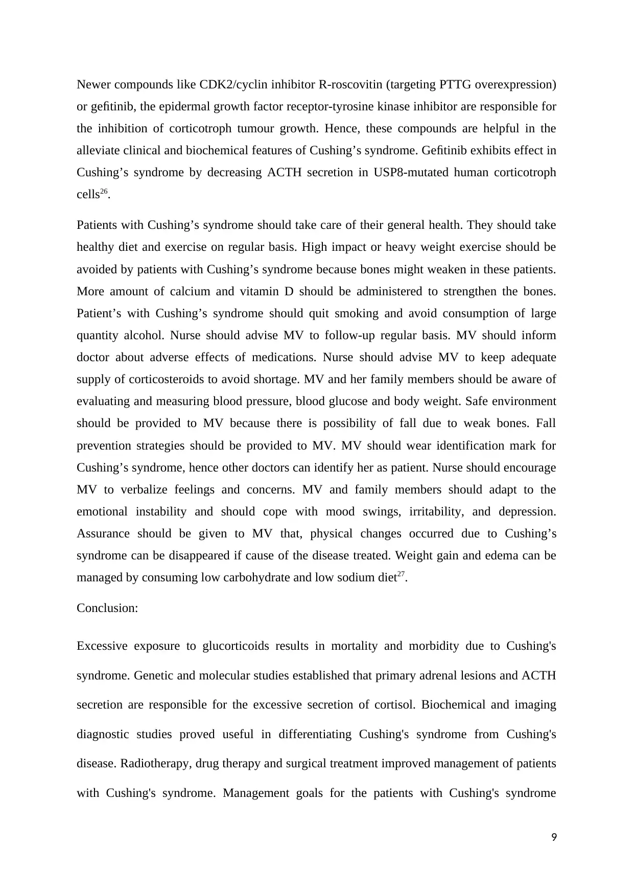
Newer compounds like CDK2/cyclin inhibitor R-roscovitin (targeting PTTG overexpression)
or gefitinib, the epidermal growth factor receptor-tyrosine kinase inhibitor are responsible for
the inhibition of corticotroph tumour growth. Hence, these compounds are helpful in the
alleviate clinical and biochemical features of Cushing’s syndrome. Gefitinib exhibits effect in
Cushing’s syndrome by decreasing ACTH secretion in USP8-mutated human corticotroph
cells26.
Patients with Cushing’s syndrome should take care of their general health. They should take
healthy diet and exercise on regular basis. High impact or heavy weight exercise should be
avoided by patients with Cushing’s syndrome because bones might weaken in these patients.
More amount of calcium and vitamin D should be administered to strengthen the bones.
Patient’s with Cushing’s syndrome should quit smoking and avoid consumption of large
quantity alcohol. Nurse should advise MV to follow-up regular basis. MV should inform
doctor about adverse effects of medications. Nurse should advise MV to keep adequate
supply of corticosteroids to avoid shortage. MV and her family members should be aware of
evaluating and measuring blood pressure, blood glucose and body weight. Safe environment
should be provided to MV because there is possibility of fall due to weak bones. Fall
prevention strategies should be provided to MV. MV should wear identification mark for
Cushing’s syndrome, hence other doctors can identify her as patient. Nurse should encourage
MV to verbalize feelings and concerns. MV and family members should adapt to the
emotional instability and should cope with mood swings, irritability, and depression.
Assurance should be given to MV that, physical changes occurred due to Cushing’s
syndrome can be disappeared if cause of the disease treated. Weight gain and edema can be
managed by consuming low carbohydrate and low sodium diet27.
Conclusion:
Excessive exposure to glucorticoids results in mortality and morbidity due to Cushing's
syndrome. Genetic and molecular studies established that primary adrenal lesions and ACTH
secretion are responsible for the excessive secretion of cortisol. Biochemical and imaging
diagnostic studies proved useful in differentiating Cushing's syndrome from Cushing's
disease. Radiotherapy, drug therapy and surgical treatment improved management of patients
with Cushing's syndrome. Management goals for the patients with Cushing's syndrome
9
⊘ This is a preview!⊘
Do you want full access?
Subscribe today to unlock all pages.

Trusted by 1+ million students worldwide

should be normal functioning of the hypothalamic-pituitary-adrenal axis, normal functioning
of the pituitary gland, and evading of tumour recurrence.
10
of the pituitary gland, and evading of tumour recurrence.
10
Paraphrase This Document
Need a fresh take? Get an instant paraphrase of this document with our AI Paraphraser
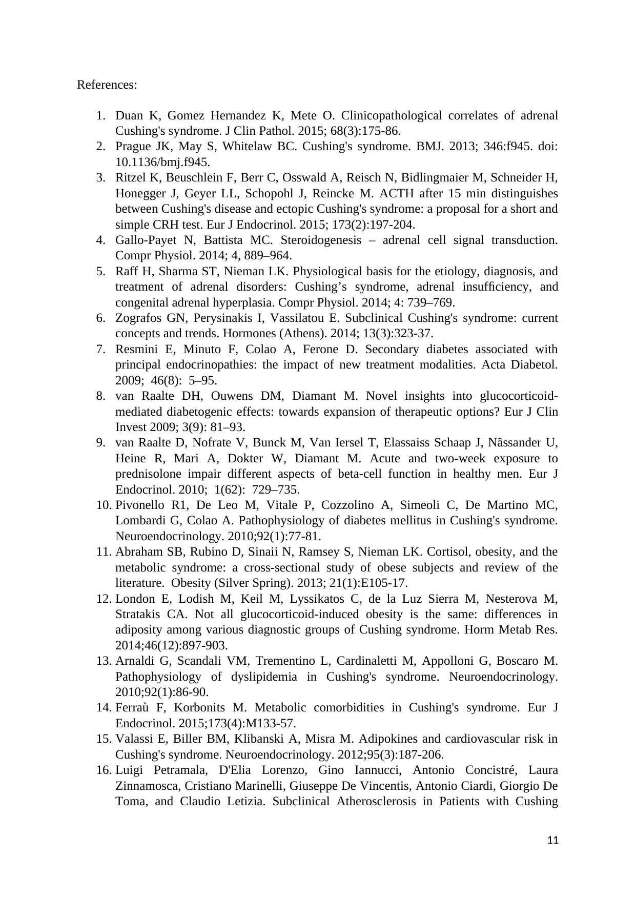
References:
1. Duan K, Gomez Hernandez K, Mete O. Clinicopathological correlates of adrenal
Cushing's syndrome. J Clin Pathol. 2015; 68(3):175-86.
2. Prague JK, May S, Whitelaw BC. Cushing's syndrome. BMJ. 2013; 346:f945. doi:
10.1136/bmj.f945.
3. Ritzel K, Beuschlein F, Berr C, Osswald A, Reisch N, Bidlingmaier M, Schneider H,
Honegger J, Geyer LL, Schopohl J, Reincke M. ACTH after 15 min distinguishes
between Cushing's disease and ectopic Cushing's syndrome: a proposal for a short and
simple CRH test. Eur J Endocrinol. 2015; 173(2):197-204.
4. Gallo-Payet N, Battista MC. Steroidogenesis – adrenal cell signal transduction.
Compr Physiol. 2014; 4, 889–964.
5. Raff H, Sharma ST, Nieman LK. Physiological basis for the etiology, diagnosis, and
treatment of adrenal disorders: Cushing’s syndrome, adrenal insufficiency, and
congenital adrenal hyperplasia. Compr Physiol. 2014; 4: 739–769.
6. Zografos GN, Perysinakis I, Vassilatou E. Subclinical Cushing's syndrome: current
concepts and trends. Hormones (Athens). 2014; 13(3):323-37.
7. Resmini E, Minuto F, Colao A, Ferone D. Secondary diabetes associated with
principal endocrinopathies: the impact of new treatment modalities. Acta Diabetol.
2009; 46(8): 5–95.
8. van Raalte DH, Ouwens DM, Diamant M. Novel insights into glucocorticoid-
mediated diabetogenic effects: towards expansion of therapeutic options? Eur J Clin
Invest 2009; 3(9): 81–93.
9. van Raalte D, Nofrate V, Bunck M, Van Iersel T, Elassaiss Schaap J, Nãssander U,
Heine R, Mari A, Dokter W, Diamant M. Acute and two-week exposure to
prednisolone impair different aspects of beta-cell function in healthy men. Eur J
Endocrinol. 2010; 1(62): 729–735.
10. Pivonello R1, De Leo M, Vitale P, Cozzolino A, Simeoli C, De Martino MC,
Lombardi G, Colao A. Pathophysiology of diabetes mellitus in Cushing's syndrome.
Neuroendocrinology. 2010;92(1):77-81.
11. Abraham SB, Rubino D, Sinaii N, Ramsey S, Nieman LK. Cortisol, obesity, and the
metabolic syndrome: a cross-sectional study of obese subjects and review of the
literature. Obesity (Silver Spring). 2013; 21(1):E105-17.
12. London E, Lodish M, Keil M, Lyssikatos C, de la Luz Sierra M, Nesterova M,
Stratakis CA. Not all glucocorticoid-induced obesity is the same: differences in
adiposity among various diagnostic groups of Cushing syndrome. Horm Metab Res.
2014;46(12):897-903.
13. Arnaldi G, Scandali VM, Trementino L, Cardinaletti M, Appolloni G, Boscaro M.
Pathophysiology of dyslipidemia in Cushing's syndrome. Neuroendocrinology.
2010;92(1):86-90.
14. Ferraù F, Korbonits M. Metabolic comorbidities in Cushing's syndrome. Eur J
Endocrinol. 2015;173(4):M133-57.
15. Valassi E, Biller BM, Klibanski A, Misra M. Adipokines and cardiovascular risk in
Cushing's syndrome. Neuroendocrinology. 2012;95(3):187-206.
16. Luigi Petramala, D'Elia Lorenzo, Gino Iannucci, Antonio Concistré, Laura
Zinnamosca, Cristiano Marinelli, Giuseppe De Vincentis, Antonio Ciardi, Giorgio De
Toma, and Claudio Letizia. Subclinical Atherosclerosis in Patients with Cushing
11
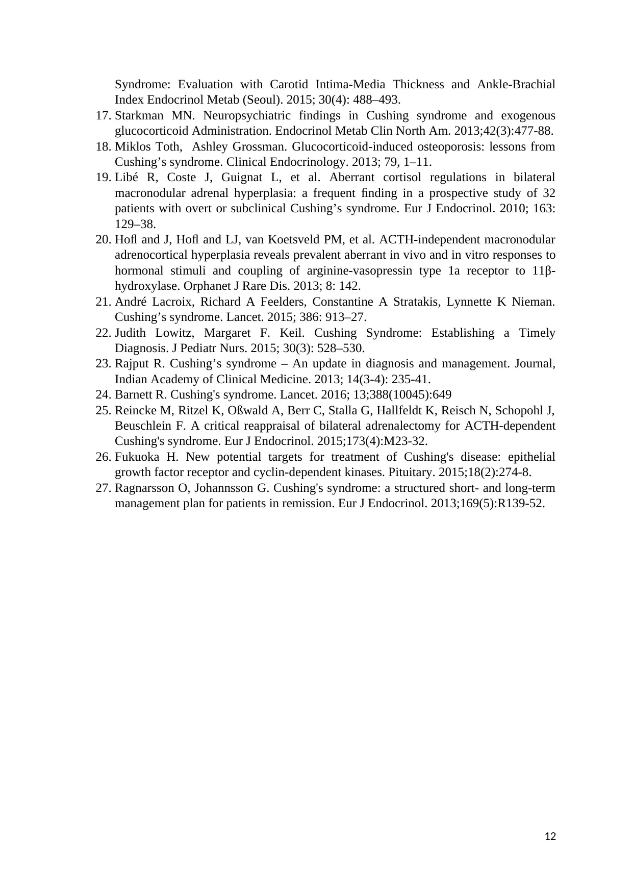
Syndrome: Evaluation with Carotid Intima-Media Thickness and Ankle-Brachial
Index Endocrinol Metab (Seoul). 2015; 30(4): 488–493.
17. Starkman MN. Neuropsychiatric findings in Cushing syndrome and exogenous
glucocorticoid Administration. Endocrinol Metab Clin North Am. 2013;42(3):477-88.
18. Miklos Toth, Ashley Grossman. Glucocorticoid-induced osteoporosis: lessons from
Cushing’s syndrome. Clinical Endocrinology. 2013; 79, 1–11.
19. Libé R, Coste J, Guignat L, et al. Aberrant cortisol regulations in bilateral
macronodular adrenal hyperplasia: a frequent finding in a prospective study of 32
patients with overt or subclinical Cushing’s syndrome. Eur J Endocrinol. 2010; 163:
129–38.
20. Hofl and J, Hofl and LJ, van Koetsveld PM, et al. ACTH-independent macronodular
adrenocortical hyperplasia reveals prevalent aberrant in vivo and in vitro responses to
hormonal stimuli and coupling of arginine-vasopressin type 1a receptor to 11β-
hydroxylase. Orphanet J Rare Dis. 2013; 8: 142.
21. André Lacroix, Richard A Feelders, Constantine A Stratakis, Lynnette K Nieman.
Cushing’s syndrome. Lancet. 2015; 386: 913–27.
22. Judith Lowitz, Margaret F. Keil. Cushing Syndrome: Establishing a Timely
Diagnosis. J Pediatr Nurs. 2015; 30(3): 528–530.
23. Rajput R. Cushing’s syndrome – An update in diagnosis and management. Journal,
Indian Academy of Clinical Medicine. 2013; 14(3-4): 235-41.
24. Barnett R. Cushing's syndrome. Lancet. 2016; 13;388(10045):649
25. Reincke M, Ritzel K, Oßwald A, Berr C, Stalla G, Hallfeldt K, Reisch N, Schopohl J,
Beuschlein F. A critical reappraisal of bilateral adrenalectomy for ACTH-dependent
Cushing's syndrome. Eur J Endocrinol. 2015;173(4):M23-32.
26. Fukuoka H. New potential targets for treatment of Cushing's disease: epithelial
growth factor receptor and cyclin-dependent kinases. Pituitary. 2015;18(2):274-8.
27. Ragnarsson O, Johannsson G. Cushing's syndrome: a structured short- and long-term
management plan for patients in remission. Eur J Endocrinol. 2013;169(5):R139-52.
12
⊘ This is a preview!⊘
Do you want full access?
Subscribe today to unlock all pages.

Trusted by 1+ million students worldwide
1 out of 12
Related Documents
Your All-in-One AI-Powered Toolkit for Academic Success.
+13062052269
info@desklib.com
Available 24*7 on WhatsApp / Email
![[object Object]](/_next/static/media/star-bottom.7253800d.svg)
Unlock your academic potential
Copyright © 2020–2026 A2Z Services. All Rights Reserved. Developed and managed by ZUCOL.


