Deep Learning Approach for Lung Cancer Detection and Analysis
VerifiedAdded on 2023/06/14
|18
|5075
|459
Report
AI Summary
This report explores the application of deep learning techniques, particularly YOLOv5, for the detection of lung cancer nodules in CT scans. The study addresses the growing prevalence of lung cancer and the importance of early diagnosis for improved patient survival rates. It reviews existing literature on cancer detection through deep learning, highlighting the limitations of traditional CAD systems and the advantages of CNNs. The methodology focuses on using YOLOv5 for lung nodule detection, leveraging publicly available datasets for testing and validation. The results indicate a high level of accuracy (93%) for unseen data, suggesting that the proposed model is more effective than other deep learning models for lung nodule detection. The research aims to assist radiologists in making quicker, more reliable decisions, detect lesions at an early stage, and improve the accuracy and sensitivity of monitoring capabilities. Desklib offers this solved assignment and many other study resources to aid students in their academic pursuits.
Lung Cancer Detection By Using Deep Learning
1
1
Paraphrase This Document
Need a fresh take? Get an instant paraphrase of this document with our AI Paraphraser
Table of Contents
3. Abstract
4. Introduction
5.Introduction
6.Introduction
7.Problem Statement
8. Contributions
9.Literature Review
13. Methodology
14. Lung Nodule Detection by Using Yolov5
15. Conclusions
16. References
2
3. Abstract
4. Introduction
5.Introduction
6.Introduction
7.Problem Statement
8. Contributions
9.Literature Review
13. Methodology
14. Lung Nodule Detection by Using Yolov5
15. Conclusions
16. References
2
Lung Cancer Detection by using Deep learning
Abstract:
Biggest challenge within lung disease within recent time has been found to be growing
quickly among people, where America faces additional increased 25,000 incidents in 2016.
China has a rising ratio with almost 2.5 million new death with growing lung cancer related
healthcare diseases, The events are found to be similar in final phases for health disease lung
cancer where it may be removed. The lung cancer is first diagnosed early and then treated
adequately. The specific deep learning technique for improving lung cancer has been
implemented within recent project, for locating the lung nodule in order to increase likelihood of
a patient survival. It can be also analyzed that there are various new techniques viably increasing,
where the Yolov5 technique is used within advanced neural network. The data set was publicly
accessible within testing upon on Kaggle website within specific aspects. It enables to gain
specific working vision towards maintaining specific working efficacy constantly. Accuracy was
completely sufficient to for the unseen data, as it further enabled to prioritize keen findings with
best working vision for gaining optimum rise diversely. Within recent findings , accuracy is
about 93% for the unseen data, where conclusion about proposed model has been found to be
accurate than other deep learning models regarding lung nodules detection.
3
⊘ This is a preview!⊘
Do you want full access?
Subscribe today to unlock all pages.
Trusted by 1+ million students worldwide
Introduction:
As per the concept of Machine learning, a series approaches which automatically make detection
of pattern of data with the use of unknown pattern in order to identify the future data or make
decision in respect to unknown circumstances. ML can be termed as a subset of Artificial
intelligence. There are usually three method of AI i.e. connectivity which is connection and
network based, symbolically which include regulatory like IBM Cloud and within probabilistic
reasoning. Most representative trait of ML is that it is guided by the varied numbers and here
person takes specific low action in the instant decision-making. The learning of algorithm is
made by reviewing specific training data, it will be forecasted with reference to introduction of
new data. In the multi layered cognition method, the major component would be counted as deep
learning and the specific form of neutral network. With respect to deep learning coverage of
large health results can be made.
The ANN was launched during 1950, where implementation of ANN for resolving real
specific dilemmas has been found to be severely limited by disappearing gradient and over-
connection issues. Within recent new aspects of functional research, there are varied new
findings being found to be emerged on where this further adds to machine learning based
specific parameters. Recent working research will determine in depth focus towards digital
4
Paraphrase This Document
Need a fresh take? Get an instant paraphrase of this document with our AI Paraphraser
network based resources, based on competent specific working vision for larger competent
pathways dynamically rising functional competencies.
Majority of limitation has already been overcome which provide the digital network in
association with large data, improvement of computer capacity with GPU and the modern
algorithm with respect to the formation of the deep learning model. The concerned model of
profound learning has shown a high human imitation performance in varied areas like the
medical imaging. As per the standard function of radiology the classification and detection of
anatomical anomalies in the diseases group can be made. Since the 1980, various ML algorithms
have been executed for classification task with various application of logical theories and math.
As per these various CAD programmers were made in the early 2000s and integrated into
clinical workflow. Also, during the clinical trials various harmful effect have been identified. It
is also found that CAD system make generation of more false positive results in comparison of
human reader which results in extra biopsies and more evaluation time (2). The main advantage
in reference to the use of CAD is still uncertain (3). But with the presence of deep learning
technologies would lead to help in the removing of shortcoming of CAD programmers along
with the achievement of great precision in detection. It also allows human readers to make
transformation of humdrum, repeated activities of radiology to AI in more effective mode.
Valuable data from medical images can be derived from the deep learning. With this latest
platform the proposing of varied diagnostic, detection of lesions and composition of preliminary
analysis can be made. In real the international company i.e. IBM is designing the radiology
application of Dr Watson.
All the features including automated identification and quantitative lesion analysis of the
medical imaging has been found to be specifically covered under this method. With an
exponentiation increase in the AI technologies, radiologist enable to provide know how of
technology along with order to consider varied new potential and effect within near future. It is
also assumed that the MOL based analytical instruments will soon be adopted in the field of
radiology. It is also assumed that although it will not substitute the radiologist but some basic
human function will be substituted. The concerned replacement is not really a last substitute but
5
it will make an overall increase in the whole practice associated with radiology as they make
supplement of extraordinary and irreplaceable human abilities. With aspect of
convolutional neural network (CNN), the computer vision and the machine learning field would
be improved significantly which would comprise of several layers of the neuron like neuronal
relationship with the step by step minimum processing.
The overall CNN is new learning method, as per stimulation found in learning method
stimulates organization within animal cerebral cortex which successfully qualifies CNN. It can
be also analyzed that developing proper competent rise on hierarchy knowledge during per-
processing, e.g. such as image recognition edge-shaped component-object layout are some of the
most crucial aspects. Also, CNN learning method has been found to be highly innovative for
strengthening learning based stimulation, working towards effective keen advancement which
has been also found to be critically essential.
The design of CNN consists of wholly linked layers and the convolutional layers as in fig 1.
Identification, distinguishing lines, local motif-like borders and interactive images is counted as
one of the main aim of the convolutional layer. Parameters which are known as convolutions for
progressive filter operator are learned. Array of acquired parameters which is also known as
kernel which multiply local neighbor of given pixel, is defined with the procedure of this
mathematical model. Retrieval of sensory objects including the borders and colors, which are
close to one that are noted for visual system with the study of meaningful kernels can be imitated
by this operation. The accomplishment of this method can be made with filter bank. Every
filtered operator is a square object which can be passed through specified image. The picture
value of the travelling grid can be summed up with the use of filter weights. The
convolutional layer applies various filters along with the production of various characteristic
charts. The central component of CNN is Convolutions. It is essential for performance in terms
of distributing the images, which also include the classification and segmentation.
6
⊘ This is a preview!⊘
Do you want full access?
Subscribe today to unlock all pages.
Trusted by 1+ million students worldwide
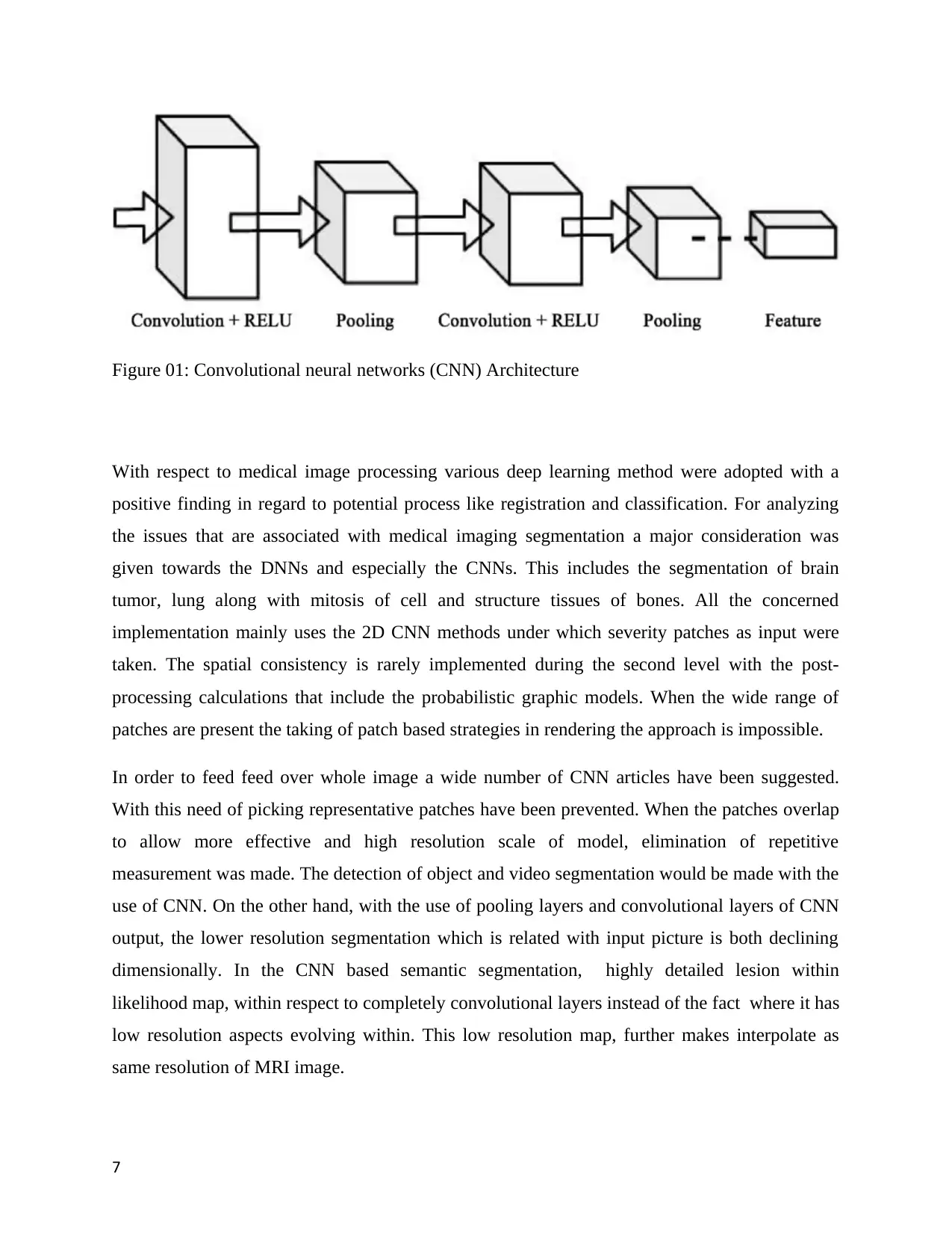
Figure 01: Convolutional neural networks (CNN) Architecture
With respect to medical image processing various deep learning method were adopted with a
positive finding in regard to potential process like registration and classification. For analyzing
the issues that are associated with medical imaging segmentation a major consideration was
given towards the DNNs and especially the CNNs. This includes the segmentation of brain
tumor, lung along with mitosis of cell and structure tissues of bones. All the concerned
implementation mainly uses the 2D CNN methods under which severity patches as input were
taken. The spatial consistency is rarely implemented during the second level with the post-
processing calculations that include the probabilistic graphic models. When the wide range of
patches are present the taking of patch based strategies in rendering the approach is impossible.
In order to feed feed over whole image a wide number of CNN articles have been suggested.
With this need of picking representative patches have been prevented. When the patches overlap
to allow more effective and high resolution scale of model, elimination of repetitive
measurement was made. The detection of object and video segmentation would be made with the
use of CNN. On the other hand, with the use of pooling layers and convolutional layers of CNN
output, the lower resolution segmentation which is related with input picture is both declining
dimensionally. In the CNN based semantic segmentation, highly detailed lesion within
likelihood map, within respect to completely convolutional layers instead of the fact where it has
low resolution aspects evolving within. This low resolution map, further makes interpolate as
same resolution of MRI image.
7
Paraphrase This Document
Need a fresh take? Get an instant paraphrase of this document with our AI Paraphraser
This project will target new deep learning approaches which are used for detecting lung cancer
nodules within recent CT Scan. The learning model, within recent aspect will be used in recent
project, where YOLOV5 is also known as single shot detector.
Before making a detailed discussion about YOLOV5, let’s have some literature regarding the
various type of cancer detection through deep learning.
Problem Statement:
The cause for development of lung cancer is yet not clear, prevention of lung diseases remains a
test. Thus, it would be said that the potential for full recovery would be made if in early stage a
performance of proficient diagnostic of lung cancer would be performed. If the lung cancer can
be detect at the early stage then it will help in reducing the hardness and mortality rate. In this
area innovation has been made in term of use of diagnostic instrument and mammography for the
identification of lung anomalies and lesions. Due to this only 10-30% of aggressive cancer have
been misclassified which is related with other reasons and factors. For example, deviation from
standard which did not specify, special problem associated with imaging and wrong reading of
abnormalities. With the process of mammography screening, a lot of cases of cancer have been
misclassified. Due to this, there is a high need to make development of deep learning based
system which use propelled image research method so that distinguishment of deviation from
standard in lung cancer can be improved. The situation is unique and common as a starting step
to support the radiologist in making decision and discern normal deviation.
Contributions:
The aim of this research is to develop a digital imaging CAD diagnostic for lung cancer,
classification method and assessment of function concept. Wirth this process the detection of
lung cancer can be made. The objectives are as follows:
To assist the radiologist and physicians to make quick, reliable and efficient decision.
8
Detection of lesions at preliminary phase which will lead to high rate of survival.
Improvement in accuracy and sensitivity monitoring capabilities.
Literature Review:
During the current time physicians and radiologist make testing through X-rays and CXR. Also
the clinics in underdeveloped areas have a small number of radiologist which are skilled. For
identification of unexplained lung nodules CADe device is used. While in case of developing
nation, in a limited time, X-ray picture must be checked during routine health inspection. As per
the research of one radiologist review only 68% of lung cancer node has been correctly identified
while within recent two radiologists have been adequately diagnosed with specific 82%.
Diagnosis of harmful pulmonary nots at early stage must be diagnose by radiologist. Although it
is tiring, vital and repetitive. Making a deep analysis of certain sample took a lot of time by
radiologist which may often lead to many mistakes with detection of tiny nodules. During this
scenario, doctors seem to get bored because of varied unconscious nodules [1]. The life
expectancy in lung cancer is not short if it will be detected in early diagnostics and unresectable.
Because of the unclear tumor characteristics, the treatment made in association with computed
tomography images is controversial.
Thus, it can be said that in order to accomplish descriptive cancer distinction result, a
standard computer aided diagnostic system (CAD) which contain recognition of pattern
measures and multiple picture analysis. Every step depends heavily on the success with regard to
the previous stage in the concerned ad-hoc image processing-pipeline. In the standard CAD
scheme the designation efficiency tuning is highly challenging and difficult. The benefit in
relation with output tuning and smooth autonomous operations can be obtained from deep
learning technologies. This research will make the simplification of profound learning technique
and traditional CAD image analytics pipeline. In the same way, the nodule classification of
computed tomography videos, conventional neutral network and the model of deep belief
network has been presented. For making contrast, the two baseline approaches of the function
computing measures have been introduced. As per the studies more discriminatory outcome are
9
⊘ This is a preview!⊘
Do you want full access?
Subscribe today to unlock all pages.
Trusted by 1+ million students worldwide
produced by deep learning approaches along with promising in the CAD domain program [2].
Cancer is counted as one of the most leading cause which lead to death. Checking in relation
with lung Cancer is difficult. With regard to this approach, a breath test in relation with the
diagnosis of the lung Cancer was developed under which a chemical sensor array is used along
with a machine learning technique. A prospective analysis in relation to lung cancer was
performed by author between 2016 and 2018 for recording instances in relation with lung Cancer
and non-tumor control. This was performed with the analysis of alveolar air sample which use
the carbon nanotube sensor arrays. During this there have been 199 monitors and 117 reports
with a removal of 72 individuals because of the reason that Cancer is located in certain location
or minimally intrusive adenocarcinoma, in situ carcinoma chemotherapy or other illnesses.
During 2016 and 2017, subject for internal validation and model derivation were used. In 2018
this model was make publically tested. With the reference of pathological studies, the evaluation
of diagnostic precision was made [3]. With the highly efficient diagnostic of lung nodule lead
majorly to the risk estimation of lung Cancer. It is an essential as well as difficult job of finding
the precise nodule of lung. An extensive study in this field has been made by the various
researchers for two decades. As building of whole CAD device require more image analysis
module, under which the previous computerized detection system (CADe) is often found time
consuming and difficult. For example, lung nodule segmentation, functionality extraction and
computed tomography image transformation. With a rise in medical photos this scheme finds it
challenging to manage and make interpretation of huge data.
Likewise, state-of-the-art deep specific learning schemes are counted as rigid aspect
within the database standard. Under this recent research the author provides a successful method,
for analysingidentification of lung nodule which is depended within multi group patches which
are cut from pulmonary photos and improved with Frangi sensor.
With significant merging of two classes of picture, there is four channel neutral
networking paradigm innovativly developed which acquire the knowledge of radiologist for
detecting the four nodules. 80% sensitivity is gained from this CADe system. Finding also within
recent aspect showed that multi group patch has based working within learning method make
improvement of accuracy of identification.
10
Paraphrase This Document
Need a fresh take? Get an instant paraphrase of this document with our AI Paraphraser
Lung nodules along with significantly reduced false positive aspect, under large volume of
picture images, where an early diagnosis of lung cancer helps in reducing specific mortality rate
of lung cancer which is accounted for 17% of the cancer related death. With the initial evaluation
by radiologist a vast number of cases found on every day. Here the CAD system help the
radiologist in terms of providing second view along with speeding up the entire operation.
Therefore, it is suggested that CAD method which use profound features which are derived from
automative encoder to make identification of lung nodules as malignant or benign. 4303 cases
which comprises LIDC dataset with a total accuracy of 75.01% are used by authors [5]. Early
treatment of cancer is the only method by which the survival of patient.
A computer-aided method of classification in CT scan for analyzing lung images by the use of
artificial neutral network was introduced by the author. The division of whole lung into CT
pictures and the parameter of the segmented picture is identified. Here the classification is made
on the basis of the statistical criteria that include skewedness, average, standard deviation, sixth
central moment, turtles and fifth central moment. The carrying of classification is made through
forwards and backward feeding of neutral network. Greater characterization is provided by
feedback back propagation network while comparing with feedback networks. Highest
classification precision was provided by the skewedness of the parameter. The Traingdx consist
max accuracy rate i.e. 91.1%, where the 13 backward propagation neural network function which
is further found to be unstable.s
A number of identification technique for the CT scan images have been tested. On the other
hand, no identification method has been performed in case of MR images. For thoracic MR
image, a detection approach which focus on neutral network is proposed. Fast-moving R-
convolution neural net (CNN) is programme so that the area of lung nodule can be located with
optimizing parameter, learning transfer and spatial 3 different channel construction.
FP Mitigation scheme planning has been done to minimize FPS, retaining real nodules
depends on anatomic characteristics. Procedure is further also checked on 142 MR scan of
Guangzhou Medical University, which is known to be the first affiliated hospital. Efficiency of
proposed approach is 3.47 Fps per scan which has been found to be 85.2 percent specifically,
where experimental findings show specific working planned rapid R-CNN network and FP
reduction hold successful detached lunch nodule, which reduces FP. The lung cancer is most
11
serious threatening life disease where early interventions and therapies have been evolved in
recent time. Though CT scan imagery has been found to be the strongest medical technology,
where doctors have been found to have challenging for reading and distinguishing CT scan
cancer photographs. Also, computer aided diagnosis has been found to be useful for physicians
for correctly classifying the cancer cells, where image recognition and specific machine learning
computer aided strategies which are further been found to be researched.
Key objective of study is to analyze varied computer aided methods and analyze best
approaches for detecting limits to propose implementation of the latest approach for developing
available best model. Model based specific advaned CAD algorithm has been built by detailed
specific mathematical models, which collects the specific scanner details in physical and
anatomical format. Recent research specifies about working aspects, where it further utilizes
multi-segmented algorithms within removing remarkable lung structures and calculates
likelihood within varied anatomical incidents around pulmonary system in body. It has been
validated on 50 low dose CT screening cases within lung cancer, where the ground was founded
by three specialist chest radiologists. The analysis of model based CAD algorithm within 50 CT
situations, in which two radiologists have the potential sensitive enhancements within both
nodules, solid and solid, exceeding 5 mm in diameter. It has been found to be calculated of 7
and 5 percent. There were no nodules, which are missed by third radiologist in working ground
reality, where 8.3 false positives were generated in CAD algorithm.
Lung disease being world main causes of death from cancer, has been found to have
effective for mitigating death and CAD has evolved quickly as vital way. It can be also analyzed
that lung cancer disease, has high level impact on healthcare structure among people based on
specific functional efficacy parametrs. Automated identification of pulmonary nodules in ct scan
(CT) image has been found to be a particular critical for CAD method. It is difficult challenge,
found to be within precise locations of the lung nodule easily. Automated 2D constitutional
neural network pulmonary, nodule detection system is proposed for supporting the specific
mechanism within CT readings. Modification of configuration in faster R-CNN with two
proposals network, within deconvolutionary layer is developed, for detecting nodule candidates
and three templates for three kinds of subsequent outcome fusion slices. Boosting architecture
built on 2D CNN is optimized for false positive elimination, where this is comparatively
12
⊘ This is a preview!⊘
Do you want full access?
Subscribe today to unlock all pages.
Trusted by 1+ million students worldwide
1 out of 18
Related Documents


Your All-in-One AI-Powered Toolkit for Academic Success.
+13062052269
info@desklib.com
Available 24*7 on WhatsApp / Email
![[object Object]](/_next/static/media/star-bottom.7253800d.svg)
Unlock your academic potential
Copyright © 2020–2026 A2Z Services. All Rights Reserved. Developed and managed by ZUCOL.