Comprehensive Report: Dental Anatomy and Oral Health Assessment

Verified

Added on  2023/01/19

|20
|3385
|60
Report
AI Summary
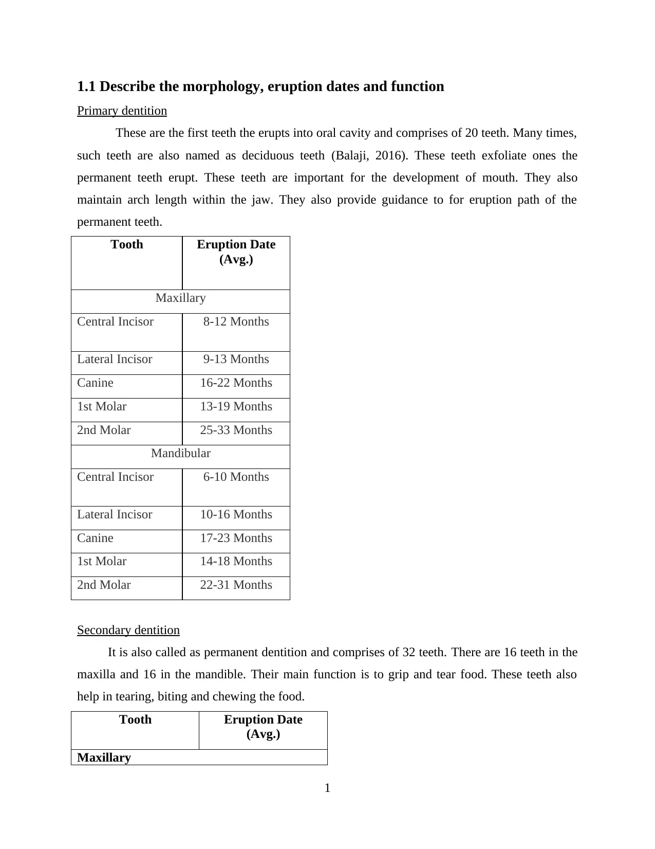
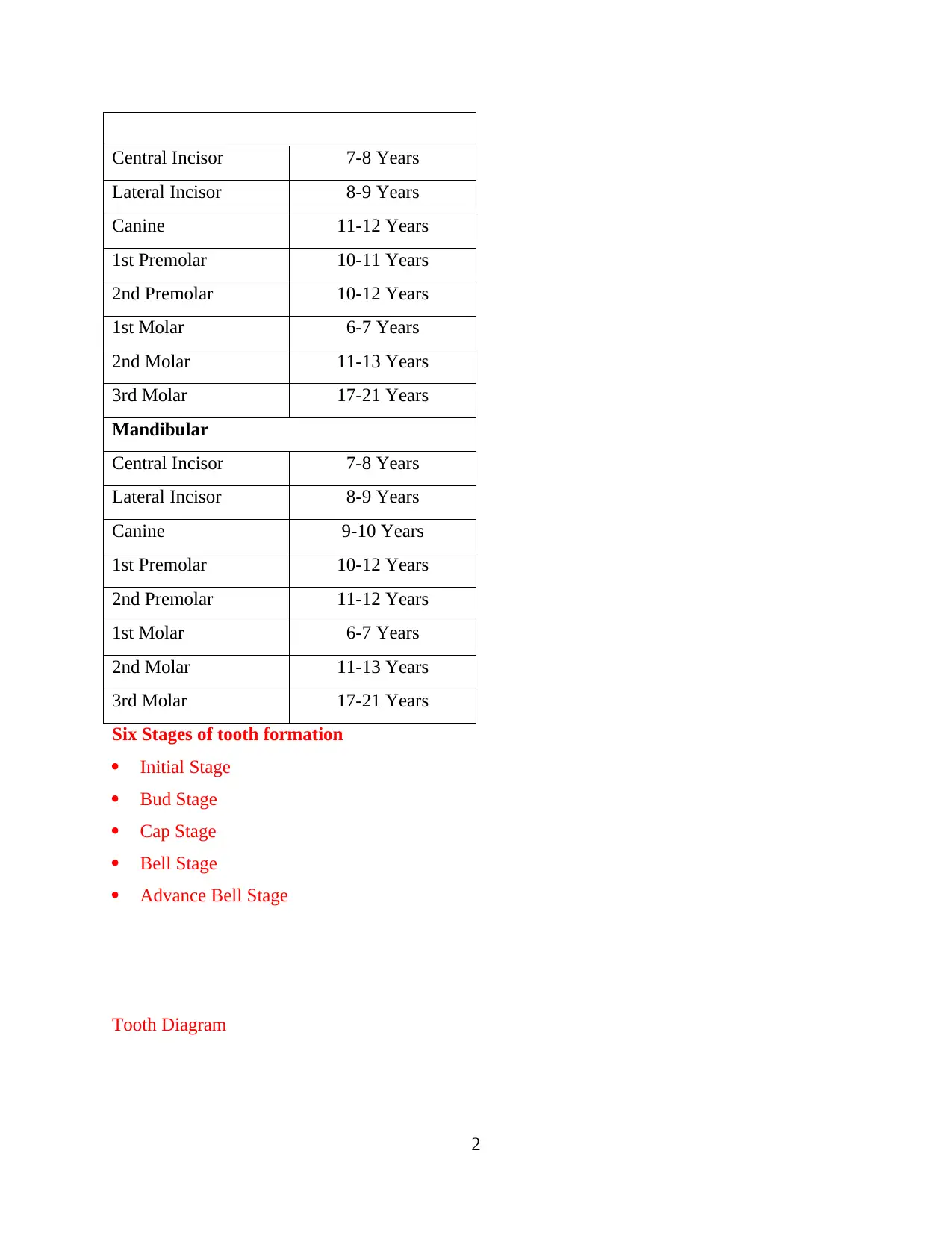
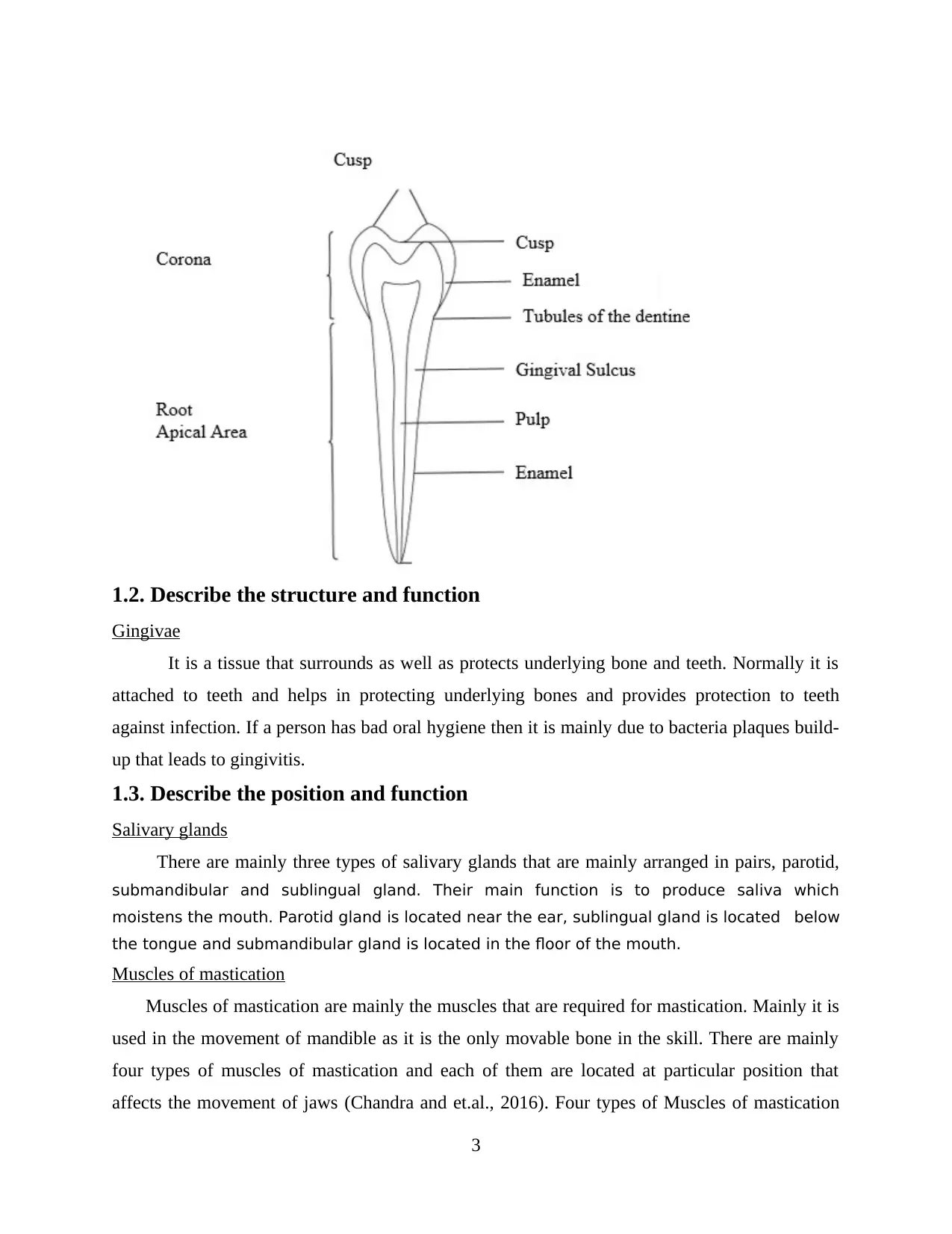
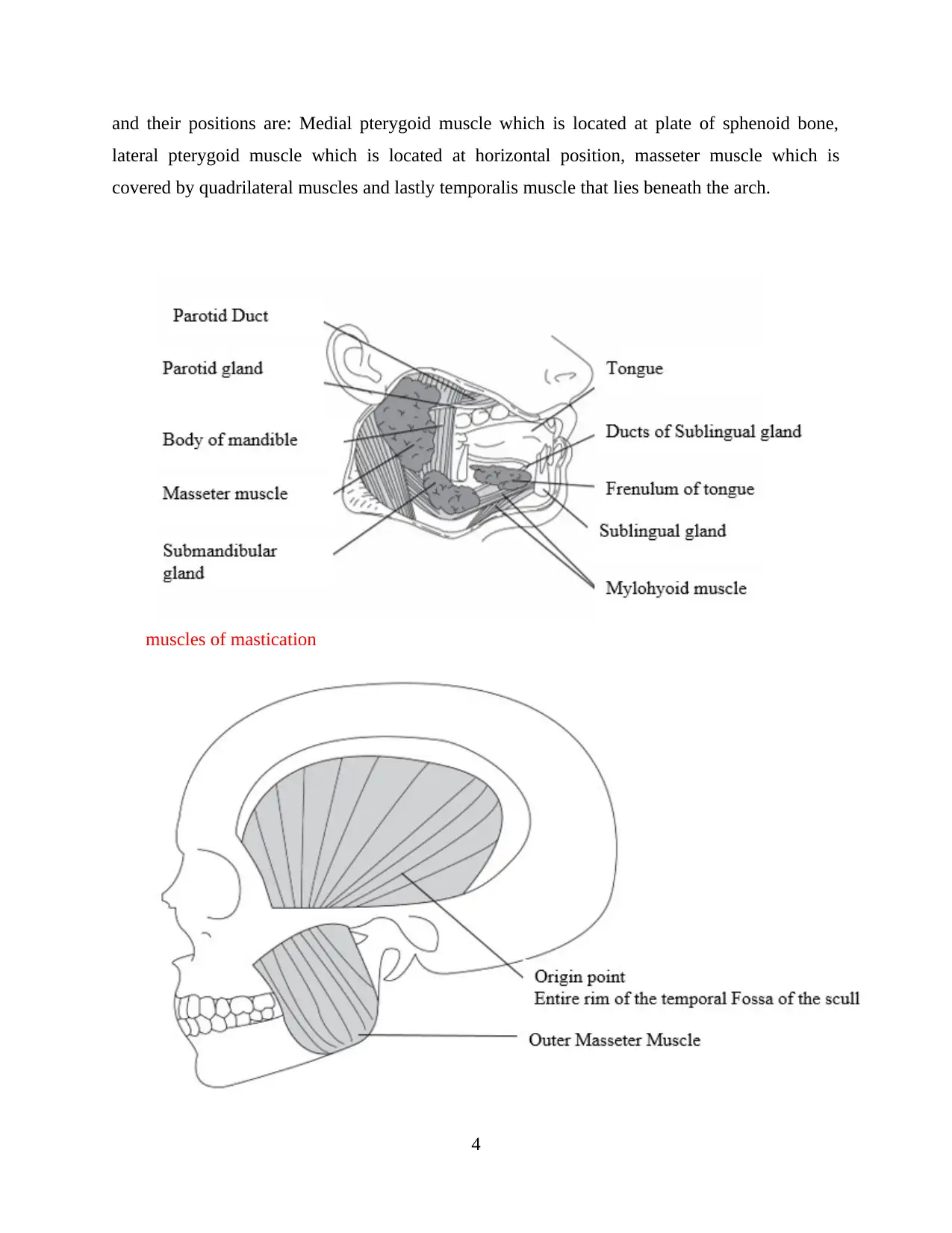
This report provides a comprehensive overview of dental anatomy and oral health assessment. It begins by describing the morphology, eruption dates, and functions of primary and secondary dentition. The report then details the structure and function of gingivae, salivary glands, and muscles of mastication, along with the structure of the maxilla and mandible. It explains the movements of the temporo-mandibular joint and the nerve and blood supply to teeth and supporting structures. The assessment section covers the main purpose of oral health assessment, the reasons for radiographs and photographs, methods for assessing soft and hard tissues, periodontal conditions, and pulp vitality measurement. It also includes materials used in dental assessment and the importance of informed consent. The report further explores malocclusion classifications, orthodontic appliances, and pre- and post-operative instructions. It describes the role of a dental nurse in orthodontics, diseases of the oral mucosa, and the effects of aging on soft tissue. Finally, it addresses medical conditions affecting oral tissues, diagnosis, prevention, and management of oral lesions, and the role of drugs in dentistry, including medical emergencies.
Document Page
DENTAL ANATOMY AND
ASSESSMENT OF ORAL
HEALTH
tabler-icon-diamond-filled.svg

Paraphrase This Document

Need a fresh take? Get an instant paraphrase of this document with our AI Paraphraser
Document Page
TABLE OF CONTENTS
Table of Contents.............................................................................................................................2
1.1 Describe the morphology, eruption dates and function.............................................................1
Primary dentition.........................................................................................................................1
Secondary dentition.....................................................................................................................1
1.2. Describe the structure and function..........................................................................................2
Gingivae.......................................................................................................................................2
1.3. Describe the position and function...........................................................................................2
Salivary glands.............................................................................................................................2
Muscles of mastication................................................................................................................3
1.4. Describe the structure...............................................................................................................3
Maxilla.........................................................................................................................................3
Mandible......................................................................................................................................3
1.5. Describe the movements of the temporo-mandibular joint......................................................3
1.6. Describe the nerve and blood supply to the teeth and supporting structures............................4
2.1. Explain the main purpose of oral health assessment................................................................4
2.2. Explain the reasons for taking radiographs and photographs during assessment and treatment
planning...........................................................................................................................................4
2.3. Describe the methods of assessing and recording soft and hard tissue conditions...................4
2.4. Explain the methods of assessing and recording periodontal conditions using periodontal
charts................................................................................................................................................5
2.5. Evaluate the methods of measuring pulp vitality.....................................................................5
2.6. Describe materials used in dental assessment..........................................................................5
2.7. Explain the relevance of obtaining written informed consent prior to any treatment being
undertaken........................................................................................................................................5
3.1. Describe the classifications of malocclusion............................................................................6
3.2 Types of orthodontic appliances................................................................................................6
3.3. Explain pre and post- operative instructions for orthodontic procedures.................................6
Document Page
3.4. Explain the role of the Dental Nurse in providing support during orthodontic........................6
4.1 Explain diseases of the oral mucosa..........................................................................................6
4.2. Describe the effects of ageing on the soft tissue.......................................................................7
4.3. Identify medical conditions that may affect the oral tissues.....................................................7
5.1. Explain the methods of diagnosis, prevention and management of malignant lesions and
potentially malignant lesions...........................................................................................................7
5.2 diagnosis and management of disorder......................................................................................7
5.3. List the classes of drugs that are used in dentistry....................................................................8
5.4. Explain the role of drugs used in dentistry...............................................................................8
6.1 Identify medical emergencies....................................................................................................8
6.2. Explain action to take in response to medical emergencies.....................................................9
CONCLUSION................................................................................................................................9
REFERENCES..............................................................................................................................10
Document Page
1.1 Describe the morphology, eruption dates and function
Primary dentition
These are the first teeth the erupts into oral cavity and comprises of 20 teeth. Many times,
such teeth are also named as deciduous teeth (Balaji, 2016). These teeth exfoliate ones the
permanent teeth erupt. These teeth are important for the development of mouth. They also
maintain arch length within the jaw. They also provide guidance to for eruption path of the
permanent teeth.
Tooth Eruption Date
(Avg.)
Maxillary
Central Incisor 8-12 Months
Lateral Incisor 9-13 Months
Canine 16-22 Months
1st Molar 13-19 Months
2nd Molar 25-33 Months
Mandibular
Central Incisor 6-10 Months
Lateral Incisor 10-16 Months
Canine 17-23 Months
1st Molar 14-18 Months
2nd Molar 22-31 Months
Secondary dentition
It is also called as permanent dentition and comprises of 32 teeth. There are 16 teeth in the
maxilla and 16 in the mandible. Their main function is to grip and tear food. These teeth also
help in tearing, biting and chewing the food.
Tooth Eruption Date
(Avg.)
Maxillary
1
tabler-icon-diamond-filled.svg

Paraphrase This Document

Need a fresh take? Get an instant paraphrase of this document with our AI Paraphraser
Document Page
Central Incisor 7-8 Years
Lateral Incisor 8-9 Years
Canine 11-12 Years
1st Premolar 10-11 Years
2nd Premolar 10-12 Years
1st Molar 6-7 Years
2nd Molar 11-13 Years
3rd Molar 17-21 Years
Mandibular
Central Incisor 7-8 Years
Lateral Incisor 8-9 Years
Canine 9-10 Years
1st Premolar 10-12 Years
2nd Premolar 11-12 Years
1st Molar 6-7 Years
2nd Molar 11-13 Years
3rd Molar 17-21 Years
Six Stages of tooth formation
Initial Stage
Bud Stage
Cap Stage
Bell Stage
Advance Bell Stage
Tooth Diagram
2
Document Page
1.2. Describe the structure and function
Gingivae
It is a tissue that surrounds as well as protects underlying bone and teeth. Normally it is
attached to teeth and helps in protecting underlying bones and provides protection to teeth
against infection. If a person has bad oral hygiene then it is mainly due to bacteria plaques build-
up that leads to gingivitis.
1.3. Describe the position and function
Salivary glands
There are mainly three types of salivary glands that are mainly arranged in pairs, parotid,
submandibular and sublingual gland. Their main function is to produce saliva which
moistens the mouth. Parotid gland is located near the ear, sublingual gland is located below
the tongue and submandibular gland is located in the floor of the mouth.
Muscles of mastication
Muscles of mastication are mainly the muscles that are required for mastication. Mainly it is
used in the movement of mandible as it is the only movable bone in the skill. There are mainly
four types of muscles of mastication and each of them are located at particular position that
affects the movement of jaws (Chandra and et.al., 2016). Four types of Muscles of mastication
3
Document Page
and their positions are: Medial pterygoid muscle which is located at plate of sphenoid bone,
lateral pterygoid muscle which is located at horizontal position, masseter muscle which is
covered by quadrilateral muscles and lastly temporalis muscle that lies beneath the arch.
muscles of mastication
4
tabler-icon-diamond-filled.svg

Paraphrase This Document

Need a fresh take? Get an instant paraphrase of this document with our AI Paraphraser
Document Page
5
Document Page
6
Parotid duct
Superior pharyngeal
constrictor muscle
Buccinator muscle
Lateral pterygoid
muscle
Document Page
7
tabler-icon-diamond-filled.svg

Paraphrase This Document

Need a fresh take? Get an instant paraphrase of this document with our AI Paraphraser
Document Page
8
Frontal bone
Orbit
Zygomodic bone
Interior nostril
Mandible
Nasal bone
Temporal bone
Bone below eye
Mandibular forman
Mandible
Parietal bone
Zygomodic bone
Occiptal bone
Frontal bone
Greater wing of spheroid bone
Nostril bone
temporal
Arbicular
Masseter muscle
Mental Forman
Mandible law Temporomandibular
joint
Document Page
1.4. Describe the structure
Maxilla
Maxilla
forms
the
upper
fixed
bone of
the jaw
by
9
chevron_up_icon
1 out of 20
circle_padding
hide_on_mobile
zoom_out_icon
[object Object]