Dental Anatomy, Physiology, and Nerve Supply Homework, Biology

Verified

Added on  2021/01/01

|7
|671
|244
Homework Assignment
AI Summary
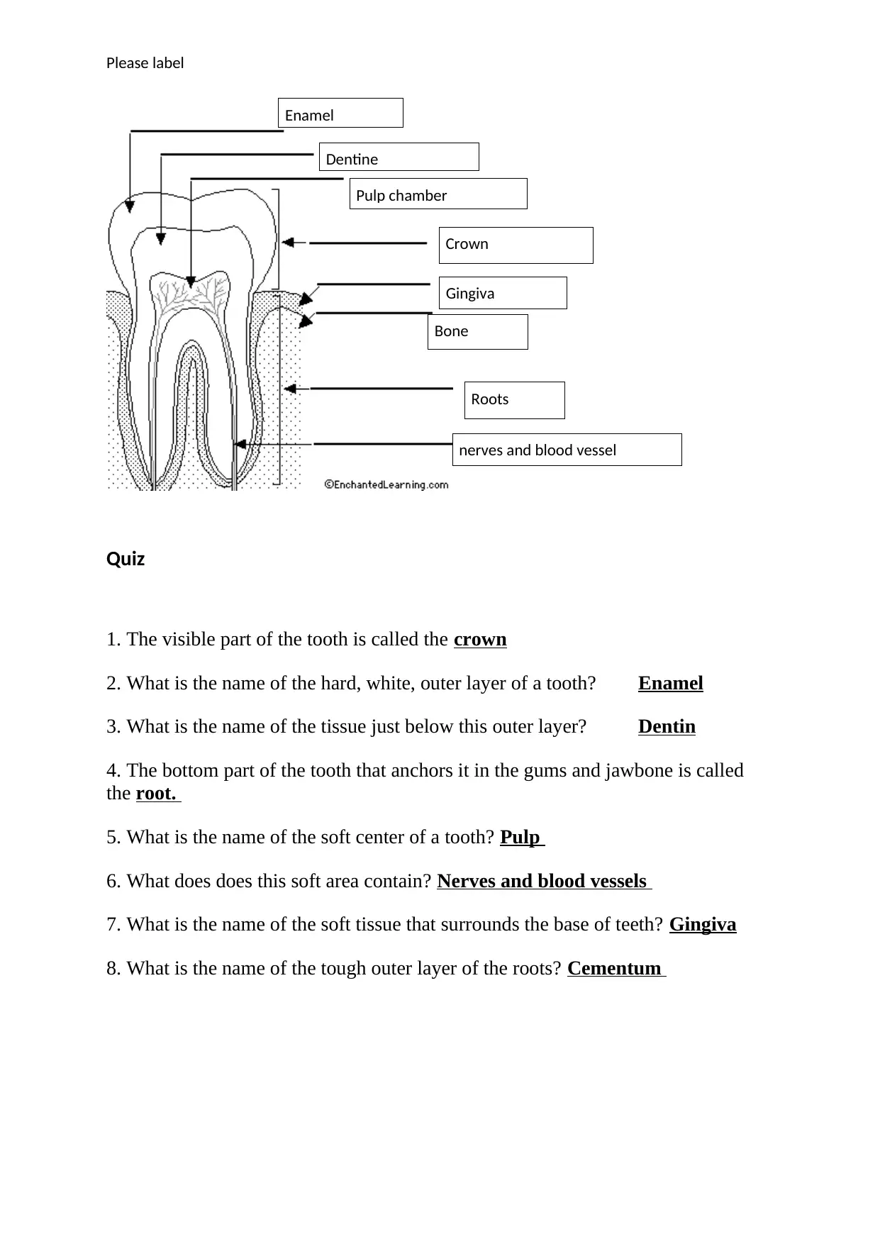
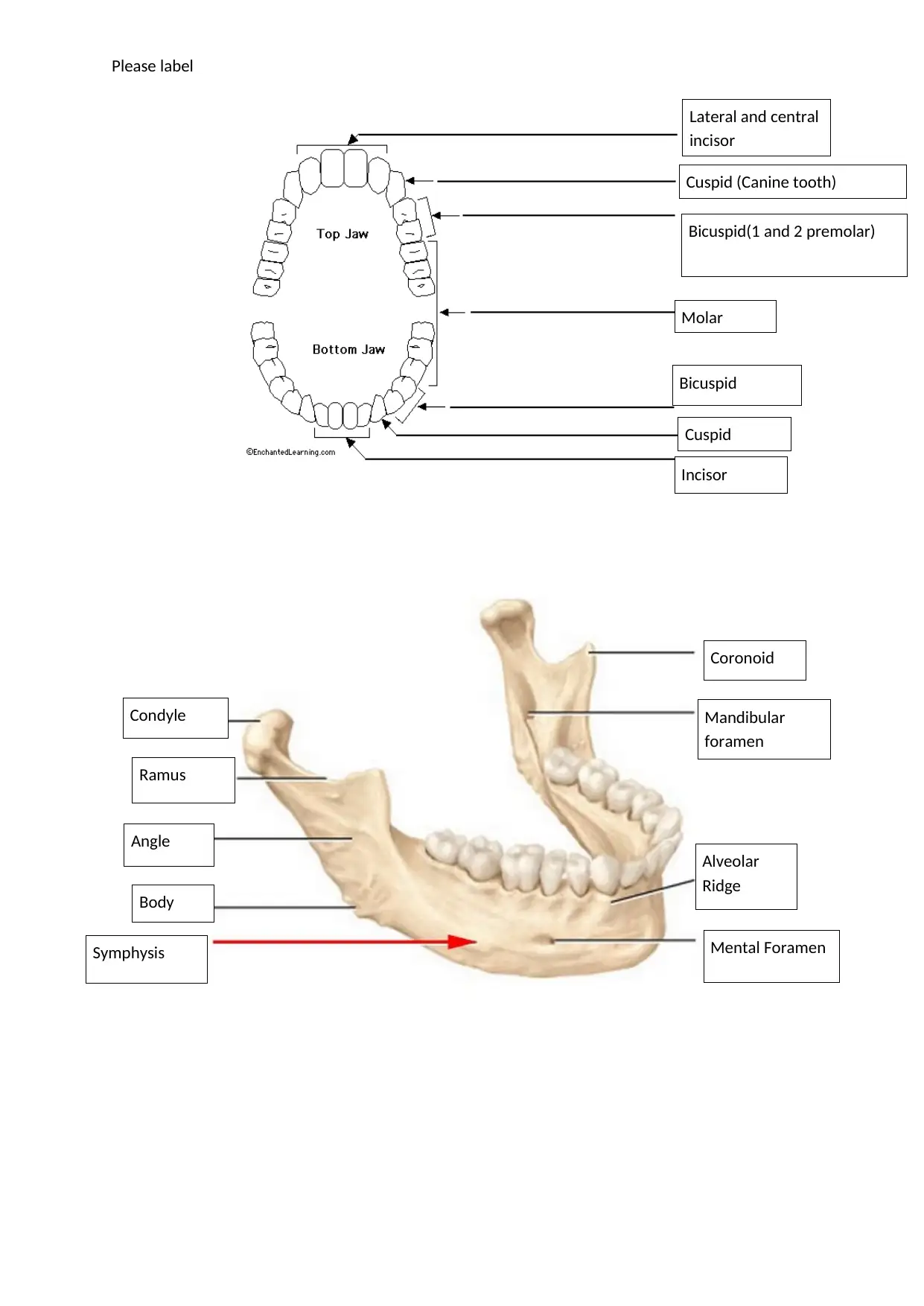
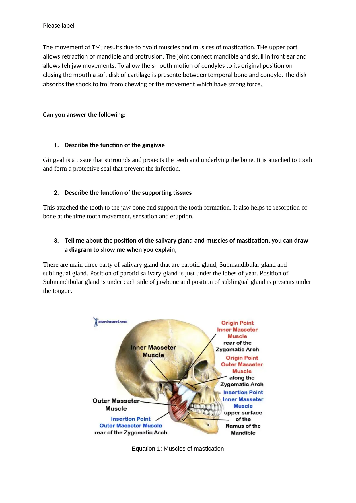
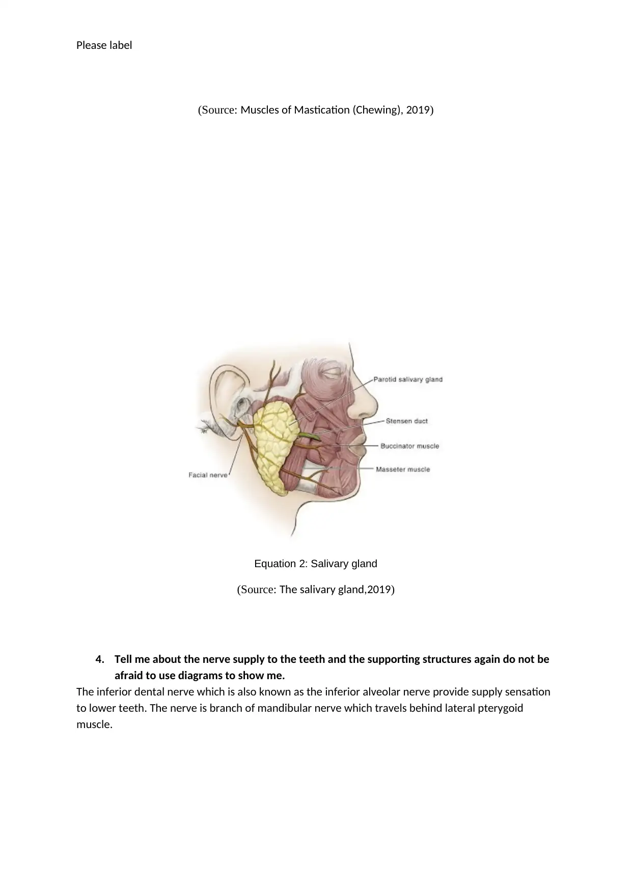
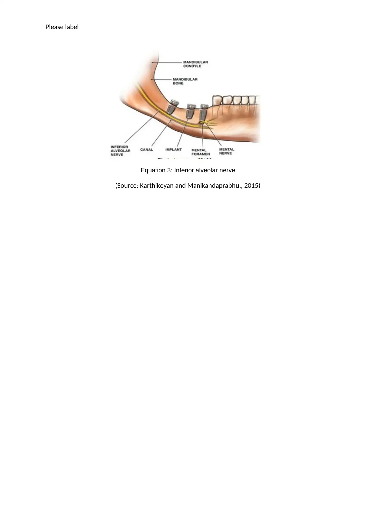
This assignment provides a comprehensive overview of dental anatomy and physiology. It begins with a quiz testing knowledge of tooth structure, including the crown, enamel, dentin, root, pulp, nerves, blood vessels, and gingiva. The assignment then delves into the anatomy of the mandible, including the TMJ (temporomandibular joint) and its movements, the function of the gingivae and supporting tissues. The assignment also explores the position and function of salivary glands and muscles of mastication. Finally, it discusses the nerve supply to the teeth and supporting structures. The assignment includes diagrams and references to support the explanations.
tabler-icon-diamond-filled.svg

Contribute Materials

Your contribution can guide someone’s learning journey. Share your documents today.
Document Page
Please label
Quiz
1. The visible part of the tooth is called the crown
2. What is the name of the hard, white, outer layer of a tooth? Enamel
3. What is the name of the tissue just below this outer layer? Dentin
4. The bottom part of the tooth that anchors it in the gums and jawbone is called
the root.
5. What is the name of the soft center of a tooth? Pulp
6. What does does this soft area contain? Nerves and blood vessels
7. What is the name of the soft tissue that surrounds the base of teeth? Gingiva
8. What is the name of the tough outer layer of the roots? Cementum
Enamel
Dentine
Crown
Roots
Pulp chamber
Gingiva
Bone
nerves and blood vessel
tabler-icon-diamond-filled.svg

Secure Best Marks with AI Grader

Need help grading? Try our AI Grader for instant feedback on your assignments.
Document Page
Please label
Coronoid
Mandibular
foramen
Alveolar
Ridge
Condyle
Ramus
Angle
Mental Foramen
Body
Symphysis
Cuspid (Canine tooth)
Bicuspid(1 and 2 premolar)
Molar
Bicuspid
Cuspid
Incisor
Lateral and central
incisor
Document Page
Please label
Now do the same for me and list the primary dates of eruption
Can you look at this diagram of the TMJ then tell me what you understand about it including the
movements in your own words,
7-8 yrs
8-9 years
11-12 yrs
10-11 yrs
10-12 yrs
6-7 yrs
12-13 yrs
17-21 yrs
17-21 yrs
11-13 yrs
6-7 yrs
11-12 yrs
10-12 yrs
9-10 yrs
7-8 yrs
6-7 yrs
Document Page
Please label
The movement at TMJ results due to hyoid muscles and muslces of mastication. THe upper part
allows retraction of mandible and protrusion. The joint connect mandible and skull in front ear and
allows teh jaw movements. To allow the smooth motion of condyles to its original position on
closing the mouth a soft disk of cartilage is presente between temporal bone and condyle. The disk
absorbs the shock to tmj from chewing or the movement which have strong force.
Can you answer the following:
1. Describe the function of the gingivae
Gingval is a tissue that surrounds and protects the teeth and underlying the bone. It is attached to tooth
and form a protective seal that prevent the infection.
2. Describe the function of the supporting tissues
This attached the tooth to the jaw bone and support the tooth formation. It also helps to resorption of
bone at the time tooth movement, sensation and eruption.
3. Tell me about the position of the salivary gland and muscles of mastication, you can draw
a diagram to show me when you explain,
There are main three party of salivary gland that are parotid gland, Submandibular gland and
sublingual gland. Position of parotid salivary gland is just under the lobes of year. Position of
Submandibular gland is under each side of jawbone and position of sublingual gland is presents under
the tongue.
Equation 1: Muscles of mastication
tabler-icon-diamond-filled.svg

Secure Best Marks with AI Grader

Need help grading? Try our AI Grader for instant feedback on your assignments.
Document Page
Please label
(Source: Muscles of Mastication (Chewing), 2019)
Equation 2: Salivary gland
(Source: The salivary gland,2019)
4. Tell me about the nerve supply to the teeth and the supporting structures again do not be
afraid to use diagrams to show me.
The inferior dental nerve which is also known as the inferior alveolar nerve provide supply sensation
to lower teeth. The nerve is branch of mandibular nerve which travels behind lateral pterygoid
muscle.
Document Page
Please label
Equation 3: Inferior alveolar nerve
(Source: Karthikeyan and Manikandaprabhu., 2015)
Document Page
Please label
REFERENCES
Karthikeyan,. T, and Manikandaprabhu., P., 2015. A novel approach for inferior Alveolar Nerve (IAN)
injury identification using Panormaic radiographic image. [Online]. Accessed through
<http://biomedpharmajournal.org/vol8no1/a-novel-approach-for-inferior-alveolar-nerve-ian-injury-
identification-using-panoramic-radiographic-image/>
Muscles of Mastication (Chewing). 2019. [Online]. Accessed through
<http://www.musclesused.com/muscles-mastication-chewin/>
The salivary gland.2019. [Online]. Accessed through
<https://sites.google.com/site/thedigestivesystems/home/1-the-oral-cavity/the-parotid-salivary-
gland>
chevron_up_icon
1 out of 7
circle_padding
hide_on_mobile
zoom_out_icon
logo.png

Your All-in-One AI-Powered Toolkit for Academic Success.

Available 24*7 on WhatsApp / Email

[object Object]