MRI Critique: Double Inversion Recovery for Paediatric Epilepsy MRI
VerifiedAdded on 2021/04/16
|14
|3010
|63
Report
AI Summary
This report critically reviews the application of double inversion recovery (DIR) MRI techniques in the diagnosis of paediatric epilepsy. The study emphasizes the importance of imaging in guiding medical pathways and addresses the challenges associated with imaging children, including the need for high-quality diagnostic images to detect subtle abnormalities like cortical dysplasia and mesial temporal sclerosis. The report highlights the use of DIR in detecting epileptic foci and its advantages over conventional techniques, particularly in visualizing cortical lesions and white matter changes. It discusses the MRI protocols, hardware components, and factors influencing sequence choice, including the superiority of 3T MRI. The report also delves into the normal anatomy display, including the appearance of the periolandix cortex and the myelination process, and illustrates how DIR aids in identifying mesial temporal sclerosis and other abnormalities. The discussion section emphasizes the potential of DIR in enabling lesion detection and lateralization in patients with epilepsy, and its role in evaluating malformations of cortical development and neoplasms. The report concludes with a comparative analysis of paediatric and adult epilepsy imaging protocols and the importance of high-resolution, multi-planar imaging techniques.
Running Header; MRI Review
UNIVERSITY:
NAME :
Title
Double Inversion recovery for paediatric Epilepsy
MRI Critique
UNIVERSITY:
NAME :
Title
Double Inversion recovery for paediatric Epilepsy
MRI Critique
Paraphrase This Document
Need a fresh take? Get an instant paraphrase of this document with our AI Paraphraser
2
MRI Critique
Summary
Clinical practice has emphasised the need for usage of imaging guides in informing
pathways for medical diagnosis. The benefit of MRI usage of magnet time is often limited
coupled with the multiple sequences generated. The challenging of conducting imaging
among children diagnosed with epilepsy has been encountered. Associated challenges include
paediatric imaging where there is acquisition of high quality diagnostic images. Detection of
focal abnormalities is often an uphill task, coupled with the subtle appearances often
associated with cortical dysplasia, lesions and messial temporal sclerosis. This study reviews
the use of double invasion recovery technique on detecting paediatric epilepsy.
3
MRI Critique
Epileptic Brain Review
The utilization of imaging protocols is key in ensuring that procedures are flowing and
consistent with appropriate image quality. Imaging protocols provide key guidance for
radiologists and radiographers for sharing secondary and tertiary care for patients.
The identification of structural abnormalities often corresponds to epiletpogenic focus
among the children which is often a challenging task, (Berg & Millichapp, 2013). Advances
made in spatial contrasts and resolutions are key factors making the detection subtle findings
on patients with epilepsy. With the cortical location of the lesions and the blurring of the inert
white matter, the MRI sequence highlights cortical and sub cortical pathology which increase
the conspicuousness of the white matter which occurs making it suitable candidate for
clinical care.
Epilepsy among children is characterised by seizures which has excessive burst on the
synchronised neuronal activity which affects the small and large neural networks resulting in
clinical manifestations which are sudden, brief and transient, (Cendes, 2013). The
characteristics of the epilepsy normally have effects on secondary predisposition which
generate abnormal electrical discharges emanating from the cortical grey area, which is often
complicated by subsequent neurobiological, psychosocial, occupational and cognitive effects.
Among children, there is a high incidence of epilepsy compared to the other population,
. However 70% are being managed medially while the rest of 30% have drug resistant
seizures. For this case functional surgeries offers best avenue for treatment which focuses on
the localized safe resection thus with this imaging provides critical in identifying aetiology
and overall seizure activity and to form guidance during therapy.
Epileptic seizures are often referred to as generalized or partial. Generalized seizures
have the onset of the global while the partial its onset is focal. Both have are common among
children, (Agarwal & Fox, 2013). The incidence of partial seizures is often greater than
primary generalized seizures. It is characterised by immediate loss of consciousness, with
convulsions which are not localized to any specific anatomic region. Partial seizures are an
indication of the onset focal motor symptoms which maps to specific anatomic areas, (Lee &
Salmon, 2009).
Magnetic resonance imaging is often the best modality to evaluate the structural
aetiology and to assess the need for surgery. For these undertaking patient demographics is
essential. The designing of MRI protocol is critical in recognizing the importance of the
superiority of 3T to 1.5 TMR imaging. This incorporates the increased contrast to noise ratio.
However the expected pathologic entity which has certain MRI sequences. With new imaging
⊘ This is a preview!⊘
Do you want full access?
Subscribe today to unlock all pages.
Trusted by 1+ million students worldwide
4
MRI Critique
priority is key to focus MRI sequence. In epilepsy cases, thin section of 3D coronal obliqueTI
gradient with echo and coronial oblique T2 series which are used for assessment of subtle
abnormalities, (McDonald, Hummer & Dunn, 2013).
MRI hardware components
MRI can either use magnetic resonance or radio frequency waves. The radiofrequency
waves are the MRI system which broadcasts the RF signals on the patient to the receiving
antennae. Surface coils used are a simple design which is placed on the focus region, with its
depth being 1 radius.
Factors which influence sequence choice
For specific imaging protocol, the combination of sequences is key to demonstrate
diagnostic efficacy of the examination. Various imaging technologies exist depending on the
institutions and manufacturers.
The advantages that MRI offers incorporate the imaging modality which has the ability
to demonstrate different tissue contrast which are T1-W, T2-W and density spin, which have
flow and diffusion, while in multiple images there are principally sagital, coronal and axial.
The prevalent disadvantaged is MRI artefacts that is generated in every image.
The sequence choice resonance is seen as a reflection of the multi contrast and multi
planner abilities of the NRI. Application of generic principle of combining T2-W in two
planes with support from T1-W in two planes often serve as a basis for imaging protocols
which optimizes MRI while reducing the impacts of artefacts, (Fahoum et al, 2013).
The usage of double inversion recovery MRI is key in assessment of central nervous
system imaging, with improved lesion in relation to background contrast, through
simultaneous suppression of signal in the cerebrospinal fluid and the white matter, (Hong et
al., 2014). The technique involved is useful in the inversion of recovery pulses. The pulses
timing is often set on the longitudinal magnetization in the cerebrospinal fluid with white
maters which passes through the null point.
DIR technique has been previously been used in the evaluation of scleroses an
demostarted sensitivity in the depiction of the cortical lesions having both 1.5 T and at 3.0 T,
(Wang et al, 2014).DIR is beneficial in characterizing epileptogenic foci which is linked to
the congenital and acquired neocortical pathology. The temporal lobe epilepsy, DIR has
demonstrated high sensitivity which is comparable to T2 which has superior sensitivity which
is compared to T2 fluid attenuated recovery inversion.
MRI protocols for epilepsy
UK guidelines have established imaging protocols which can be used effectively for
Paraphrase This Document
Need a fresh take? Get an instant paraphrase of this document with our AI Paraphraser
5
MRI Critique
children who have either multiple or focal seizures. Among children with epilepsy, it is
important to focus and detect on focal cortical abnormities. The majority being the children
with epilepsy, detecting the focal cortical is key. The majority have extra temporal and
smaller proportion have mesial temporal. The cortical abnormalities are easily diagnosed with
conventional brain imaging techniques and using the standard braibn protocol. The usage of
T-W sequence or either the STIR, T2-W or FLAIR, is key in ensuring visualizations of the
mesial temporal lobe, (So & Lee, 2014).
Children with intractable epilepsy, like seizures which are managed for by surgery
procedure. A rigorous epilepsy protocol is undertaken, which includes the 3D- volume, T1-W
acquisition with hippocampal T2 relaxometry. In identification of mesial temporal
abnormalities, the coronal studies have planned scout image. The 3-D, T1-W eco gradient
data set is acquired through isometric means found in the sargital plane found on the
hippocampus.
3-D failure is often reconstructed on same lines, an acquired sequence measures the
true values of T2 of the hippocampi which has the mesial temporal sclerosis. The T2 values
have shown sensitivity in the presence of MTS compared to visual and the T2 relaxometry in
bilateral disease, (Hong et al, 2016).
Image analysis
The implementation of the DIR, being employed entails coronal 3D acquisition which
is whole head which utilizes body transmits and local signal reception which has a dedicated
32 channel coil. The imaging in this review was performed under 3.0-T clinical systems, with
the DIR systems, modified through the 3DT2 acquisition, which allows the permeability of
the in the k-space which echoes trains and flip angle schemes of evolution. Optimization is
enhanced through simulation apparatus which are provided in line with the spatial non
selective mode of preparing suppression of white mater.
The sequence parameters used entailed Tr 7500ms, TI1 3000MS, Excitations 1, voxel
size 1-mm isotropic BW 789 Hz/pixel; field of view 200 × 173 mm; parallel acceleration
factor (iPAT) 2; acquisition time 7 min 24 s.
Pulse sequence diagrams
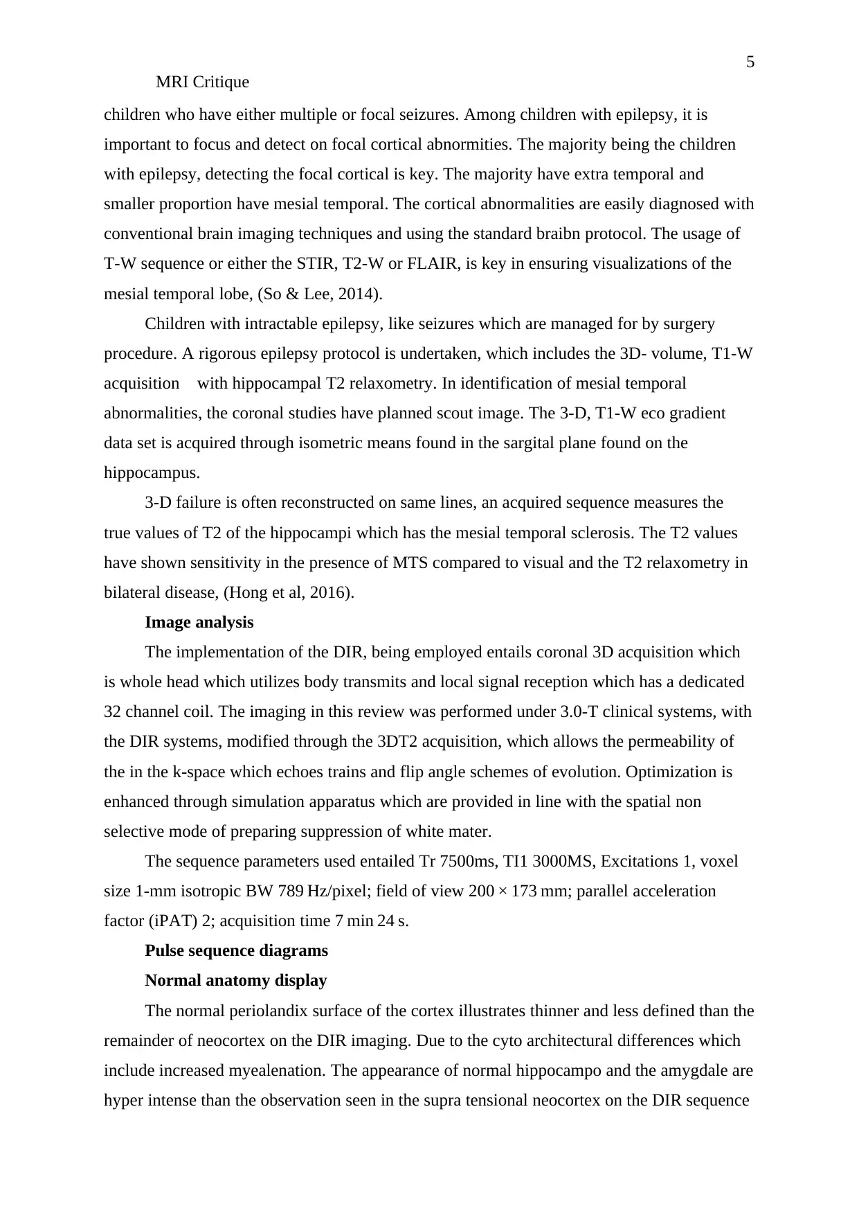
Normal anatomy display
The normal periolandix surface of the cortex illustrates thinner and less defined than the
remainder of neocortex on the DIR imaging. Due to the cyto architectural differences which
include increased myealenation. The appearance of normal hippocampo and the amygdale are
hyper intense than the observation seen in the supra tensional neocortex on the DIR sequence
MRI Critique
children who have either multiple or focal seizures. Among children with epilepsy, it is
important to focus and detect on focal cortical abnormities. The majority being the children
with epilepsy, detecting the focal cortical is key. The majority have extra temporal and
smaller proportion have mesial temporal. The cortical abnormalities are easily diagnosed with
conventional brain imaging techniques and using the standard braibn protocol. The usage of
T-W sequence or either the STIR, T2-W or FLAIR, is key in ensuring visualizations of the
mesial temporal lobe, (So & Lee, 2014).
Children with intractable epilepsy, like seizures which are managed for by surgery
procedure. A rigorous epilepsy protocol is undertaken, which includes the 3D- volume, T1-W
acquisition with hippocampal T2 relaxometry. In identification of mesial temporal
abnormalities, the coronal studies have planned scout image. The 3-D, T1-W eco gradient
data set is acquired through isometric means found in the sargital plane found on the
hippocampus.
3-D failure is often reconstructed on same lines, an acquired sequence measures the
true values of T2 of the hippocampi which has the mesial temporal sclerosis. The T2 values
have shown sensitivity in the presence of MTS compared to visual and the T2 relaxometry in
bilateral disease, (Hong et al, 2016).
Image analysis
The implementation of the DIR, being employed entails coronal 3D acquisition which
is whole head which utilizes body transmits and local signal reception which has a dedicated
32 channel coil. The imaging in this review was performed under 3.0-T clinical systems, with
the DIR systems, modified through the 3DT2 acquisition, which allows the permeability of
the in the k-space which echoes trains and flip angle schemes of evolution. Optimization is
enhanced through simulation apparatus which are provided in line with the spatial non
selective mode of preparing suppression of white mater.
The sequence parameters used entailed Tr 7500ms, TI1 3000MS, Excitations 1, voxel
size 1-mm isotropic BW 789 Hz/pixel; field of view 200 × 173 mm; parallel acceleration
factor (iPAT) 2; acquisition time 7 min 24 s.
Pulse sequence diagrams
Normal anatomy display
The normal periolandix surface of the cortex illustrates thinner and less defined than the
remainder of neocortex on the DIR imaging. Due to the cyto architectural differences which
include increased myealenation. The appearance of normal hippocampo and the amygdale are
hyper intense than the observation seen in the supra tensional neocortex on the DIR sequence
6
MRI Critique
and also as observed on the other weighted T2 sequence .
This display of normal anatomical view is shown below;
Diagram a; Reflects the normal periolandic cortex, with axial T1 weighted 3
dimensional magnetization which is appearing on the echo gradient
Diagram b; shows the reformatted double inversion recovery images , (DRI).
⊘ This is a preview!⊘
Do you want full access?
Subscribe today to unlock all pages.
Trusted by 1+ million students worldwide
7
MRI Critique
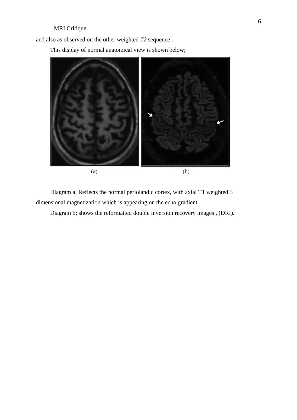
Figure 2;
a- Normal hippocampus of the coronal T2 weigted
b- Shows the fluid attenuated inversion recovery diagram
MRI Critique
Figure 2;
a- Normal hippocampus of the coronal T2 weigted
b- Shows the fluid attenuated inversion recovery diagram
Paraphrase This Document
Need a fresh take? Get an instant paraphrase of this document with our AI Paraphraser
8
MRI Critique
c- Double inversion images of the recovery , (DRI)
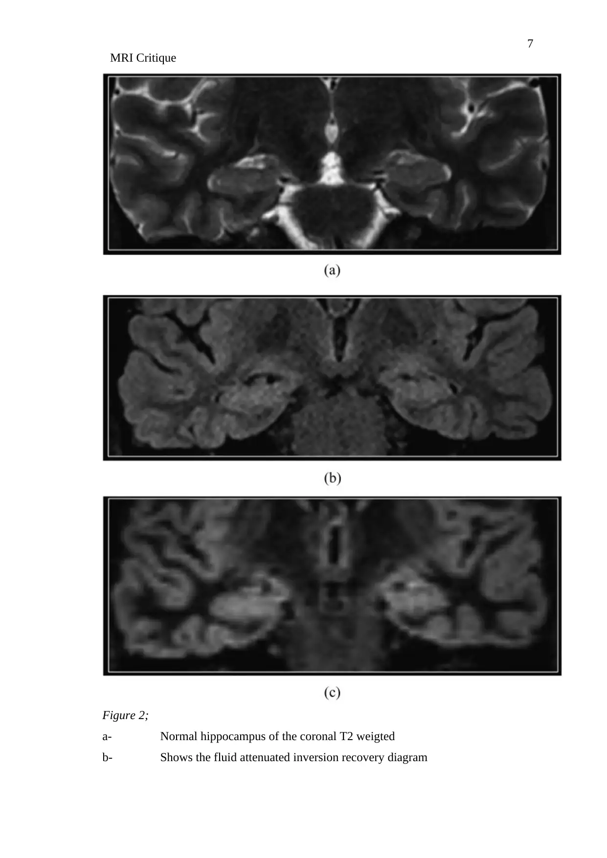
The normal area of the myelinisation is often conspicuous when displayed on the DIR
imaging, as it appears having brighter signal when compared to the myealined white matter.
This is attributed Ti the diminished T2 shortening of the white matter in the un myealeanated
phase, as illustrated below on figure 3. The distinction can be done carefully from the
abnormal white matter through careful analysis in the anterior temporal poles of the white
matter among children between the ages of 1-2 years.
Figure 3
Diagram ;
a- Shows incomplete myelination of a twenty month child, it displays the sagittal
refracted double invasersion. , (DRI)
b- While this image shows the sagittal T2 for fluid attenuated inversion recovery
The images displayed above shows the relative increased signal intensity on the white
matter of the temporal lobes which is compared the frontal; white matter. This occurrence is
due to the presence of incomplete myelinisation which is visible by the arrow.
Mesial temporal sclerosis
The temporal sclerosis often represent a diagnostic challenge when using MRI due to
hippo campo atrophy, the hypertense signal of T2 and the disruption of the internal
architecture like the early subtle. The DIR imaging is beneficial in order to demonstrate the
increasing conspicuity on the on the signal intensity of the asymmetry on the hippocampi as
MRI Critique
c- Double inversion images of the recovery , (DRI)
The normal area of the myelinisation is often conspicuous when displayed on the DIR
imaging, as it appears having brighter signal when compared to the myealined white matter.
This is attributed Ti the diminished T2 shortening of the white matter in the un myealeanated
phase, as illustrated below on figure 3. The distinction can be done carefully from the
abnormal white matter through careful analysis in the anterior temporal poles of the white
matter among children between the ages of 1-2 years.
Figure 3
Diagram ;
a- Shows incomplete myelination of a twenty month child, it displays the sagittal
refracted double invasersion. , (DRI)
b- While this image shows the sagittal T2 for fluid attenuated inversion recovery
The images displayed above shows the relative increased signal intensity on the white
matter of the temporal lobes which is compared the frontal; white matter. This occurrence is
due to the presence of incomplete myelinisation which is visible by the arrow.
Mesial temporal sclerosis
The temporal sclerosis often represent a diagnostic challenge when using MRI due to
hippo campo atrophy, the hypertense signal of T2 and the disruption of the internal
architecture like the early subtle. The DIR imaging is beneficial in order to demonstrate the
increasing conspicuity on the on the signal intensity of the asymmetry on the hippocampi as
9
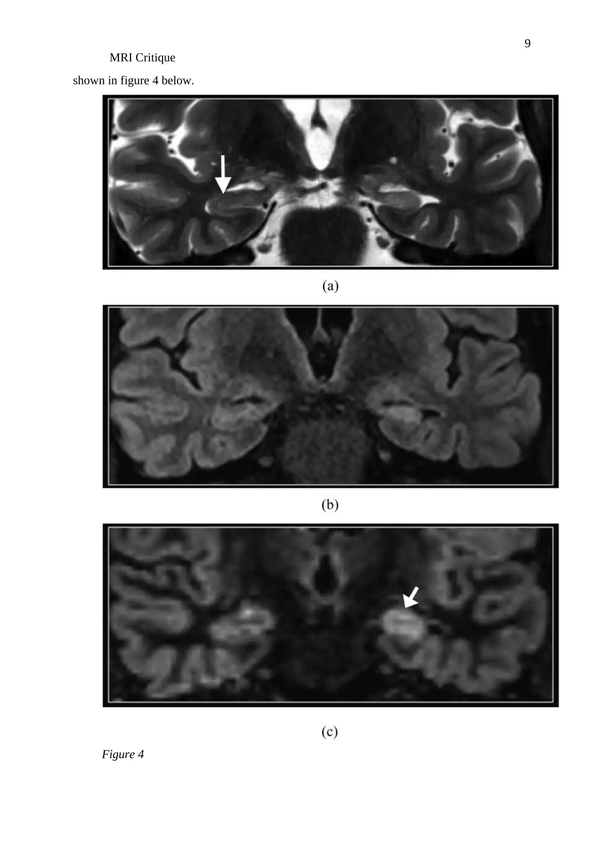
MRI Critique
shown in figure 4 below.
Figure 4
MRI Critique
shown in figure 4 below.
Figure 4
⊘ This is a preview!⊘
Do you want full access?
Subscribe today to unlock all pages.
Trusted by 1+ million students worldwide
10
MRI Critique
Diagram;
a- Showing the mesial temporal sclerosis on the cornla T1 weighted.
b- T2 fluid attenuated inversion recovery, (DRI)
c- Hypertense atrophyic on the left hippocampus.
Seizures have shown to have and causes swelling accompanied with T2 hyper tense
signal which has effects on the affected hippocampus. The Dir imaging shows the
asymmetrical hyper intensity of the located hippocampus. The DRI imaging in this case
shows the asymmetric signal hyper intensity of the hippocampus as illustrated on the figure
below, figure 5;
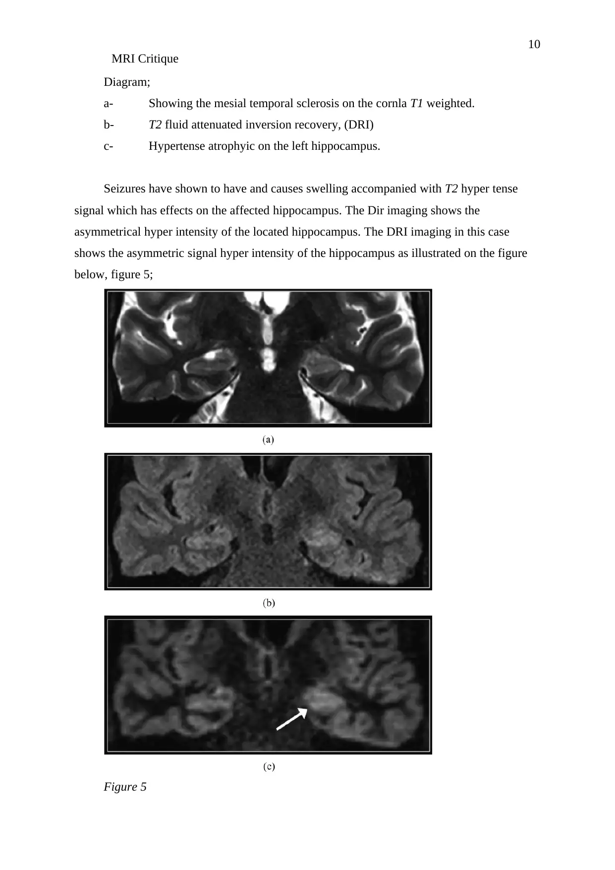
Figure 5
MRI Critique
Diagram;
a- Showing the mesial temporal sclerosis on the cornla T1 weighted.
b- T2 fluid attenuated inversion recovery, (DRI)
c- Hypertense atrophyic on the left hippocampus.
Seizures have shown to have and causes swelling accompanied with T2 hyper tense
signal which has effects on the affected hippocampus. The Dir imaging shows the
asymmetrical hyper intensity of the located hippocampus. The DRI imaging in this case
shows the asymmetric signal hyper intensity of the hippocampus as illustrated on the figure
below, figure 5;
Figure 5
Paraphrase This Document
Need a fresh take? Get an instant paraphrase of this document with our AI Paraphraser
11
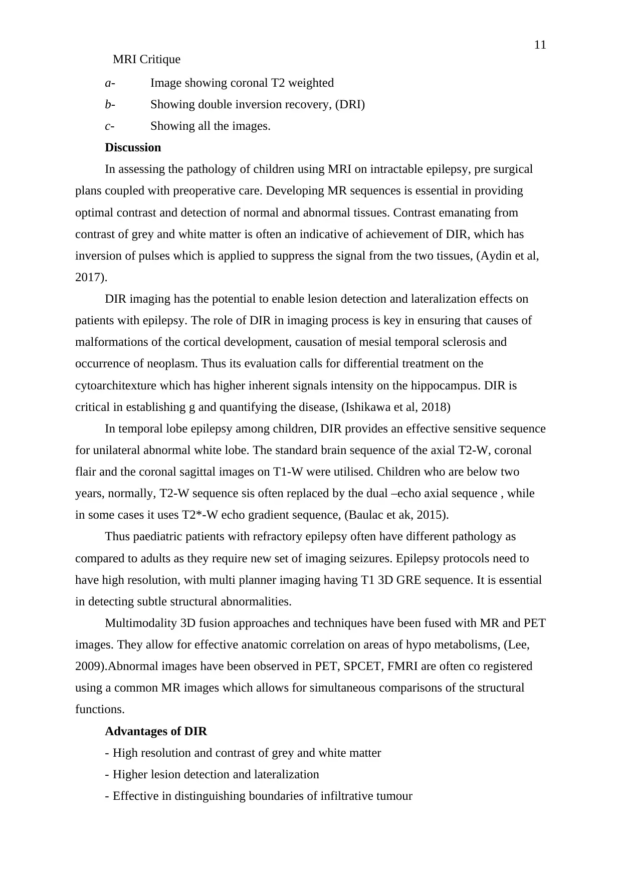
MRI Critique
a- Image showing coronal T2 weighted
b- Showing double inversion recovery, (DRI)
c- Showing all the images.
Discussion
In assessing the pathology of children using MRI on intractable epilepsy, pre surgical
plans coupled with preoperative care. Developing MR sequences is essential in providing
optimal contrast and detection of normal and abnormal tissues. Contrast emanating from
contrast of grey and white matter is often an indicative of achievement of DIR, which has
inversion of pulses which is applied to suppress the signal from the two tissues, (Aydin et al,
2017).
DIR imaging has the potential to enable lesion detection and lateralization effects on
patients with epilepsy. The role of DIR in imaging process is key in ensuring that causes of
malformations of the cortical development, causation of mesial temporal sclerosis and
occurrence of neoplasm. Thus its evaluation calls for differential treatment on the
cytoarchitexture which has higher inherent signals intensity on the hippocampus. DIR is
critical in establishing g and quantifying the disease, (Ishikawa et al, 2018)
In temporal lobe epilepsy among children, DIR provides an effective sensitive sequence
for unilateral abnormal white lobe. The standard brain sequence of the axial T2-W, coronal
flair and the coronal sagittal images on T1-W were utilised. Children who are below two
years, normally, T2-W sequence sis often replaced by the dual –echo axial sequence , while
in some cases it uses T2*-W echo gradient sequence, (Baulac et ak, 2015).
Thus paediatric patients with refractory epilepsy often have different pathology as
compared to adults as they require new set of imaging seizures. Epilepsy protocols need to
have high resolution, with multi planner imaging having T1 3D GRE sequence. It is essential
in detecting subtle structural abnormalities.
Multimodality 3D fusion approaches and techniques have been fused with MR and PET
images. They allow for effective anatomic correlation on areas of hypo metabolisms, (Lee,
2009).Abnormal images have been observed in PET, SPCET, FMRI are often co registered
using a common MR images which allows for simultaneous comparisons of the structural
functions.
Advantages of DIR
- High resolution and contrast of grey and white matter
- Higher lesion detection and lateralization
- Effective in distinguishing boundaries of infiltrative tumour
12
MRI Critique
Disadvantages
- Limited diagnostic vale in the myelinated brain
Conclusion
Thus the application of DIR sequence protocol in paediatric epilepsy shows how
accurate are they effective in detecting epileptogenic abnormalities. With the increased
conspiscusty and depiction of the subtle abnormalities, usage of DIR imaging g among
children having refractory epilepsy is crucial. DRI is associated with increased sensitivity of
the depiction of cortical lesions. It is beneficial characterizing epileptogenic foci which are
related to neocortical pathology.
⊘ This is a preview!⊘
Do you want full access?
Subscribe today to unlock all pages.
Trusted by 1+ million students worldwide
1 out of 14

Your All-in-One AI-Powered Toolkit for Academic Success.
+13062052269
info@desklib.com
Available 24*7 on WhatsApp / Email
![[object Object]](/_next/static/media/star-bottom.7253800d.svg)
Unlock your academic potential
Copyright © 2020–2026 A2Z Services. All Rights Reserved. Developed and managed by ZUCOL.