BIOC311 - EMSA: Analysis of Liver Nuclear Extract Experiment Report

Verified

Added on  2022/09/02

|5
|851
|27
Homework Assignment
AI Summary
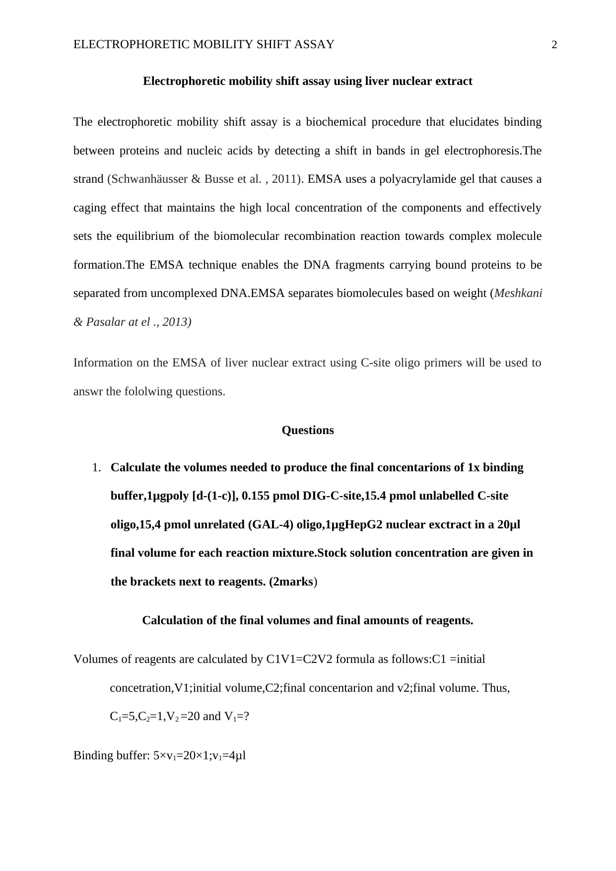
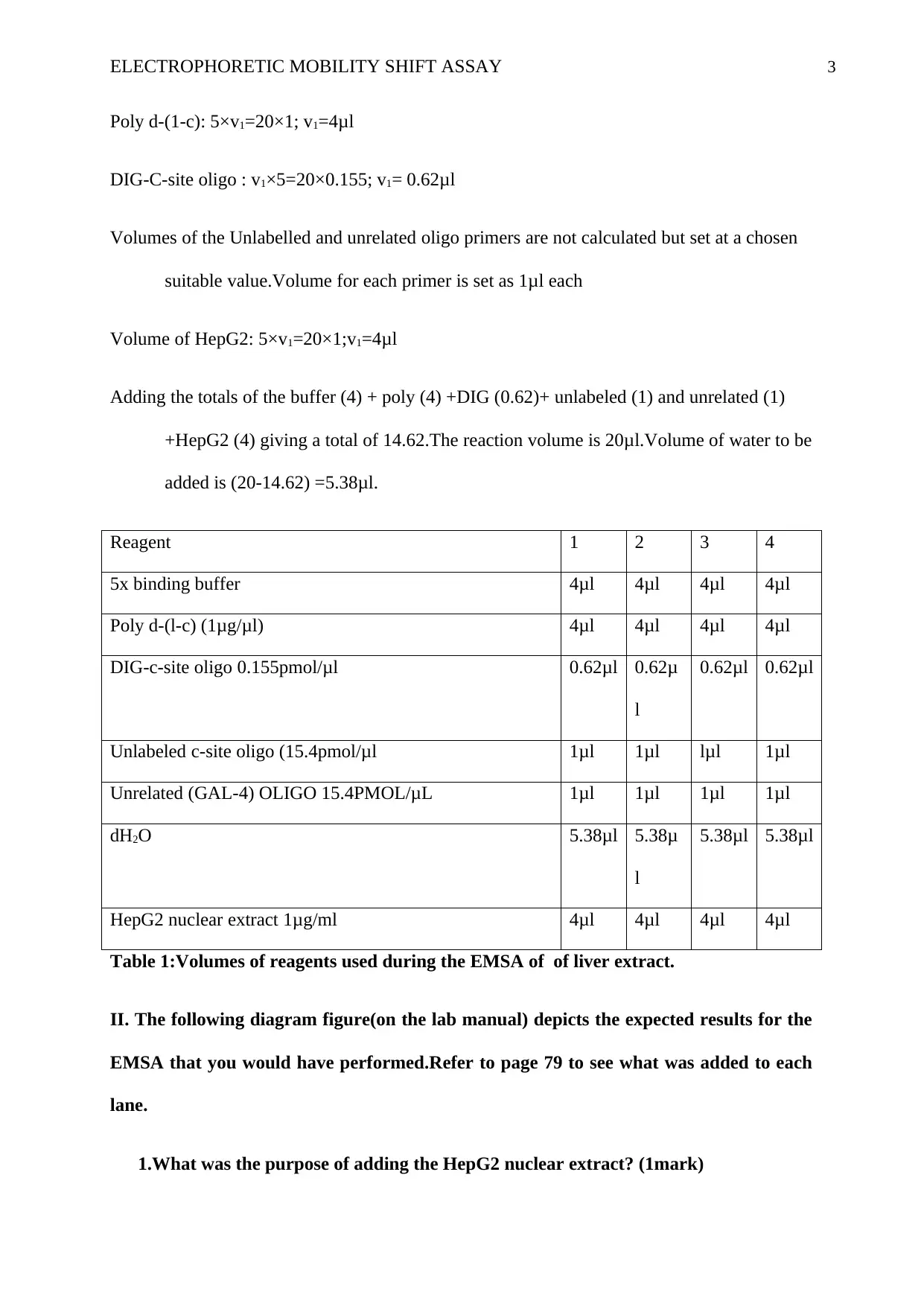
This EMSA lab report details an experiment investigating DNA-protein interactions using a liver nuclear extract. The report includes calculations for reagent volumes, based on provided stock concentrations, to achieve specific final concentrations of binding buffer, poly [d-(I-C)], DIG-C-site oligo, unlabeled C-site oligo, unrelated (GAL-4) oligo, and HepG2 nuclear extract. The core of the experiment revolves around the electrophoretic mobility shift assay (EMSA), a technique used to detect the binding of proteins to DNA fragments. The report answers questions regarding the purpose of adding HepG2 nuclear extract, the presence of unbound DIG-C-site oligo in lane 1, the effect of unlabeled C-site oligo on binding, the role of unrelated GAL-4 oligo, the function of the stratalinker, and what the antibody detected. The EMSA technique relies on the principle that DNA-protein complexes migrate differently in a gel compared to free DNA, allowing for the visualization and analysis of these interactions. The experiment utilizes the DIG Gel Shift Kit, 2nd Generation, to label DNA oligos with digoxigenin for detection. The results of the experiment are analyzed by comparing the migration of bound and unbound DIG-C-site oligo, revealing the presence and specificity of DNA-binding proteins in the liver nuclear extract.
Document Page
Running head: ELECTROPHORETIC MOBILITY SHIFT ASSAY 1
Electrophoretic mobility shift assay using liver nuclear extract
Name
Instituttional Affiliations
tabler-icon-diamond-filled.svg

Paraphrase This Document

Need a fresh take? Get an instant paraphrase of this document with our AI Paraphraser
Document Page
ELECTROPHORETIC MOBILITY SHIFT ASSAY 2
Electrophoretic mobility shift assay using liver nuclear extract
The electrophoretic mobility shift assay is a biochemical procedure that elucidates binding
between proteins and nucleic acids by detecting a shift in bands in gel electrophoresis.The
strand (Schwanhäusser & Busse et al. , 2011). EMSA uses a polyacrylamide gel that causes a
caging effect that maintains the high local concentration of the components and effectively
sets the equilibrium of the biomolecular recombination reaction towards complex molecule
formation.The EMSA technique enables the DNA fragments carrying bound proteins to be
separated from uncomplexed DNA.EMSA separates biomolecules based on weight (Meshkani
& Pasalar at el ., 2013)
Information on the EMSA of liver nuclear extract using C-site oligo primers will be used to
answr the fololwing questions.
Questions
1. Calculate the volumes needed to produce the final concentarions of 1x binding
buffer,1μgpoly [d-(1-c)], 0.155 pmol DIG-C-site,15.4 pmol unlabelled C-site
oligo,15,4 pmol unrelated (GAL-4) oligo,1μgHepG2 nuclear exctract in a 20μl
final volume for each reaction mixture.Stock solution concentration are given in
the brackets next to reagents. (2marks)
Calculation of the final volumes and final amounts of reagents.
Volumes of reagents are calculated by C1V1=C2V2 formula as follows:C1 =initial
concetration,V1;initial volume,C2;final concentarion and v2;final volume. Thus,
C1=5,C2=1,V2 =20 and V1=?
Binding buffer: 5×v1=20×1;v1=4μl
Document Page
ELECTROPHORETIC MOBILITY SHIFT ASSAY 3
Poly d-(1-c): 5×v1=20×1; v1=4μl
DIG-C-site oligo : v1×5=20×0.155; v1= 0.62μl
Volumes of the Unlabelled and unrelated oligo primers are not calculated but set at a chosen
suitable value.Volume for each primer is set as 1μl each
Volume of HepG2: 5×v1=20×1;v1=4μl
Adding the totals of the buffer (4) + poly (4) +DIG (0.62)+ unlabeled (1) and unrelated (1)
+HepG2 (4) giving a total of 14.62.The reaction volume is 20μl.Volume of water to be
added is (20-14.62) =5.38μl.
Reagent 1 2 3 4
5x binding buffer 4μl 4μl 4μl 4μl
Poly d-(l-c) (1μg/μl) 4μl 4μl 4μl 4μl
DIG-c-site oligo 0.155pmol/μl 0.62μl 0.62μ
l
0.62μl 0.62μl
Unlabeled c-site oligo (15.4pmol/μl 1μl 1μl lμl 1μl
Unrelated (GAL-4) OLIGO 15.4PMOL/μL 1μl 1μl 1μl 1μl
dH2O 5.38μl 5.38μ
l
5.38μl 5.38μl
HepG2 nuclear extract 1μg/ml 4μl 4μl 4μl 4μl
Table 1:Volumes of reagents used during the EMSA of of liver extract.
II. The following diagram figure(on the lab manual) depicts the expected results for the
EMSA that you would have performed.Refer to page 79 to see what was added to each
lane.
1.What was the purpose of adding the HepG2 nuclear extract? (1mark)
Document Page
ELECTROPHORETIC MOBILITY SHIFT ASSAY 4
HepG2 nuclear extract was added as a source of PEPCK promoter binding proteins that
would bind to the C-site oligos primers to show the presence of C/EBP protein in the liver.
2. Why is only unbound DIG-C-site oligo present in lane 1? (1mark)
Unbound DIG-C-site oligo is a free DNA fragment which has not formed any complex
with the C/EBP.The unbound DIG-C-site oligo is thus electrophoresed faster due to low
molecular weight.
3. Why does adding unlabeled C-site oligo(lane 3) reduce the amount of bound
DIG-C-site? (1mark)
Unlabeled C-site oligo has codons complementary to the C/EBP from the HepG2 liver
nuclear.The unlabeled C-site oligo therefore binds to a portion of the C/EBP reducing
the portion of binding for the labeled C-site oligo hence reducing the amount of DIG-
C-site.
4. What effect does adding an unrelated GAL-4 oligo have on binding? (i.e.,
compare lanes 2 and 4) (1mark)
Unrelated GAL-4oligo is added to differentiate between non-specific and specific
DNA binding of proteins.Unrelated GAL-4 ensures that specific binding is well
recognised from non-specific binding.
5. Why was the stratalinker used? (1mark)
Stratalinker were used to aid crosslinking of DNA on the nylon membrane in
readiness to observe the degree of flow of bands autoradiographically.
6. What did the antibody detect? (1mark)
tabler-icon-diamond-filled.svg

Paraphrase This Document

Need a fresh take? Get an instant paraphrase of this document with our AI Paraphraser
Document Page
ELECTROPHORETIC MOBILITY SHIFT ASSAY 5
Antibody solution was used as an anti-DIG antibody to detect labelled DNA-oligos.
References
Schwanhäusser, B., Busse, D., Li, N., Dittmar, G., Schuchhardt, J., Wolf, J., ... & Selbach, M.
(2011). Global quantification of mammalian gene expression
control. Nature, 473(7347), 337.
Mani, M., Golmohammadi, T., Khaghani, S., Zamani, Z., Azadmanesh, K., Meshkani, R., &
Pasalar, P. (2013). Homocysteine Induces Heme Oxygenase-1 Expression via
Transcription Factor Nrf2 Activation in HepG2 Cells. Iranian biomedical
journal, 17(2), 93.
chevron_up_icon
1 out of 5
circle_padding
hide_on_mobile
zoom_out_icon
[object Object]