Bioelectromagnetics and Reproduction: ELF-EMF and RFR Exposure Effects
VerifiedAdded on 2021/06/30
|37
|16339
|105
Report
AI Summary
This report, prepared for the BioInitiative Working Group by researchers from Jawaharlal Nehru University (JNU), comprehensively reviews the effects of Extremely Low Frequency Electromagnetic Fields (ELF-EMF) and Radiofrequency Radiation (RFR) on fertility and reproduction. It presents evidence from human and animal studies, focusing on the impact of ELF-EMF and RFR on male sperm function, including sperm count, motility, viability, and DNA damage. The report explores the biophysics of ELF-EMF and RFR, detailing how these fields interact with the human body and induce electric fields and currents. It examines various factors influencing the effects of radiation, such as frequency, body size, and tissue properties. The review highlights studies on both male and female reproductive health, including oxidative stress, DNA damage in spermatozoa, and potential risks of miscarriage. The report also discusses specific research findings, such as the impact of ELF-EMF on testosterone levels, sperm chromatin condensation, and germ cell apoptosis, emphasizing the need for further investigation into the long-term effects of electromagnetic field exposure on reproductive health.
SECTION 18
Electromagnetic Field Exposure Effects
(ELF-EMF and RFR)
on Fertility and Reproduction
Prof. Jitendra Behari, PhD
Bioelectromagnetics Laboratory
School of Environmental Sciences
Jawaharlal Nehru University
New Delhi, India
Dr. Paulraj Rajamani, PhD
Bioelectromagnetics Laboratory
School of Environmental Sciences
Jawaharlal Nehru University
New Delhi, India
Prepared for the BioInitiative Working Group
November 2012
Paraphrase This Document
Need a fresh take? Get an instant paraphrase of this document with our AI Paraphraser
2
I. INTRODUCTION
Electromagnetic fields and radiofrequency radiation (RFR) interact with human tissues and
may have adverse effects on fertility and reproduction. This review presents evidence for
ELF-EMF and RFR effects on many parameters of male sperm function; leading to questions
about the genotoxicity and carcinogenicity of such exposures on fertility and reproduction in
men. Much of the evidence comes from human and animal studies on sperm and male
fertility factors, but there are also studies showing adverse effects on fertility and miscarriage
in women.
During the last four decades or so there has been a growing concern on the effects of
electromagnetic radiations on biological systems in general. This is because of the global
introduction of electronic devices on a massive level for communications and data
transmission, personal wireless devices, air surveillance systems, industry applications,
medical/diagnostic and therapeutic purposes that are now new sources of electromagnetic
fields (ELF-EMF) and radiofrequency microwave radiation (RFR). This has added another
layer of pollutant (electropollution) to a growing list of environmental contaminants in air,
water, soil and from noise pollution which can adversely affect human health.
There are many sources of EMF in our environment and this non-ionizing radiation interacts
with the human body. Use of electronic household items and cell phones are reported to
decrease fertility potential in men by decreasing sperm count, motility, viability, inducing
pathological changes in sperm and testes morphology, and so on (Erogul et al. 2006). In
accordance with this, several authors (Agarwal et al. 2008, 2009; Kumar et al. 2010, 2011a;
Pourlis 2009; Kesari et al. 2010, 2011, 2012) focused mainly on the male reproduction
patterns. It involves the development from undifferentiated diploid stem cells to highly
differentiated haploid stem cells. Spermatogenesis is a complex process and it is influenced
by many genes and hormones. It takes place in the testis, which may be exposed to various
microwave frequencies which are currently in use (Behari and Kesari 2006). Among various
factors of infertility, oxidative stress has become the main focus of interest as a potential
cause of male infertility (Agarwal and Said 2003; Aitken and Roman, 2008; Kumar et al,
2010, 2011a). Male infertility is commonly associated with high rates of DNA
(deoxyribonucleic acid) damage in the spermatozoa and such damage is correlated with a
wide range of adverse clinical outcomes. Several studies, especially at power frequency 50/60
3
Hz magnetic field have found an association of exposure to human health, with emphasis on a
range of clinical conditions including childhood leukaemia, brain tumours, genotoxicity and
neurodegenerative disease, infertility, birth defects, increased risk of miscarriage, childhood
morbidity and de novo mutations (Hardell and Sage 2008; Gharagozloo and Aitken 2011;
Garcia et al. 2008; Huss et al. 2008; O’Carroll and Henshaw 2008; International Agency for
Research on Cancer (IARC) Monographs of the Evaluation of Carcinogenic Risks to Human
2002; California Health Department Services (CHDS) Report 2002). Sperm DNA damage is
therefore regarded as a potential risk factor to the development of normal human embryos
leading to impaired embryonic development.
II. THE BIOPHYSICS OF EXTREMELY LOW FREQUENCY FIELDS
Whenever a body having finite conductivity (biological body) is intercepted by EMF it
induces electric fields and circulating electric currents, which in turn competes with
endogenous current and voltages, thus disturbing normal physiological balance. The depth of
penetration within the body depends upon its frequency and the electric properties of the
exposed portion in the body. If the current density exceeds a certain threshold value,
excitation of muscles and nerves due to membrane depolarization is possible. The mode of
interaction of non-ionizing radiation with biological systems can be broadly divided into two
parts: extremely low frequency and radiofrequency/microwaves.
Whenever an electric field interacts with a biological body the incident field will be distorted,
such that the external field will be nearly perpendicular to the boundary surface. At 60 Hz
Einternal / Eexternal ≈ 4(10-8 ). (1)
Thus a 60 Hz external field of 100 kV/m will produce an average internal E field of the order
of 4mV/m.
As far as the magnetic components of the extremely low frequency fields are concerned,
magnetic permeability of most biological materials is practically equal to that of free space
(4.10-7) H/m. This signifies that ELF H field ‘inside’ will be practically equal to the H field
‘outside’. Only exceptions could be those biological materials that have magnetic particles
inside. A time varying magnetic field (also electric field) can also induce electric currents
into stationary conducting objects. Thus, all modes of interaction of time varying E fields
with living matter may be triggered by time-varying (not by static) magnetic field. According
to Faraday’s law of electromagnetic induction time varying magnetic flux will induce E fields
with resulting electrical potential differences and “eddy” currents through available
⊘ This is a preview!⊘
Do you want full access?
Subscribe today to unlock all pages.
Trusted by 1+ million students worldwide
4
conducting paths. Sources generating low frequency electric and magnetic fields are more
likely to produce physiologically significant internal E fields through the mechanism of
magnetic induction. If an erect person is targeted by a vertical electric field it will be
considerably “enhanced” at the top of the person’s head and shoulder, and one would predict
therefore that the field in the tissue would also be enhanced above that of a flat slice exposed
to the same field (Deon, 1982). In a 60 Hz electric field of 1kV/m in air, the current densities
(Am/m2) in neck, waist and ankle turn out to be 0.591x10 -3, 0.427 x-3 and 3.35x10⁻³
respectively (Polk 1986).
III. THE BIOPHYSICS OF RADIOFREQUENCY AND MICROWAVE FIELDS
The biological bodies are inhomogeneous, having tissue-specific dielectric properties and the
complexity of the shape; which make the computations of the induced field difficult. The
fields induced inside the body act differently depending upon the frequency and more
particularly on (L/λ), (where L is the length of the biological body and λ the wavelength of
the incident field) upon, but are not limited to the following parameters:
(i) The location of the field with respect to the surroundings, e.g. if there are metallic
objects around, the person is grounded or otherwise.
(ii) Polarisation of the incident wave with respect to the orientation of the human
body.
(iii) Size of the human body (L) with respect to the wavelength (λ) of the incident
radiations (L/λ).
(iv) The portion of the human body.
(v) The electrical properties of the tissue in question.
In free space propagation of electromagnetic field the power density is given by
Power density = E2/1200 Π mW/cm2 (1)
Where, E is the electric field strength.
The frequency in the radio frequency-microwave region are somewhat penetrated inside the
biological body interacting with the tissues inside.
Paraphrase This Document
Need a fresh take? Get an instant paraphrase of this document with our AI Paraphraser
5
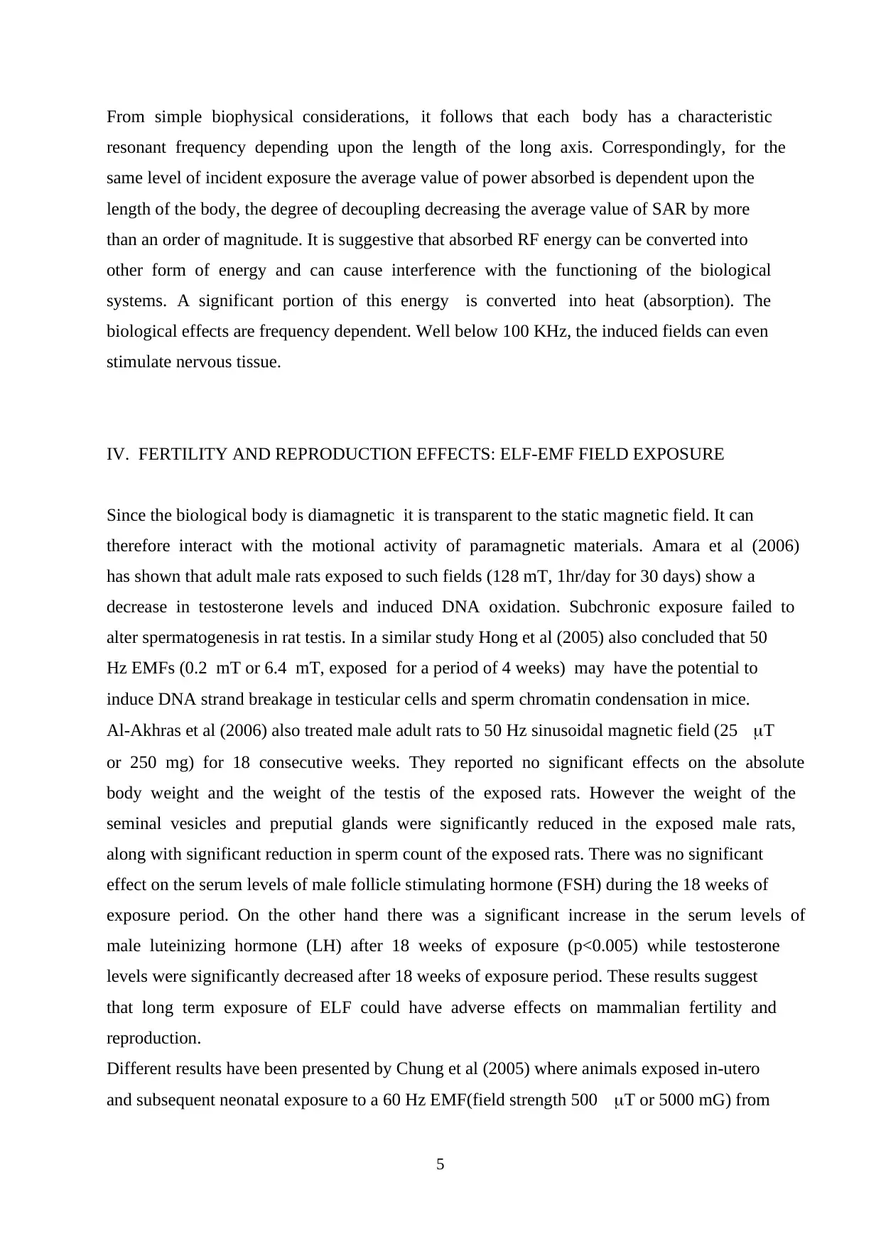
From simple biophysical considerations, it follows that each body has a characteristic
resonant frequency depending upon the length of the long axis. Correspondingly, for the
same level of incident exposure the average value of power absorbed is dependent upon the
length of the body, the degree of decoupling decreasing the average value of SAR by more
than an order of magnitude. It is suggestive that absorbed RF energy can be converted into
other form of energy and can cause interference with the functioning of the biological
systems. A significant portion of this energy is converted into heat (absorption). The
biological effects are frequency dependent. Well below 100 KHz, the induced fields can even
stimulate nervous tissue.
IV. FERTILITY AND REPRODUCTION EFFECTS: ELF-EMF FIELD EXPOSURE
Since the biological body is diamagnetic it is transparent to the static magnetic field. It can
therefore interact with the motional activity of paramagnetic materials. Amara et al (2006)
has shown that adult male rats exposed to such fields (128 mT, 1hr/day for 30 days) show a
decrease in testosterone levels and induced DNA oxidation. Subchronic exposure failed to
alter spermatogenesis in rat testis. In a similar study Hong et al (2005) also concluded that 50
Hz EMFs (0.2 mT or 6.4 mT, exposed for a period of 4 weeks) may have the potential to
induce DNA strand breakage in testicular cells and sperm chromatin condensation in mice.
Al-Akhras et al (2006) also treated male adult rats to 50 Hz sinusoidal magnetic field (25 T
or 250 mg) for 18 consecutive weeks. They reported no significant effects on the absolute
body weight and the weight of the testis of the exposed rats. However the weight of the
seminal vesicles and preputial glands were significantly reduced in the exposed male rats,
along with significant reduction in sperm count of the exposed rats. There was no significant
effect on the serum levels of male follicle stimulating hormone (FSH) during the 18 weeks of
exposure period. On the other hand there was a significant increase in the serum levels of
male luteinizing hormone (LH) after 18 weeks of exposure (p<0.005) while testosterone
levels were significantly decreased after 18 weeks of exposure period. These results suggest
that long term exposure of ELF could have adverse effects on mammalian fertility and
reproduction.
Different results have been presented by Chung et al (2005) where animals exposed in-utero
and subsequent neonatal exposure to a 60 Hz EMF(field strength 500 T or 5000 mG) from
6
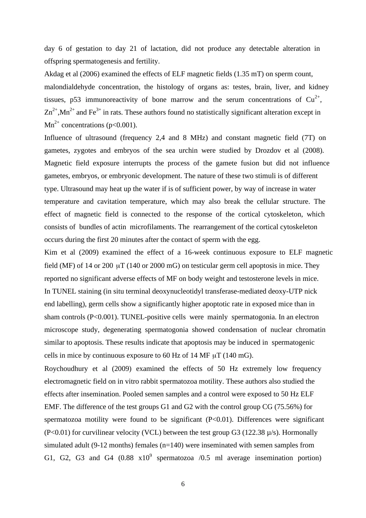
day 6 of gestation to day 21 of lactation, did not produce any detectable alteration in
offspring spermatogenesis and fertility.
Akdag et al (2006) examined the effects of ELF magnetic fields (1.35 mT) on sperm count,
malondialdehyde concentration, the histology of organs as: testes, brain, liver, and kidney
tissues, p53 immunoreactivity of bone marrow and the serum concentrations of Cu2+,
Zn2+,Mn2+ and Fe3+ in rats. These authors found no statistically significant alteration except in
Mn2+ concentrations (p<0.001).
Influence of ultrasound (frequency 2,4 and 8 MHz) and constant magnetic field (7T) on
gametes, zygotes and embryos of the sea urchin were studied by Drozdov et al (2008).
Magnetic field exposure interrupts the process of the gamete fusion but did not influence
gametes, embryos, or embryonic development. The nature of these two stimuli is of different
type. Ultrasound may heat up the water if is of sufficient power, by way of increase in water
temperature and cavitation temperature, which may also break the cellular structure. The
effect of magnetic field is connected to the response of the cortical cytoskeleton, which
consists of bundles of actin microfilaments. The rearrangement of the cortical cytoskeleton
occurs during the first 20 minutes after the contact of sperm with the egg.
Kim et al (2009) examined the effect of a 16-week continuous exposure to ELF magnetic
field (MF) of 14 or 200 T (140 or 2000 mG) on testicular germ cell apoptosis in mice. They
reported no significant adverse effects of MF on body weight and testosterone levels in mice.
In TUNEL staining (in situ terminal deoxynucleotidyl transferase-mediated deoxy-UTP nick
end labelling), germ cells show a significantly higher apoptotic rate in exposed mice than in
sham controls (P<0.001). TUNEL-positive cells were mainly spermatogonia. In an electron
microscope study, degenerating spermatogonia showed condensation of nuclear chromatin
similar to apoptosis. These results indicate that apoptosis may be induced in spermatogenic
cells in mice by continuous exposure to 60 Hz of 14 MF T (140 mG).
Roychoudhury et al (2009) examined the effects of 50 Hz extremely low frequency
electromagnetic field on in vitro rabbit spermatozoa motility. These authors also studied the
effects after insemination. Pooled semen samples and a control were exposed to 50 Hz ELF
EMF. The difference of the test groups G1 and G2 with the control group CG (75.56%) for
spermatozoa motility were found to be significant (P<0.01). Differences were significant
(P<0.01) for curvilinear velocity (VCL) between the test group G3 (122.38 μ/s). Hormonally
simulated adult (9-12 months) females (n=140) were inseminated with semen samples from
G1, G2, G3 and G4 (0.88 x10⁹ spermatozoa /0.5 ml average insemination portion)
⊘ This is a preview!⊘
Do you want full access?
Subscribe today to unlock all pages.
Trusted by 1+ million students worldwide
7
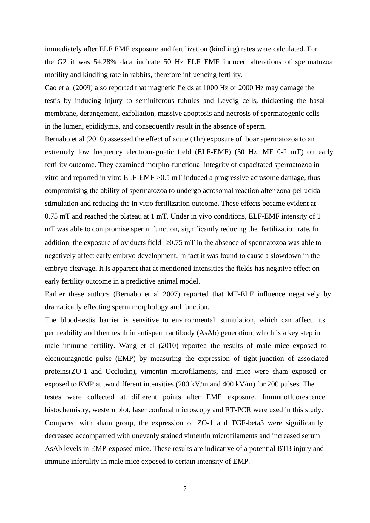
immediately after ELF EMF exposure and fertilization (kindling) rates were calculated. For
the G2 it was 54.28% data indicate 50 Hz ELF EMF induced alterations of spermatozoa
motility and kindling rate in rabbits, therefore influencing fertility.
Cao et al (2009) also reported that magnetic fields at 1000 Hz or 2000 Hz may damage the
testis by inducing injury to seminiferous tubules and Leydig cells, thickening the basal
membrane, derangement, exfoliation, massive apoptosis and necrosis of spermatogenic cells
in the lumen, epididymis, and consequently result in the absence of sperm.
Bernabo et al (2010) assessed the effect of acute (1hr) exposure of boar spermatozoa to an
extremely low frequency electromagnetic field (ELF-EMF) (50 Hz, MF 0-2 mT) on early
fertility outcome. They examined morpho-functional integrity of capacitated spermatozoa in
vitro and reported in vitro ELF-EMF >0.5 mT induced a progressive acrosome damage, thus
compromising the ability of spermatozoa to undergo acrosomal reaction after zona-pellucida
stimulation and reducing the in vitro fertilization outcome. These effects became evident at
0.75 mT and reached the plateau at 1 mT. Under in vivo conditions, ELF-EMF intensity of 1
mT was able to compromise sperm function, significantly reducing the fertilization rate. In
addition, the exposure of oviducts field 0.75 mT in the absence of spermatozoa was able to
negatively affect early embryo development. In fact it was found to cause a slowdown in the
embryo cleavage. It is apparent that at mentioned intensities the fields has negative effect on
early fertility outcome in a predictive animal model.
Earlier these authors (Bernabo et al 2007) reported that MF-ELF influence negatively by
dramatically effecting sperm morphology and function.
The blood-testis barrier is sensitive to environmental stimulation, which can affect its
permeability and then result in antisperm antibody (AsAb) generation, which is a key step in
male immune fertility. Wang et al (2010) reported the results of male mice exposed to
electromagnetic pulse (EMP) by measuring the expression of tight-junction of associated
proteins(ZO-1 and Occludin), vimentin microfilaments, and mice were sham exposed or
exposed to EMP at two different intensities (200 kV/m and 400 kV/m) for 200 pulses. The
testes were collected at different points after EMP exposure. Immunofluorescence
histochemistry, western blot, laser confocal microscopy and RT-PCR were used in this study.
Compared with sham group, the expression of ZO-1 and TGF-beta3 were significantly
decreased accompanied with unevenly stained vimentin microfilaments and increased serum
AsAb levels in EMP-exposed mice. These results are indicative of a potential BTB injury and
immune infertility in male mice exposed to certain intensity of EMP.
Paraphrase This Document
Need a fresh take? Get an instant paraphrase of this document with our AI Paraphraser
8
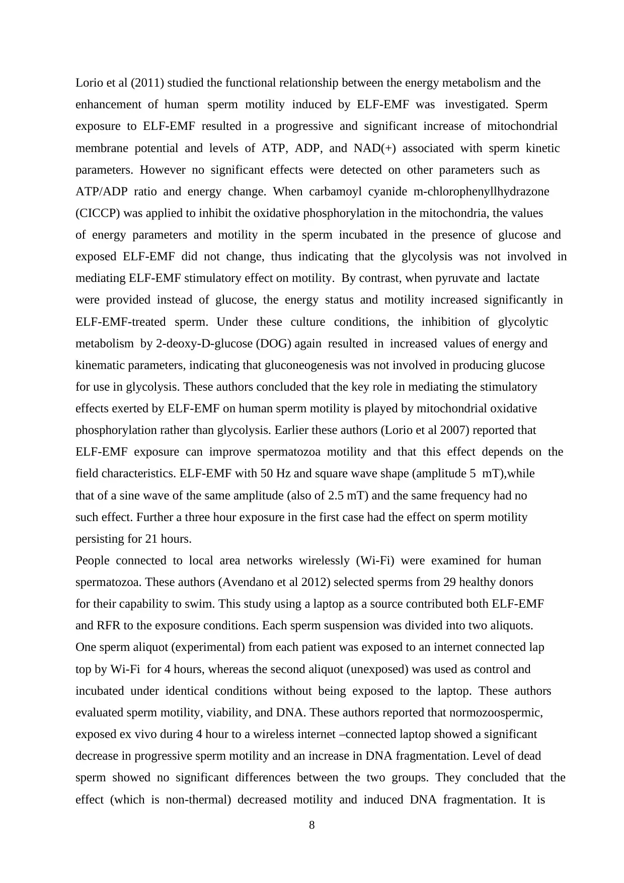
Lorio et al (2011) studied the functional relationship between the energy metabolism and the
enhancement of human sperm motility induced by ELF-EMF was investigated. Sperm
exposure to ELF-EMF resulted in a progressive and significant increase of mitochondrial
membrane potential and levels of ATP, ADP, and NAD(+) associated with sperm kinetic
parameters. However no significant effects were detected on other parameters such as
ATP/ADP ratio and energy change. When carbamoyl cyanide m-chlorophenyllhydrazone
(CICCP) was applied to inhibit the oxidative phosphorylation in the mitochondria, the values
of energy parameters and motility in the sperm incubated in the presence of glucose and
exposed ELF-EMF did not change, thus indicating that the glycolysis was not involved in
mediating ELF-EMF stimulatory effect on motility. By contrast, when pyruvate and lactate
were provided instead of glucose, the energy status and motility increased significantly in
ELF-EMF-treated sperm. Under these culture conditions, the inhibition of glycolytic
metabolism by 2-deoxy-D-glucose (DOG) again resulted in increased values of energy and
kinematic parameters, indicating that gluconeogenesis was not involved in producing glucose
for use in glycolysis. These authors concluded that the key role in mediating the stimulatory
effects exerted by ELF-EMF on human sperm motility is played by mitochondrial oxidative
phosphorylation rather than glycolysis. Earlier these authors (Lorio et al 2007) reported that
ELF-EMF exposure can improve spermatozoa motility and that this effect depends on the
field characteristics. ELF-EMF with 50 Hz and square wave shape (amplitude 5 mT),while
that of a sine wave of the same amplitude (also of 2.5 mT) and the same frequency had no
such effect. Further a three hour exposure in the first case had the effect on sperm motility
persisting for 21 hours.
People connected to local area networks wirelessly (Wi-Fi) were examined for human
spermatozoa. These authors (Avendano et al 2012) selected sperms from 29 healthy donors
for their capability to swim. This study using a laptop as a source contributed both ELF-EMF
and RFR to the exposure conditions. Each sperm suspension was divided into two aliquots.
One sperm aliquot (experimental) from each patient was exposed to an internet connected lap
top by Wi-Fi for 4 hours, whereas the second aliquot (unexposed) was used as control and
incubated under identical conditions without being exposed to the laptop. These authors
evaluated sperm motility, viability, and DNA. These authors reported that normozoospermic,
exposed ex vivo during 4 hour to a wireless internet –connected laptop showed a significant
decrease in progressive sperm motility and an increase in DNA fragmentation. Level of dead
sperm showed no significant differences between the two groups. They concluded that the
effect (which is non-thermal) decreased motility and induced DNA fragmentation. It is
9
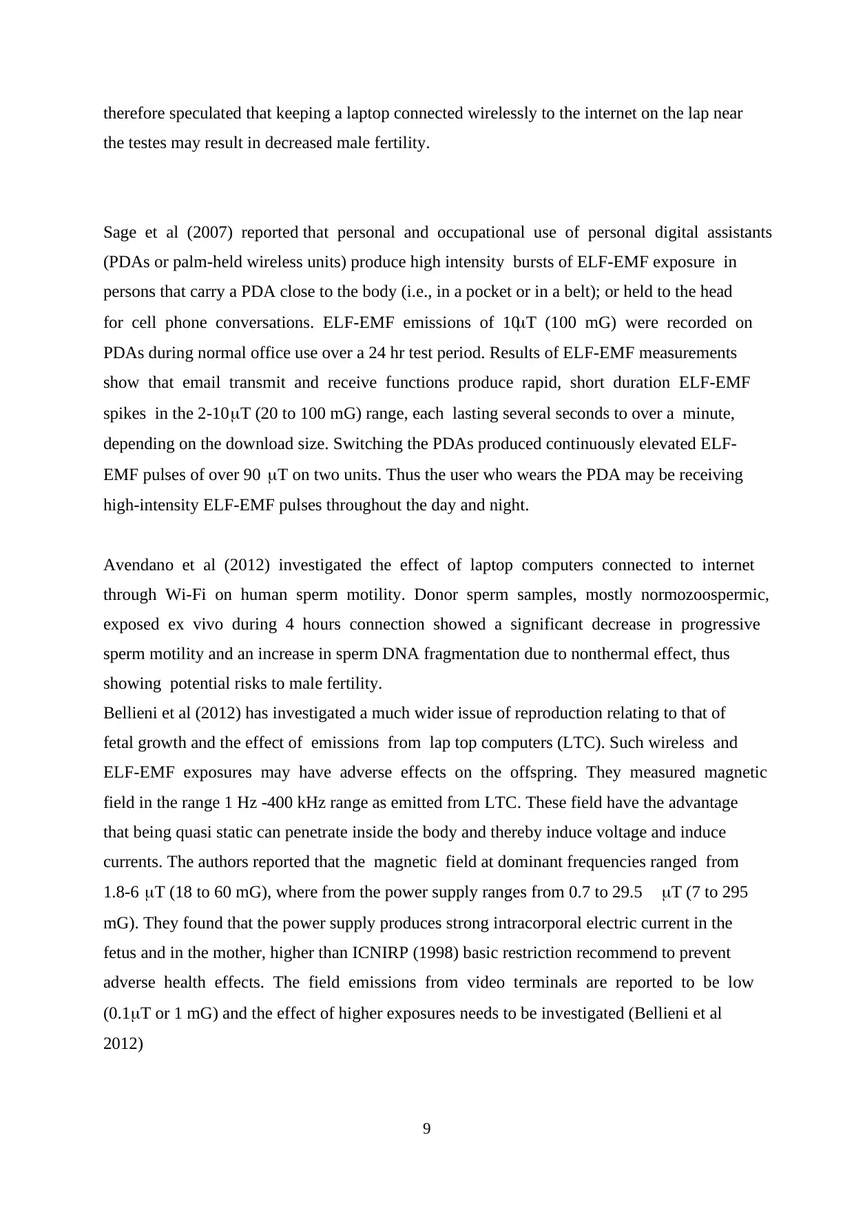
therefore speculated that keeping a laptop connected wirelessly to the internet on the lap near
the testes may result in decreased male fertility.
Sage et al (2007) reported that personal and occupational use of personal digital assistants
(PDAs or palm-held wireless units) produce high intensity bursts of ELF-EMF exposure in
persons that carry a PDA close to the body (i.e., in a pocket or in a belt); or held to the head
for cell phone conversations. ELF-EMF emissions of 10T (100 mG) were recorded on
PDAs during normal office use over a 24 hr test period. Results of ELF-EMF measurements
show that email transmit and receive functions produce rapid, short duration ELF-EMF
spikes in the 2-10T (20 to 100 mG) range, each lasting several seconds to over a minute,
depending on the download size. Switching the PDAs produced continuously elevated ELF-
EMF pulses of over 90 T on two units. Thus the user who wears the PDA may be receiving
high-intensity ELF-EMF pulses throughout the day and night.
Avendano et al (2012) investigated the effect of laptop computers connected to internet
through Wi-Fi on human sperm motility. Donor sperm samples, mostly normozoospermic,
exposed ex vivo during 4 hours connection showed a significant decrease in progressive
sperm motility and an increase in sperm DNA fragmentation due to nonthermal effect, thus
showing potential risks to male fertility.
Bellieni et al (2012) has investigated a much wider issue of reproduction relating to that of
fetal growth and the effect of emissions from lap top computers (LTC). Such wireless and
ELF-EMF exposures may have adverse effects on the offspring. They measured magnetic
field in the range 1 Hz -400 kHz range as emitted from LTC. These field have the advantage
that being quasi static can penetrate inside the body and thereby induce voltage and induce
currents. The authors reported that the magnetic field at dominant frequencies ranged from
1.8-6 T (18 to 60 mG), where from the power supply ranges from 0.7 to 29.5 T (7 to 295
mG). They found that the power supply produces strong intracorporal electric current in the
fetus and in the mother, higher than ICNIRP (1998) basic restriction recommend to prevent
adverse health effects. The field emissions from video terminals are reported to be low
(0.1T or 1 mG) and the effect of higher exposures needs to be investigated (Bellieni et al
2012)
⊘ This is a preview!⊘
Do you want full access?
Subscribe today to unlock all pages.
Trusted by 1+ million students worldwide
10
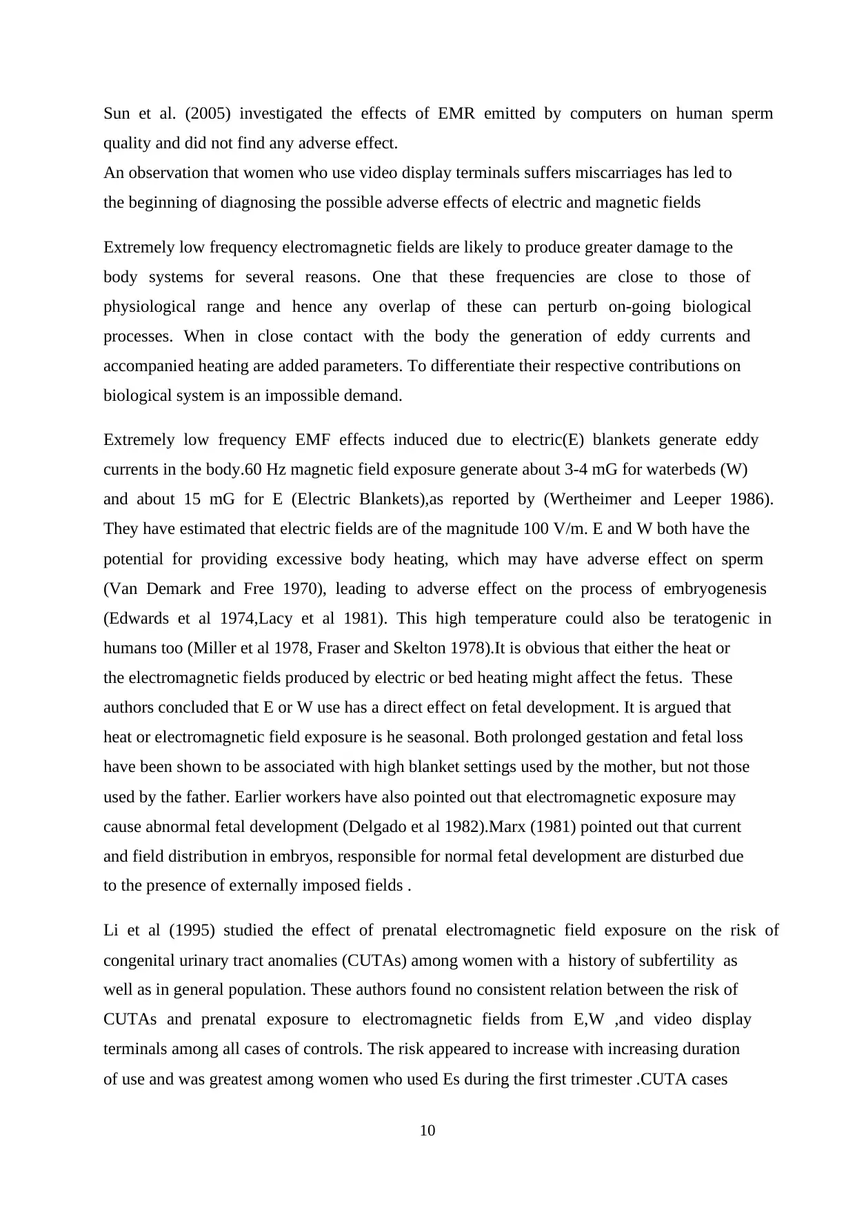
Sun et al. (2005) investigated the effects of EMR emitted by computers on human sperm
quality and did not find any adverse effect.
An observation that women who use video display terminals suffers miscarriages has led to
the beginning of diagnosing the possible adverse effects of electric and magnetic fields
Extremely low frequency electromagnetic fields are likely to produce greater damage to the
body systems for several reasons. One that these frequencies are close to those of
physiological range and hence any overlap of these can perturb on-going biological
processes. When in close contact with the body the generation of eddy currents and
accompanied heating are added parameters. To differentiate their respective contributions on
biological system is an impossible demand.
Extremely low frequency EMF effects induced due to electric(E) blankets generate eddy
currents in the body.60 Hz magnetic field exposure generate about 3-4 mG for waterbeds (W)
and about 15 mG for E (Electric Blankets),as reported by (Wertheimer and Leeper 1986).
They have estimated that electric fields are of the magnitude 100 V/m. E and W both have the
potential for providing excessive body heating, which may have adverse effect on sperm
(Van Demark and Free 1970), leading to adverse effect on the process of embryogenesis
(Edwards et al 1974,Lacy et al 1981). This high temperature could also be teratogenic in
humans too (Miller et al 1978, Fraser and Skelton 1978).It is obvious that either the heat or
the electromagnetic fields produced by electric or bed heating might affect the fetus. These
authors concluded that E or W use has a direct effect on fetal development. It is argued that
heat or electromagnetic field exposure is he seasonal. Both prolonged gestation and fetal loss
have been shown to be associated with high blanket settings used by the mother, but not those
used by the father. Earlier workers have also pointed out that electromagnetic exposure may
cause abnormal fetal development (Delgado et al 1982).Marx (1981) pointed out that current
and field distribution in embryos, responsible for normal fetal development are disturbed due
to the presence of externally imposed fields .
Li et al (1995) studied the effect of prenatal electromagnetic field exposure on the risk of
congenital urinary tract anomalies (CUTAs) among women with a history of subfertility as
well as in general population. These authors found no consistent relation between the risk of
CUTAs and prenatal exposure to electromagnetic fields from E,W ,and video display
terminals among all cases of controls. The risk appeared to increase with increasing duration
of use and was greatest among women who used Es during the first trimester .CUTA cases
Paraphrase This Document
Need a fresh take? Get an instant paraphrase of this document with our AI Paraphraser
11
exposed to Es prenatally appeared more likely to have anomalies of the ureter, bladder than
unexposed cases. However there is an absence of association with the risk of electrically
heated water beds and video display terminals and demands further investigations. They
further pointed out that only women with a history of subfertility were subject to said
exposure ,since the positive association between potential E use and risk of CUTAs was
observed in this group. They concluded that out of the three E,W and video terminals, E has
the maximum capacity,keeping in view the proximity with all parts of the body and duration
of exposure. Women with subfertility history are more prone to adverse pregnancy outcome.
Juutilainen et al (1993) carried out case control study, although on a small number ,on
women .They measured magnetic field at the front door and reported a five-fold increase in
preclinical miscarriage. Lee et al (2001) conducted a case control study nested in a
miscarriage study. They defined cases as women who had a clinical miscarriage before 20
weeks of gestation and controls as women who had a live birth. They observed a gradient in
miscarriage risk as the number of environmental parameters increased above the 50th
percentile. Their findings are not consistent with the results of mechanistic and mammalian
studies (Portiere and Wolfe 1987) ,while some laboratory results supports alterations in the
development of chick embryos exposed to EMF.(Farrell et al 1997). While numerous data
have been generated but are inconclusive and the possibility of more funding seems remote.
In summary the possibility of immediate abortion has not found favour with the researchers.
However a weak link is possible. A temperature rise causing adverse effect on sperm is
possible and certainly avoidance is recommended more so for pregnant women. Another
point of interest would be to see if any adverse effects are reversible.
The area certainly demands more investigations.
A summary of these data is presented in Table 1 (Studies on Effects of ELF-EMF on Fertility
and Reproduction).
12
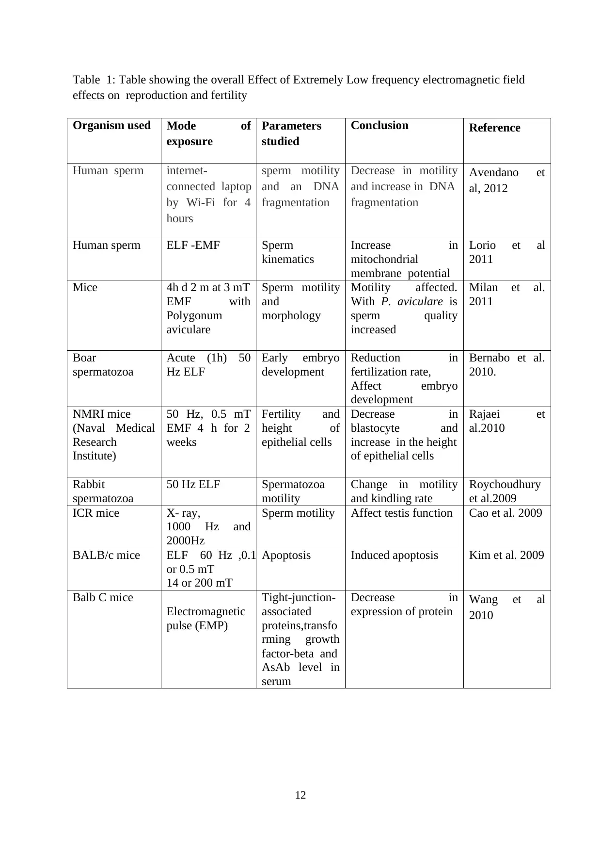
Table 1: Table showing the overall Effect of Extremely Low frequency electromagnetic field
effects on reproduction and fertility
Organism used Mode of
exposure
Parameters
studied
Conclusion Reference
Human sperm internet-
connected laptop
by Wi-Fi for 4
hours
sperm motility
and an DNA
fragmentation
Decrease in motility
and increase in DNA
fragmentation
Avendano et
al, 2012
Human sperm ELF -EMF Sperm
kinematics
Increase in
mitochondrial
membrane potential
Lorio et al
2011
Mice 4h d 2 m at 3 mT
EMF with
Polygonum
aviculare
Sperm motility
and
morphology
Motility affected.
With P. aviculare is
sperm quality
increased
Milan et al.
2011
Boar
spermatozoa
Acute (1h) 50
Hz ELF
Early embryo
development
Reduction in
fertilization rate,
Affect embryo
development
Bernabo et al.
2010.
NMRI mice
(Naval Medical
Research
Institute)
50 Hz, 0.5 mT
EMF 4 h for 2
weeks
Fertility and
height of
epithelial cells
Decrease in
blastocyte and
increase in the height
of epithelial cells
Rajaei et
al.2010
Rabbit
spermatozoa
50 Hz ELF Spermatozoa
motility
Change in motility
and kindling rate
Roychoudhury
et al.2009
ICR mice X- ray,
1000 Hz and
2000Hz
Sperm motility Affect testis function Cao et al. 2009
BALB/c mice ELF 60 Hz ,0.1
or 0.5 mT
14 or 200 mT
Apoptosis Induced apoptosis Kim et al. 2009
Balb C mice
Electromagnetic
pulse (EMP)
Tight-junction-
associated
proteins,transfo
rming growth
factor-beta and
AsAb level in
serum
Decrease in
expression of protein Wang et al
2010
⊘ This is a preview!⊘
Do you want full access?
Subscribe today to unlock all pages.
Trusted by 1+ million students worldwide
1 out of 37
Your All-in-One AI-Powered Toolkit for Academic Success.
+13062052269
info@desklib.com
Available 24*7 on WhatsApp / Email
![[object Object]](/_next/static/media/star-bottom.7253800d.svg)
Unlock your academic potential
Copyright © 2020–2026 A2Z Services. All Rights Reserved. Developed and managed by ZUCOL.