Biology Project: Eukaryotic Microbes, Classification, and Impact
VerifiedAdded on 2021/04/21
|8
|1471
|132
Project
AI Summary
This project provides a detailed overview of various eukaryotic microbes, including *Trichomonas vaginalis*, *Entamoeba histolytica*, *Plasmodium falciparum*, *Cryptococcus neoformans*, *Ascaris lumbricoides*, *Prototheca wickerhamii*, and *Pfiesteria piscicida*. The project classifies each organism, describing their characteristics, life cycles, and the diseases they cause, such as trichomoniasis, amebiasis, malaria, cryptococcosis, ascariasis, protothecosis, and the effects of *Pfiesteria piscicida*. The classification follows the categories of Archaeplastida, Excavata, Amoebozoans, Rhizaria, Chromalveolata, and Opisthokonta. The project also includes information on the symptoms, transmission, and management of each disease, providing a comprehensive understanding of these microbes and their impact on human health. References from scientific journals and websites are provided to support the information.
Name:
Instructor’s name:
Course:
Date:
Eukaryotic Microbes Project
Eukaryotes are known for their presence of nucleus and numerous membrane-bound
organelles like mitochondria, endoplasmic reticulum and Golgi apparatus and several
chromosomes (Berman). Eukaryotes comprise a large number of organisms that can be classified
into six categories namely: Archaeplastida, Excavata, Amoebozoans, Rhizaria, Chromalveolata
and Opisthokonta (Adl, Simpson and Farmer).
Trichomonas vaginalis
Classification
Kingdom: Excavata
Genus: Trichomonas
Species: Trichomonas vaginalis
T.vaginalis possesses five flagella. Four are found at the anterior while the fifth is located within
the undulating membrane that gives it a quivering motility. It is pear shaped and has a rigid costa that
supports the undulating membrane. It possesses an axostyle that anchors T.vaginalis to the epithelial of
the vagina.
Paraphrase This Document
Need a fresh take? Get an instant paraphrase of this document with our AI Paraphraser
T.vaginalis reproduces through longitudinal binary fusion. Trophozite is the only stage present in
its lifecycle. It is transmitted to the host through direct contact, especially through sexual intercourse.
T.vaginalis causes trichomoniasis that is characterized by itching and irritation in the genital
area, a frothy discharge that has a foul odor and discomfort during sex and urination. Management is by
antibiotics like metronidazole and tinidazole (Womenshealth.gov). Trichomoniasis has a mortality rate
of 1.5% globally.
Entamoeba histolytica
Classification
Kingdom: Amoebozoans
Genus: Entamoeba
Species: Entamoeba histolytica
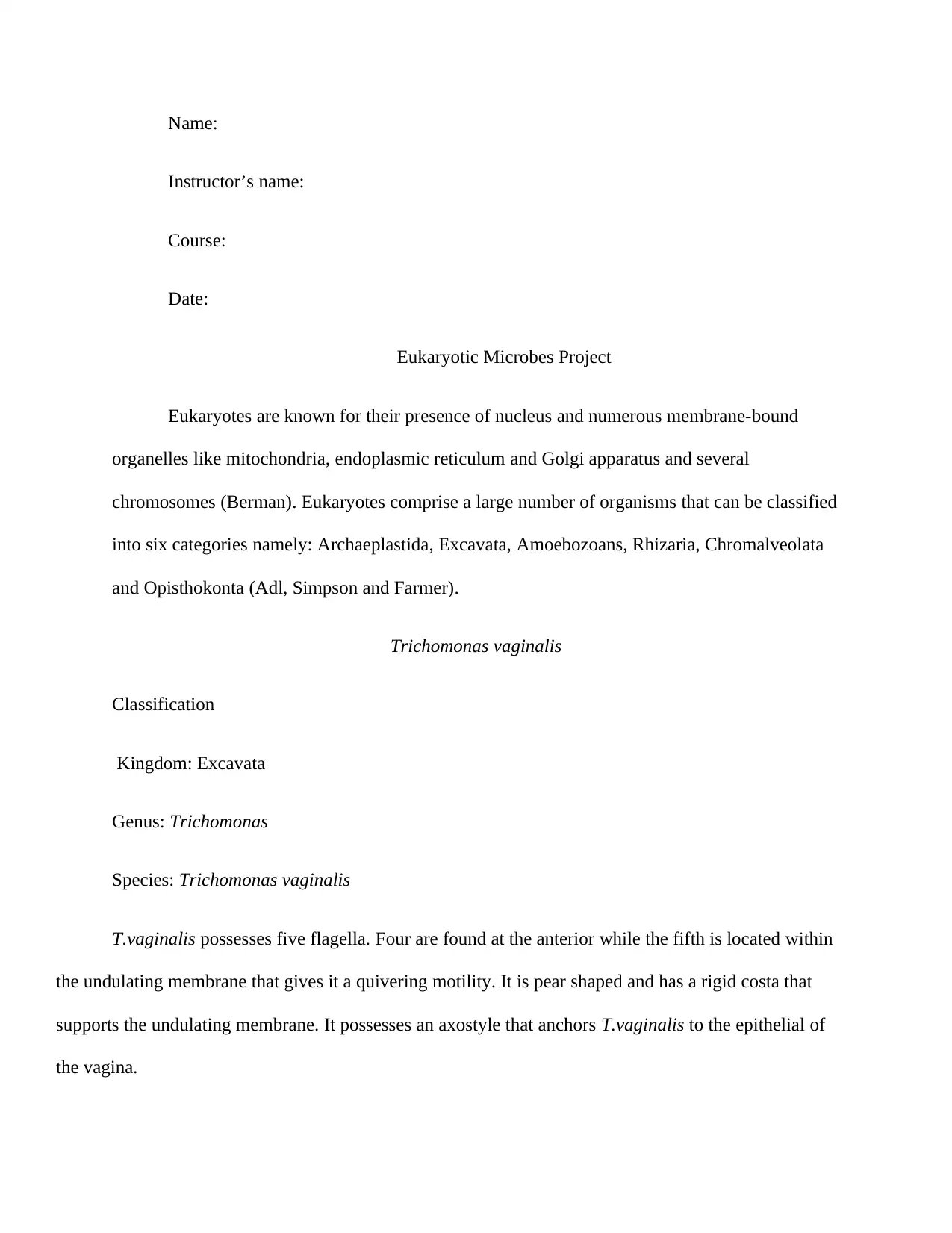
E.histolytica is anaerobic and occurs in three forms. The triphozoite has no distinct shape.
The cytoplasm is divided into a clear ectoplasm and a granulated endoplasm. A pseudopodia is
used in locomotion. The precyst is smaller in size with a round shape and projecting
pseudopodia. The cyst has a diameter of 12-15 μm. A cyst wall protects it from digestion by the
gastric juice.
its lifecycle. It is transmitted to the host through direct contact, especially through sexual intercourse.
T.vaginalis causes trichomoniasis that is characterized by itching and irritation in the genital
area, a frothy discharge that has a foul odor and discomfort during sex and urination. Management is by
antibiotics like metronidazole and tinidazole (Womenshealth.gov). Trichomoniasis has a mortality rate
of 1.5% globally.
Entamoeba histolytica
Classification
Kingdom: Amoebozoans
Genus: Entamoeba
Species: Entamoeba histolytica
E.histolytica is anaerobic and occurs in three forms. The triphozoite has no distinct shape.
The cytoplasm is divided into a clear ectoplasm and a granulated endoplasm. A pseudopodia is
used in locomotion. The precyst is smaller in size with a round shape and projecting
pseudopodia. The cyst has a diameter of 12-15 μm. A cyst wall protects it from digestion by the
gastric juice.
E.histolytica is passed in stool. One is infected with E.histolytica when he/ she ingest
mature cysts. Excystation occurs in the small intestine and trophozoite is released. These migrate
to the colon, where multiplication by binary fusion occurs and cysts are produced and passed in
the feces (CDC).
E.histolytica causes ameabiasis. It is characterized by diarrhea, stomach pain and
cramping. In severe cases, amebic dysentery occurs. Ameabiasis is treated by antibiotics.
E.histolytica is estimated at 50 million people with 100,000 deaths annually.
Plasmodium falciparum
Classification
Kingdom: Alveolata
Genus: Plasmodium
Species: Plasmodium falciparum
This is a unicellular protozoan parasite. During the ring stage, it is smaller and has
numerous rings. Young trophozoites have an amoebic shape with well defined rings and one
chromatin bead. The schizont has merozonites that help to rupture of erythrocytes. The asexual
P.falciparum are ring shaped while the sexual form’s size is 7-14μm and is banana shaped.
During the feeding, the Anopheles mosquito injects the body with sporozoites which
infect the liver and mature to schizonts. These rupture and release merozonites that infect the
blood. Asexual multiplication occurs in the erythrocytes. Some merozonites differentiate to the
sexual gametophytes. The male and female gametophytes are ingested by the Anopheles
⊘ This is a preview!⊘
Do you want full access?
Subscribe today to unlock all pages.
Trusted by 1+ million students worldwide
mosquito where the male penetrate the female producing zygotes which develop to oocytes.
These oocytes rupture and release sporozoites. (CDC)
P.falciparum causes malaria that causes fever, chills, headache, nausea and general
weakness. Infections may progress to severe malaria. Malaria is managed by antimalarial drugs
like chloroquine, mefloquine or amediaquine. Malaria infection is estimated at 212 million with
429,000 deaths (WHO)
Cryptococcus neoformans
Classification
Kingdom: Fungi
Genus: Cryptococcus
Species: Cryptococcus neoformans
Cryptococcus neoformans is an obligate aerobe. C.neoformans has a diameter of 2-8μm. It has a
unicellular nucleus that is irregular in shape. The cytoplasm has ribosomes and cell organelles.
C.neoformans reproduces through sexual means. This occurs when two haploid fuse and
mitosis follow. This results in the production of hyphae. The end of the hyphae forms a basidium
that produces spores. The fusion of the haploid cells forms one diploid nucleus. Meiosis and
mitosis follow.
Cryptococcis is caused by C.neoformans. It affects the lungs and the CNS. In the lungs, it
causes coughing, fever, chest pain and shortness of breath. Cryptococcal meningitis causes fever,
headache, nausea, vomiting, confusion and sensitivity to light. Cryptococcis is managed by
Paraphrase This Document
Need a fresh take? Get an instant paraphrase of this document with our AI Paraphraser
antifungal medication. The estimate of cryptoccocal meningitis is at 220,000 globally with a
mortality rate of 181,000 deaths per year (CDC).
Ascaris lumbricoides
Clasification
Kingdom: Animalia
Genus: Ascaris
Species: Ascaris lumbricoides
A.lumbricoides is very long ranging from 15-50m depending on the sex. They have a cream
color. Its muscle bands are longitudinal. It doesn’t have a circulatory system and the other
systems are suspended within the pseudocoelom.
A female worm produces up to 200,000 a day which are passed through the feces. Unfertilized
eggs are not infective but fertilized eggs are. When consumed, the larvae hatch and invade the
intestinal mucosa and circulate to the lungs where they mature and ascend to the throat where
they are swallowed. At the small intestines, they mature further.
A.lumbricoides causes ascariaisis which is asymptomatic. Severe infection causes intestinal
blockage. Ascariasis is managed by antihelmenthic medications such as albendazole and
mebendazole. Ascariasis is estimated at 807 million–1.2 billion people globally (CDC).
Prototheca wickerhamii
Classification
Kingdom: Archaeplastida
Genus: Prototheca
Species: Prototheca wickerhamii
They are smooth and moist and live in colonies. They vary in size from 3-30μm. They
lack mycelium. They reproduce through large sporangia that contain sporangiosphores. These
are produced through nuclear division and cleavage of the cytoplasm.
They only grow when illuminated which activates the G1 phase where cell volume of
daughter cells is doubled. The division of the first DNA replication occurs. They reach the
commitment point (CP) which allows for the beginning of the second G1 phase. The process
repeats itself and that leads to the reaching of the second CP. This process can occur again
leading to the achieving of the third CP.
P.wickerhamii causes protothecosis which is characterized by cutaneous lesions,
infections of the bursa subcutanea olecrani and systemic infections. Management is by topical
therapy with amphotericin B, fluconazole, among others. Protothecosis has a mortality rate of
2.2% (Lass-Flori and Mayr).
Pfiesteria piscicida
Classification
Kingdom: Stramenopila
Genus: Pfiesteria
Species: Pfiesteria piscicida
⊘ This is a preview!⊘
Do you want full access?
Subscribe today to unlock all pages.
Trusted by 1+ million students worldwide
It has flagella for movement. Heterotrophic and mixotrophic are their means of feeding.
They have circular chromosomes but no nucleosomes. They also have extranuclear spindles and
nuclear DNA.
They have a sexual life cycle whereby pairs of motile gamete pair to form a planozygote.
These grow through feeding of cryptophytes. They form zyogote cysts through the formation of
a uninuclear nonmotile cyst which exhibited nuclear cyclosis. A single cell division occurs after
the nuclear cyclosis.
Infected individuals exhibit irritated and burning skin, confusion, memory loss,
behavioral changes, headaches, skin rash, nausea and vomiting. Studies are ongoing to determine
its epidemiology.
This division of eukaryotes to the different categories has made their study easier and
their impact on the human health is well understood.
References
Adl, Sina M, et al. "The New Higher Level Classification of Eukaryotes with Emphasis
on the Taxonomy of Protists." J. Eukaryot. Microbiol (2005): 399-451.
Berman, Jules J. Taxonomic Guide to Infectious Diseases: Understanding the Biologic
Classes of Pathogenic Organisms. Amsterdam: Academic Press, 2012.
CDC. C. neoformans Infection Statistics. 20 July 2017. 23 February 2018
<https://www.cdc.gov/fungal/diseases/cryptococcosis-neoformans/statistics.html>.
—. Malaria. 29 December 2017. 23 February 2018
<https://www.cdc.gov/dpdx/malaria/index.html>.
Paraphrase This Document
Need a fresh take? Get an instant paraphrase of this document with our AI Paraphraser
—. Parasites- Ascariasis. 15 February 2018. 23 February 2018
<https://www.cdc.gov/parasites/ascariasis/index.html>.
—. Pathogen & Environment. 20 July 2015. 23 February 2018
<https://www.cdc.gov/parasites/amebiasis/pathogen.html>.
Lass-Flori, Cornelia and Astrid Mayr. "Human Protothecosis." Clinical Microbiology
Reviews (2007): 230-242.
WHO. 10 facts on malaria. December 2016. 23 February 2018
<http://www.who.int/features/factfiles/malaria/en/>.
Womenshealth.gov. Trichomoniasis. 12 June 2017. 23 February 2018
<https://www.womenshealth.gov/a-z-topics/trichomoniasis>.
1 out of 8
Your All-in-One AI-Powered Toolkit for Academic Success.
+13062052269
info@desklib.com
Available 24*7 on WhatsApp / Email
![[object Object]](/_next/static/media/star-bottom.7253800d.svg)
Unlock your academic potential
Copyright © 2020–2026 A2Z Services. All Rights Reserved. Developed and managed by ZUCOL.