EXSC 610: Chapter Essay Questions - Module/Week 4 Assignment
VerifiedAdded on 2022/11/14
|4
|1016
|288
Homework Assignment
AI Summary
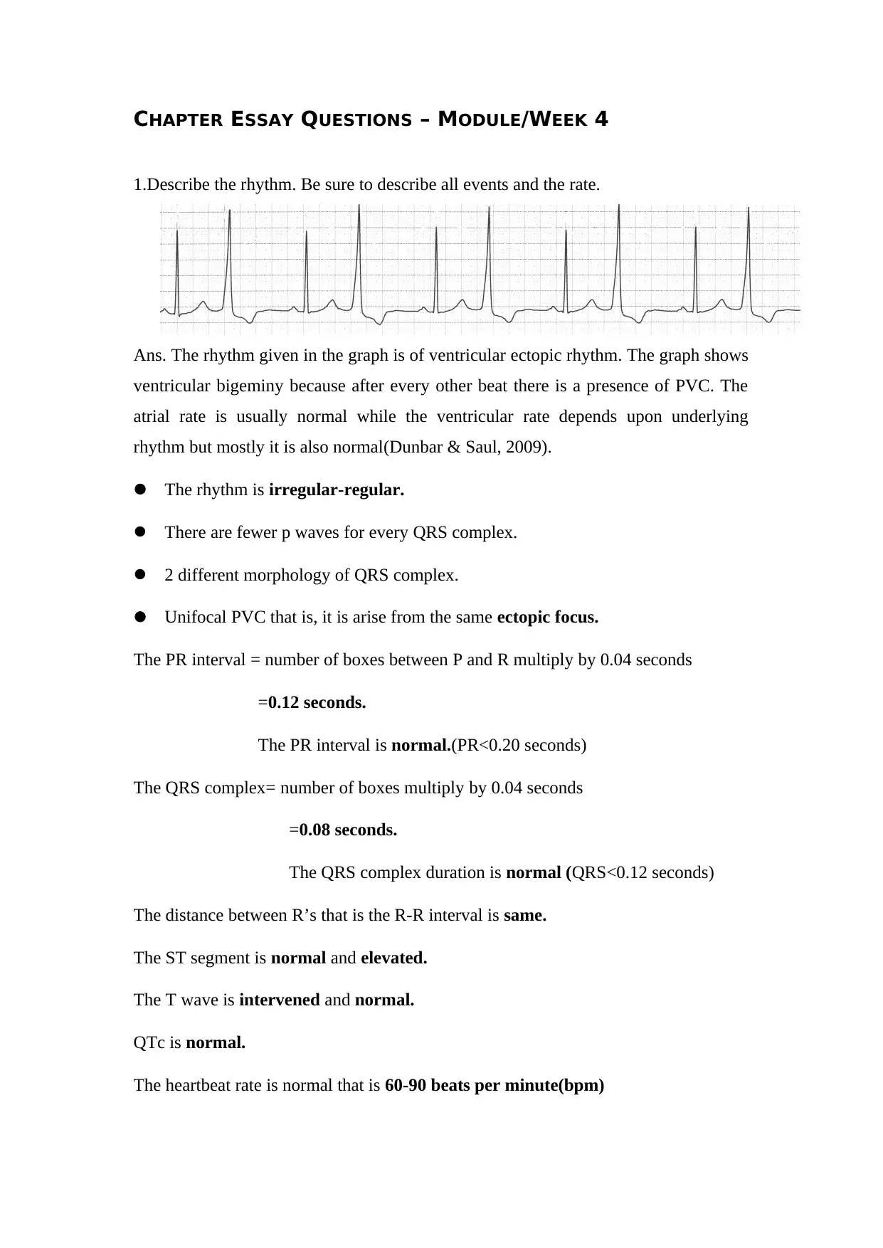
This assignment addresses four essay questions related to ECG interpretation and cardiac rhythms. The first question describes a ventricular ectopic rhythm, detailing the presence of ventricular bigeminy, PVCs, and the characteristics of the ECG graph. The second question explains the causes and variations of PVCs, including single, doublet, triplet, and multiform types. The third question explores the function of electronic pacemakers and their impact on ECG readings, including how they alter QRS complexes and maintain a stable heart rate. The final question defines third-degree atrioventricular block and distinguishes it from atrioventricular dissociation, highlighting the differences in their conditions and ECG presentations. The assignment references the course textbook and additional scholarly sources to support the explanations.

CHAPTER ESSAY QUESTIONS – MODULE/WEEK 4
1.Describe the rhythm. Be sure to describe all events and the rate.
Ans. The rhythm given in the graph is of ventricular ectopic rhythm. The graph shows
ventricular bigeminy because after every other beat there is a presence of PVC. The
atrial rate is usually normal while the ventricular rate depends upon underlying
rhythm but mostly it is also normal(Dunbar & Saul, 2009).
The rhythm is irregular-regular.
There are fewer p waves for every QRS complex.
2 different morphology of QRS complex.
Unifocal PVC that is, it is arise from the same ectopic focus.
The PR interval = number of boxes between P and R multiply by 0.04 seconds
=0.12 seconds.
The PR interval is normal.(PR<0.20 seconds)
The QRS complex= number of boxes multiply by 0.04 seconds
=0.08 seconds.
The QRS complex duration is normal (QRS<0.12 seconds)
The distance between R’s that is the R-R interval is same.
The ST segment is normal and elevated.
The T wave is intervened and normal.
QTc is normal.
The heartbeat rate is normal that is 60-90 beats per minute(bpm)
Paraphrase This Document
Need a fresh take? Get an instant paraphrase of this document with our AI Paraphraser

2.Explain what is occurring during a PVC and all possible variations of PVCs
discussed in the textbook.
Ans. The normal heartbeat is generated through the SA node that is the sino atrial
node. when instead of coming from the SA node the heartbeat comes from the
purkinje fibre it generates a PVC. There is always a pause after every regular
heartbeat, given that PVC occurs before every regular heartbeat(Lee & Deyell, n.d.).
The QRS complex in the box is generally wider and distinguished in it’s morphology
from other complexes. The beats are early and are immature.
There are 2 main types of PVC’s that can occur single or in repeated patterns. The
isolated PVC’s are the single PVC’s. Two PVC’s that occur together are called as
doublet where three PVC’s are called as triplets. Ventricular tachycardia is when 3 or
more than 3 consecutive PVC’s occur. Similarly, a doublet is when 2 alternate PVC’s
is present with one sinus beat this is called as ventricular bigeminy(Lee & Deyell,
n.d.). A trigeminy happens when 3 are alternate in a beat.
There are multiform and uniform types of PVC’s, in uniform all the PVC’s look alike
whereas in multiform the PVC’s are different in their morphology or appearance.
Uniform PVC’s look like the same because they arise from the same ectopic focus,
while multiform comes from different parts of ventricles and are called as multifocal
PVC’s(Dunbar & Saul, 2009).
3.Explain how an electronic pacemaker works and how it alters ECG.
Ans. When the natural pacemaker, the SA node does not work properly an artificial or
electronic pacemaker is implanted in the body. An electronic pacemaker works on a
battery, a generator and a microchip which transmits the signals from the heart.
The basic and the very first electronic pacemaker works on a pacing electrode which
was connected to the controlling box via a wire and is placed in the epicardial surface.
The pacemaker is placed in the left atrium because if placed in the right atrium it will
from clots(Dunbar & Saul, 2009).
The impulses from the pacemaker depolarises the area in the ventricle which is near
the pacing electrode through wires. Depolarisation is then spread all over via cells or
gap junctions.
It alters the ECG by creating QRS complexes more wider, because the depolarisation

did not spread through a normal heart condition. The heartbeat is stable around 72
beats per minute (Pacemaker biology, 2004).
The early or the old pacemakers had an already set up pace on which the basic design
of the pacemaker is established. The new and virtual pacemakers has no set up pace
but works according to the human heart and sets pace accordingly(Pacemaker
biology, 2004).
4.Define a third-degree atrioventricular block and then explain how atrioventricular
disassociation differs from a third-degree block.
Ans. Third degree atrioventricular block is the complete dissociation of the atria and
the ventricle resulting in a complete heart block. The atria and the ventricle are
independent of each other and does not work simultaneously. The p waves are never
with the QRS complexes. The third degree block is permanent and it often require an
electronic pacemaker to make AV conduction possible(Brady & Alibertis, 2013).
Both the cases are different but very much confusing. Atrioventricular disassociation
differs from a third-degree block by the fact that atrioventricular dissociation is not a
permanent condition and can happen to anyone in sleep while a third degree block is a
permanent condition. In both the cases there is no conduction between the atria and
the ventricle.
Av dissociation has almost the same ECG as the third degree block but differsin the
ventricle activity that is in AV dissociation the ventricular rate is faster(Dunbar &
Saul, 2009).
⊘ This is a preview!⊘
Do you want full access?
Subscribe today to unlock all pages.

Trusted by 1+ million students worldwide

References
Brady, W., & Alibertis, K. (2013). The ECG in prehospital emergency care.
Chichester: Wiley-Blackwell.
Dunbar, C., & Saul, B. (2009). ECG interpretation for the clinical exercise
physiologist. Philadelphia, PA: Wolters Kluwer/Lippincott Williams Wilkins.
Globe Fearon. (2004). Pacemaker biology. Parsippany N.J.
Lee, A., & Deyell, M. Premature ventricular contraction-induced cardiomyopathy.
1 out of 4
Your All-in-One AI-Powered Toolkit for Academic Success.
+13062052269
info@desklib.com
Available 24*7 on WhatsApp / Email
Unlock your academic potential
Copyright © 2020–2026 A2Z Services. All Rights Reserved. Developed and managed by ZUCOL.


