Female Reproductive System Report

Verified

Added on  2019/09/20

|5
|928
|413
Report
AI Summary
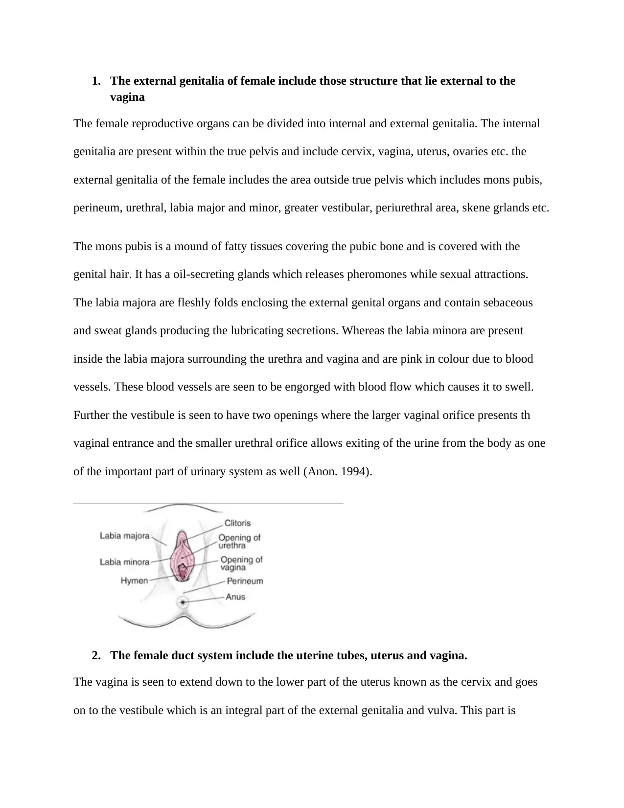
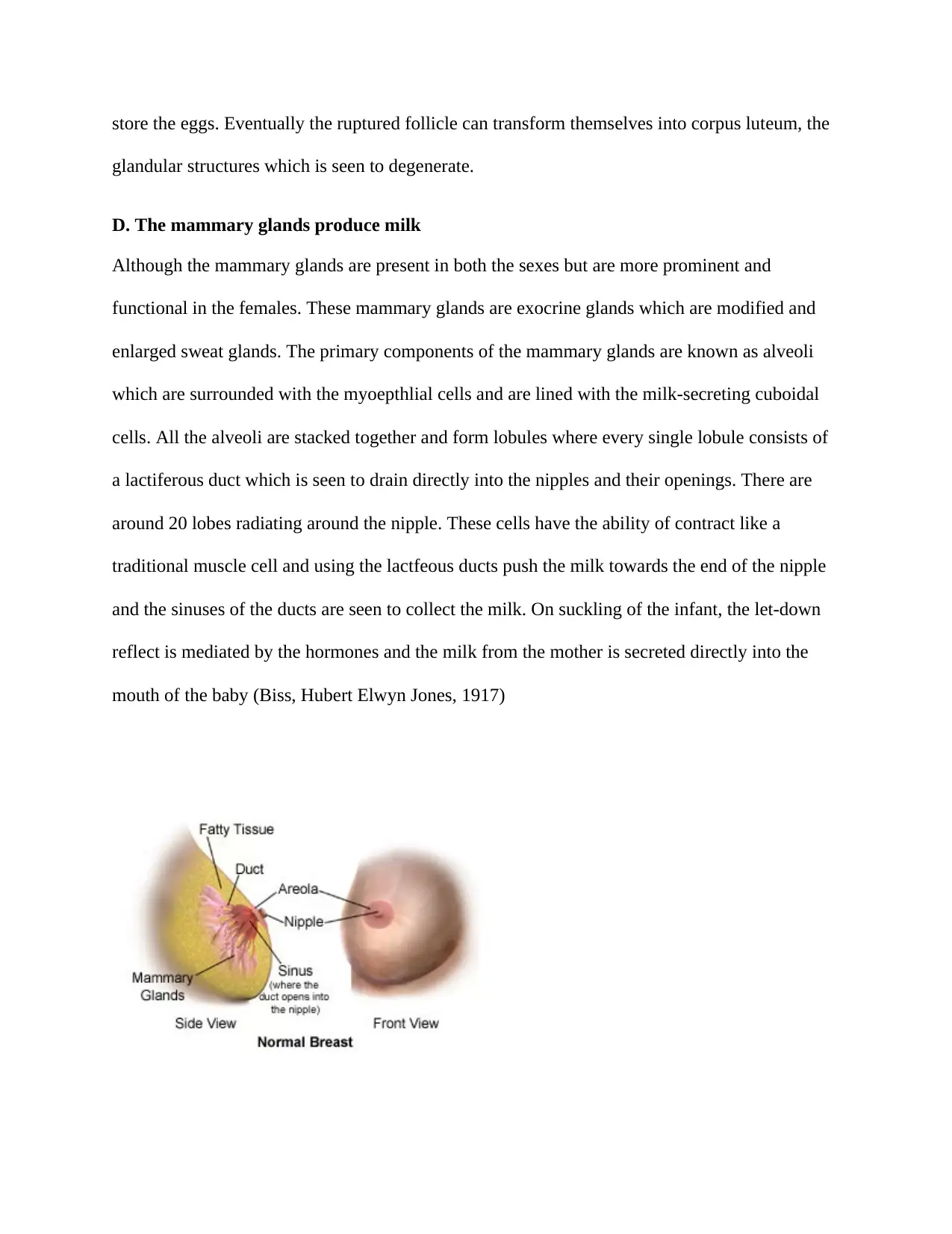
This report details the female reproductive system, dividing it into internal and external genitalia. The external genitalia include the mons pubis, labia majora and minora, and vestibule. The internal genitalia comprise the vagina, uterus, fallopian tubes, and ovaries. The report explains the functions of each organ, including the vagina's role in sexual intercourse, menstruation, and childbirth; the uterus's role in fetal development; the ovaries' role in egg production and hormone secretion; and the fallopian tubes' role in egg transport. The report also describes the mammary glands and their milk-producing function. References are provided for further reading.
tabler-icon-diamond-filled.svg

Contribute Materials

Your contribution can guide someone’s learning journey. Share your documents today.
Document Page
1. The external genitalia of female include those structure that lie external to the
vagina
The female reproductive organs can be divided into internal and external genitalia. The internal
genitalia are present within the true pelvis and include cervix, vagina, uterus, ovaries etc. the
external genitalia of the female includes the area outside true pelvis which includes mons pubis,
perineum, urethral, labia major and minor, greater vestibular, periurethral area, skene grlands etc.
The mons pubis is a mound of fatty tissues covering the pubic bone and is covered with the
genital hair. It has a oil-secreting glands which releases pheromones while sexual attractions.
The labia majora are fleshly folds enclosing the external genital organs and contain sebaceous
and sweat glands producing the lubricating secretions. Whereas the labia minora are present
inside the labia majora surrounding the urethra and vagina and are pink in colour due to blood
vessels. These blood vessels are seen to be engorged with blood flow which causes it to swell.
Further the vestibule is seen to have two openings where the larger vaginal orifice presents th
vaginal entrance and the smaller urethral orifice allows exiting of the urine from the body as one
of the important part of urinary system as well (Anon. 1994).
2. The female duct system include the uterine tubes, uterus and vagina.
The vagina is seen to extend down to the lower part of the uterus known as the cervix and goes
on to the vestibule which is an integral part of the external genitalia and vulva. This part is
tabler-icon-diamond-filled.svg

Secure Best Marks with AI Grader

Need help grading? Try our AI Grader for instant feedback on your assignments.
Document Page
present in front of the rectum and behind the bladder. The muscles wall of vagina is smooth and
is lined by the inner mucous membrane. Exactly like the inner layer of the uterine tubes, this
lining is seen to be continuous with the uterus and its mucous lining. The three primary functions
of the vagina are: receiving the male penis while experiencing the sexual intercourse, carrying
the menstrual flow outside and serving as a birth canal while experiencing the labor pain. The
uterus has a shape of upside-down pear and has muscular walls with thick linings containing the
strongest muscles which have the capacity of contracting and expanding and can accommodate
the growing fetus during pregnancy. The upper corner of uterus are connected to the ovaries
using the fallopian tubes (Avraham, Regina, 2001)
C. Immature eggs develop in follicles in the ovaries
The ovaries are two oval-shaped organs that lie to the upper right and left of the uterus. They
produce, store, and release eggs into the fallopian tubes in the process called ovulation. The
female gametes are seen to be produced from the ovaries whereas the sex hormones are secreted
rom the progesterone and the estrogen. These ovaries are filled with the follicles which are
structure with fluids where the eggs or the oocytes grow till they mature. As the females are
incapable of producing or making new eggs, a continuous decline in the eggs is seen every
month. The ovarian follicles are sac-like structures which contain oocyte that is enclosed with
the different layers. These follicles have different structures at different stages as primordial,
primary and vesicular or secondary follicles (Enger, Eldon D, Frederick C Ross, and David B
Bailey. n.d)
Every month, a large supply of eggs are present in the ovary which are either primordial or
immature in nature and come to maturation after the while. Till then, the follicles of the ovary
Document Page
store the eggs. Eventually the ruptured follicle can transform themselves into corpus luteum, the
glandular structures which is seen to degenerate.
D. The mammary glands produce milk
Although the mammary glands are present in both the sexes but are more prominent and
functional in the females. These mammary glands are exocrine glands which are modified and
enlarged sweat glands. The primary components of the mammary glands are known as alveoli
which are surrounded with the myoepthlial cells and are lined with the milk-secreting cuboidal
cells. All the alveoli are stacked together and form lobules where every single lobule consists of
a lactiferous duct which is seen to drain directly into the nipples and their openings. There are
around 20 lobes radiating around the nipple. These cells have the ability of contract like a
traditional muscle cell and using the lactfeous ducts push the milk towards the end of the nipple
and the sinuses of the ducts are seen to collect the milk. On suckling of the infant, the let-down
reflect is mediated by the hormones and the milk from the mother is secreted directly into the
mouth of the baby (Biss, Hubert Elwyn Jones, 1917)
Document Page
tabler-icon-diamond-filled.svg

Secure Best Marks with AI Grader

Need help grading? Try our AI Grader for instant feedback on your assignments.
Document Page
References
Anon. 1994. Vagina And External Female Genitalia. 1st ed. Salt Lake City, UT: Medicode.
Avraham, Regina. 2001. The Reproductive System. 1st ed. Philadelphia: Chelsea House
Publishers.
Enger, Eldon D, Frederick C Ross, and David B Bailey. n.d. Concepts In Biology. 1st ed.
Biss, Hubert Elwyn Jones. 1917. The Anatomy And Physiology Of The Female Body. 1st ed.
London: Baillière, Tindall and Cox.
chevron_up_icon
1 out of 5
circle_padding
hide_on_mobile
zoom_out_icon
logo.png

Your All-in-One AI-Powered Toolkit for Academic Success.

Available 24*7 on WhatsApp / Email

[object Object]