CBMS880/731/331: Fluorescence Microscopy Assignment - Dyes and Lasers

Verified

Added on  2019/09/21

|4
|491
|286
Homework Assignment
AI Summary
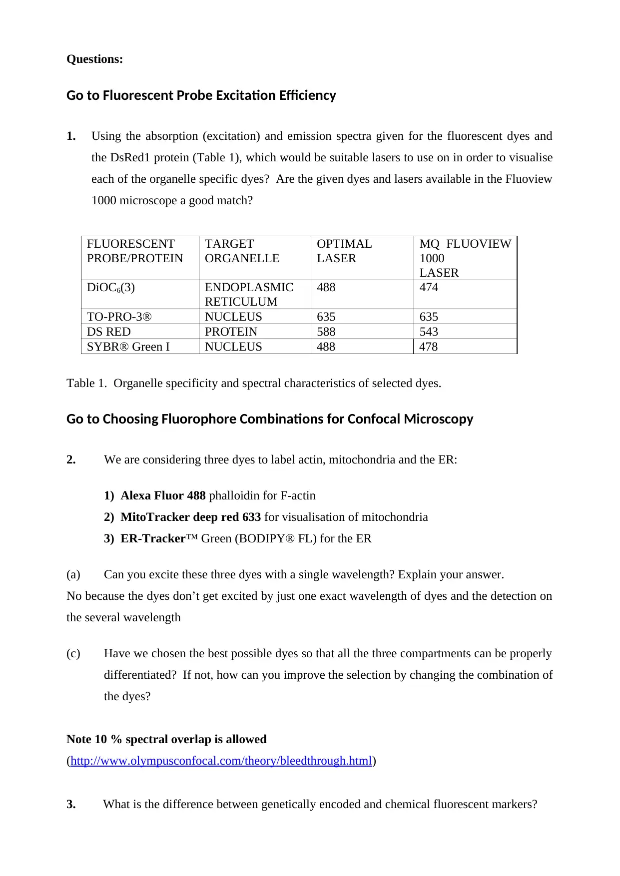
This assignment focuses on fluorescence microscopy techniques, specifically examining the use of lasers, dyes, and their applications in visualizing different cellular organelles. The assignment requires students to analyze the excitation and emission spectra of various fluorescent dyes and determine the appropriate lasers for the FLUOVIEW 1000 microscope. Students will explore the properties of dyes like DiOC6(3), TO-PRO-3, DsRED, and SYBR Green I, and evaluate the suitability of these dyes for specific organelle labeling. The assignment also delves into the selection of dye combinations for multi-labeling experiments, considering spectral overlap and the ability to differentiate between different cellular compartments. Furthermore, it explores the differences between genetically encoded and chemical fluorescent markers. The assignment uses interactive tutorials and external resources to guide students through the concepts and practical applications of fluorescence microscopy.
Document Page
Fluorescence microscopy
tutorial and quiz 2016
http:// www.olympusfluoview.com/theory/
index.html
CBMS880/731/331
tabler-icon-diamond-filled.svg

Paraphrase This Document

Need a fresh take? Get an instant paraphrase of this document with our AI Paraphraser
Document Page
Fluorescence microscopy tutorial and quiz
Provide answers to the following questions in consultation with the interactive tutorial at
www.olympusconfocal.com/java/index.html and other relevant material made available to you.
Also look into http://www.invitrogen.com/site/us/en/home/brands/Molecular-Probes.html? and
other similar websites to answer question 3. For some background, have a look at
http://micro.magnet.fsu.edu/primer/lightandcolor/fluoroexcitation.html.
Your tools:
-The FLUOVIEW 1000 system at MU has four lasers diodes: 405 nm, 473 nm, 543 nm and 635
nm
-Always go for the maximum excitation and emission possible.
-You will find many of the probes in the table under the headings “Conventional dyes”, “Alexa
dyes”, “Fluorescent proteins” and “Cyanine dyes”.
DsRED is a fluorescent protein
DiOC6(3) is similar to FITC, a traditional dye
TO-PRO-3® is a cyanine dye (Cy5)
SYBR® Green I is similar to Alexa 488
Laser types:
Start with looking at the Laser Scanning Confocal Microscope Simulator
Document Page
Questions:
Go to Fluorescent Probe Excitation Efficiency
1. Using the absorption (excitation) and emission spectra given for the fluorescent dyes and
the DsRed1 protein (Table 1), which would be suitable lasers to use on in order to visualise
each of the organelle specific dyes? Are the given dyes and lasers available in the Fluoview
1000 microscope a good match?
FLUORESCENT
PROBE/PROTEIN
TARGET
ORGANELLE
OPTIMAL
LASER
MQ FLUOVIEW
1000
LASER
DiOC6(3) ENDOPLASMIC
RETICULUM
488 474
TO-PRO-3® NUCLEUS 635 635
DS RED PROTEIN 588 543
SYBR® Green I NUCLEUS 488 478
Table 1. Organelle specificity and spectral characteristics of selected dyes.
Go to Choosing Fluorophore Combinations for Confocal Microscopy
2. We are considering three dyes to label actin, mitochondria and the ER:
1) Alexa Fluor 488 phalloidin for F-actin
2) MitoTracker deep red 633 for visualisation of mitochondria
3) ER-Tracker™ Green (BODIPY® FL) for the ER
(a) Can you excite these three dyes with a single wavelength? Explain your answer.
No because the dyes don’t get excited by just one exact wavelength of dyes and the detection on
the several wavelength
(c) Have we chosen the best possible dyes so that all the three compartments can be properly
differentiated? If not, how can you improve the selection by changing the combination of
the dyes?
Note 10 % spectral overlap is allowed
(http://www.olympusconfocal.com/theory/bleedthrough.html)
3. What is the difference between genetically encoded and chemical fluorescent markers?
Document Page
chevron_up_icon
1 out of 4
circle_padding
hide_on_mobile
zoom_out_icon
[object Object]