FA05 Functional Anatomy Assignment: Posture, Injuries, and Movement
VerifiedAdded on 2021/06/16
|28
|10026
|30
Homework Assignment
AI Summary
This functional anatomy assignment assesses the student's understanding of posture, muscle function, and common injuries. The assignment requires the identification of postural abnormalities (lordosis, kyphosis, and winged scapula), tight and weak muscles, and the suggestion of corrective exercises. It also explores the impact of poor posture on injuries like sciatica, neck pain, patellofemoral knee pain, lower back pain, and shoulder impingement. The assignment further delves into how poor posture affects muscle strength, flexibility, muscle tension, and overall function. It includes a section on joint movements, planes of movement, and range of motion. The student must also describe muscle actions (agonist, antagonist, synergist, fixator) during exercise, explain Wolff's law and its link to bone remodeling, and analyze factors affecting the center of gravity and agility. Finally, the student is asked to describe the normal spinal curves and anatomical features associated with straight-line running, including lordosis, kyphosis, and scoliosis.
FA05 – Functional anatomy
Assignment v1.4 (2018/01/25)
FA05 Functional anatomy
Name
Email address
Assessment
Assignment – Short answer
Assignments may include a variety of questions, this can include short or longer answer questions. These
questions are designed to test how you apply your knowledge into a real-world situation. All assignments
are completed as a Microsoft Word document and must be submitted through My eCampus for grading.
Your assessor is looking for how you apply your knowledge and how you think critically about the topic
area.
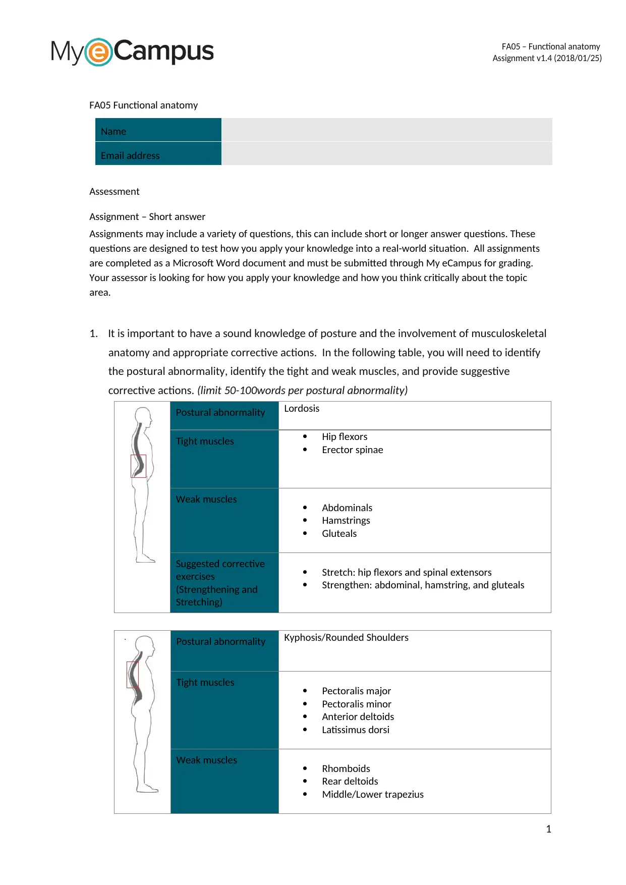
1. It is important to have a sound knowledge of posture and the involvement of musculoskeletal
anatomy and appropriate corrective actions. In the following table, you will need to identify
the postural abnormality, identify the tight and weak muscles, and provide suggestive
corrective actions. (limit 50-100words per postural abnormality)
Postural abnormality Lordosis
Tight muscles Hip flexors
Erector spinae
Weak muscles Abdominals
Hamstrings
Gluteals
Suggested corrective
exercises
(Strengthening and
Stretching)
Stretch: hip flexors and spinal extensors
Strengthen: abdominal, hamstring, and gluteals
Postural abnormality Kyphosis/Rounded Shoulders
Tight muscles Pectoralis major
Pectoralis minor
Anterior deltoids
Latissimus dorsi
Weak muscles Rhomboids
Rear deltoids
Middle/Lower trapezius
1
Assignment v1.4 (2018/01/25)
FA05 Functional anatomy
Name
Email address
Assessment
Assignment – Short answer
Assignments may include a variety of questions, this can include short or longer answer questions. These
questions are designed to test how you apply your knowledge into a real-world situation. All assignments
are completed as a Microsoft Word document and must be submitted through My eCampus for grading.
Your assessor is looking for how you apply your knowledge and how you think critically about the topic
area.
1. It is important to have a sound knowledge of posture and the involvement of musculoskeletal
anatomy and appropriate corrective actions. In the following table, you will need to identify
the postural abnormality, identify the tight and weak muscles, and provide suggestive
corrective actions. (limit 50-100words per postural abnormality)
Postural abnormality Lordosis
Tight muscles Hip flexors
Erector spinae
Weak muscles Abdominals
Hamstrings
Gluteals
Suggested corrective
exercises
(Strengthening and
Stretching)
Stretch: hip flexors and spinal extensors
Strengthen: abdominal, hamstring, and gluteals
Postural abnormality Kyphosis/Rounded Shoulders
Tight muscles Pectoralis major
Pectoralis minor
Anterior deltoids
Latissimus dorsi
Weak muscles Rhomboids
Rear deltoids
Middle/Lower trapezius
1
Paraphrase This Document
Need a fresh take? Get an instant paraphrase of this document with our AI Paraphraser
FA05 – Functional anatomy
Assignment v1.4 (2018/01/25)
Suggested corrective
exercises
(Strengthening and
Stretching)
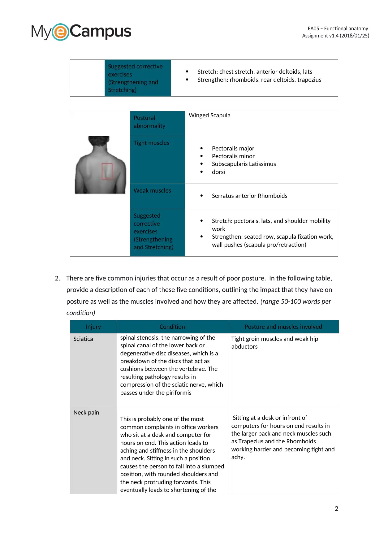
Stretch: chest stretch, anterior deltoids, lats
Strengthen: rhomboids, rear deltoids, trapezius
Postural
abnormality
Winged Scapula
Tight muscles Pectoralis major
Pectoralis minor
Subscapularis Latissimus
dorsi
Weak muscles Serratus anterior Rhomboids
Suggested
corrective
exercises
(Strengthening
and Stretching)
Stretch: pectorals, lats, and shoulder mobility
work
Strengthen: seated row, scapula fixation work,
wall pushes (scapula pro/retraction)
2. There are five common injuries that occur as a result of poor posture. In the following table,
provide a description of each of these five conditions, outlining the impact that they have on
posture as well as the muscles involved and how they are affected. (range 50-100 words per
condition)
Injury Condition Posture and muscles involved
Sciatica spinal stenosis, the narrowing of the
spinal canal of the lower back or
degenerative disc diseases, which is a
breakdown of the discs that act as
cushions between the vertebrae. The
resulting pathology results in
compression of the sciatic nerve, which
passes under the piriformis
Tight groin muscles and weak hip
abductors
Neck pain
This is probably one of the most
common complaints in office workers
who sit at a desk and computer for
hours on end. This action leads to
aching and stiffness in the shoulders
and neck. Sitting in such a position
causes the person to fall into a slumped
position, with rounded shoulders and
the neck protruding forwards. This
eventually leads to shortening of the
Sitting at a desk or infront of
computers for hours on end results in
the larger back and neck muscles such
as Trapezius and the Rhomboids
working harder and becoming tight and
achy.
2
Assignment v1.4 (2018/01/25)
Suggested corrective
exercises
(Strengthening and
Stretching)
Stretch: chest stretch, anterior deltoids, lats
Strengthen: rhomboids, rear deltoids, trapezius
Postural
abnormality
Winged Scapula
Tight muscles Pectoralis major
Pectoralis minor
Subscapularis Latissimus
dorsi
Weak muscles Serratus anterior Rhomboids
Suggested
corrective
exercises
(Strengthening
and Stretching)
Stretch: pectorals, lats, and shoulder mobility
work
Strengthen: seated row, scapula fixation work,
wall pushes (scapula pro/retraction)
2. There are five common injuries that occur as a result of poor posture. In the following table,
provide a description of each of these five conditions, outlining the impact that they have on
posture as well as the muscles involved and how they are affected. (range 50-100 words per
condition)
Injury Condition Posture and muscles involved
Sciatica spinal stenosis, the narrowing of the
spinal canal of the lower back or
degenerative disc diseases, which is a
breakdown of the discs that act as
cushions between the vertebrae. The
resulting pathology results in
compression of the sciatic nerve, which
passes under the piriformis
Tight groin muscles and weak hip
abductors
Neck pain
This is probably one of the most
common complaints in office workers
who sit at a desk and computer for
hours on end. This action leads to
aching and stiffness in the shoulders
and neck. Sitting in such a position
causes the person to fall into a slumped
position, with rounded shoulders and
the neck protruding forwards. This
eventually leads to shortening of the
Sitting at a desk or infront of
computers for hours on end results in
the larger back and neck muscles such
as Trapezius and the Rhomboids
working harder and becoming tight and
achy.
2
FA05 – Functional anatomy
Assignment v1.4 (2018/01/25)
Injury Condition Posture and muscles involved
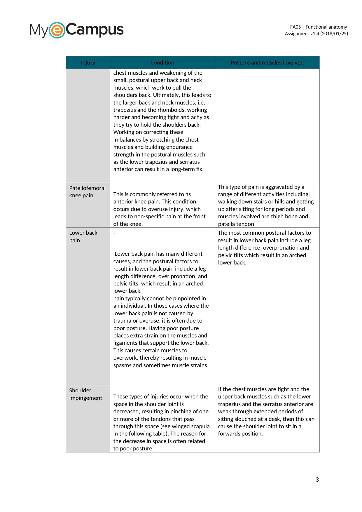
chest muscles and weakening of the
small, postural upper back and neck
muscles, which work to pull the
shoulders back. Ultimately, this leads to
the larger back and neck muscles, i.e.
trapezius and the rhomboids, working
harder and becoming tight and achy as
they try to hold the shoulders back.
Working on correcting these
imbalances by stretching the chest
muscles and building endurance
strength in the postural muscles such
as the lower trapezius and serratus
anterior can result in a long-term fix.
Patellofemoral
knee pain This is commonly referred to as
anterior knee pain. This condition
occurs due to overuse injury, which
leads to non-specific pain at the front
of the knee.
This type of pain is aggravated by a
range of different activities including:
walking down stairs or hills and getting
up after sitting for long periods and
muscles involved are thigh bone and
patella tendon
Lower back
pain
.
.
Lower back pain has many different
causes, and the postural factors to
result in lower back pain include a leg
length difference, over pronation, and
pelvic tilts, which result in an arched
lower back.
pain typically cannot be pinpointed in
an individual. In those cases where the
lower back pain is not caused by
trauma or overuse, it is often due to
poor posture. Having poor posture
places extra strain on the muscles and
ligaments that support the lower back.
This causes certain muscles to
overwork, thereby resulting in muscle
spasms and sometimes muscle strains.
The most common postural factors to
result in lower back pain include a leg
length difference, overpronation and
pelvic tilts which result in an arched
lower back.
Shoulder
impingement These types of injuries occur when the
space in the shoulder joint is
decreased, resulting in pinching of one
or more of the tendons that pass
through this space (see winged scapula
in the following table). The reason for
the decrease in space is often related
to poor posture.
If the chest muscles are tight and the
upper back muscles such as the lower
trapezius and the serratus anterior are
weak through extended periods of
sitting slouched at a desk, then this can
cause the shoulder joint to sit in a
forwards position.
3
Assignment v1.4 (2018/01/25)
Injury Condition Posture and muscles involved
chest muscles and weakening of the
small, postural upper back and neck
muscles, which work to pull the
shoulders back. Ultimately, this leads to
the larger back and neck muscles, i.e.
trapezius and the rhomboids, working
harder and becoming tight and achy as
they try to hold the shoulders back.
Working on correcting these
imbalances by stretching the chest
muscles and building endurance
strength in the postural muscles such
as the lower trapezius and serratus
anterior can result in a long-term fix.
Patellofemoral
knee pain This is commonly referred to as
anterior knee pain. This condition
occurs due to overuse injury, which
leads to non-specific pain at the front
of the knee.
This type of pain is aggravated by a
range of different activities including:
walking down stairs or hills and getting
up after sitting for long periods and
muscles involved are thigh bone and
patella tendon
Lower back
pain
.
.
Lower back pain has many different
causes, and the postural factors to
result in lower back pain include a leg
length difference, over pronation, and
pelvic tilts, which result in an arched
lower back.
pain typically cannot be pinpointed in
an individual. In those cases where the
lower back pain is not caused by
trauma or overuse, it is often due to
poor posture. Having poor posture
places extra strain on the muscles and
ligaments that support the lower back.
This causes certain muscles to
overwork, thereby resulting in muscle
spasms and sometimes muscle strains.
The most common postural factors to
result in lower back pain include a leg
length difference, overpronation and
pelvic tilts which result in an arched
lower back.
Shoulder
impingement These types of injuries occur when the
space in the shoulder joint is
decreased, resulting in pinching of one
or more of the tendons that pass
through this space (see winged scapula
in the following table). The reason for
the decrease in space is often related
to poor posture.
If the chest muscles are tight and the
upper back muscles such as the lower
trapezius and the serratus anterior are
weak through extended periods of
sitting slouched at a desk, then this can
cause the shoulder joint to sit in a
forwards position.
3
⊘ This is a preview!⊘
Do you want full access?
Subscribe today to unlock all pages.
Trusted by 1+ million students worldwide
FA05 – Functional anatomy
Assignment v1.4 (2018/01/25)
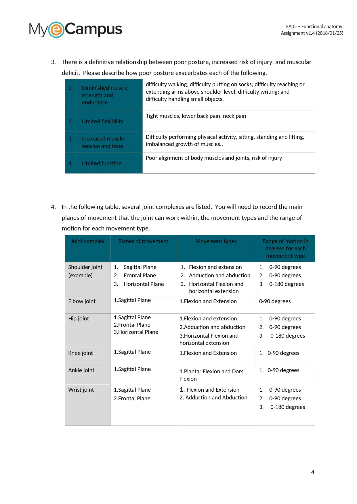
3. There is a definitive relationship between poor posture, increased risk of injury, and muscular
deficit. Please describe how poor posture exacerbates each of the following.
1. Diminished muscle
strength and
endurance
difficulty walking; difficulty putting on socks; difficulty reaching or
extending arms above shoulder level; difficulty writing; and
difficulty handling small objects.
2. Limited flexibility Tight muscles, lower back pain, neck pain
3. Increased muscle
tension and tone
Difficulty performing physical activity, sitting, standing and lifting,
imbalanced growth of muscles..
4. Limited function Poor alignment of body muscles and joints, risk of injury
4. In the following table, several joint complexes are listed. You will need to record the main
planes of movement that the joint can work within, the movement types and the range of
motion for each movement type.
Joint complex Planes of movement Movement types Range of motion in
degrees for each
movement type
Shoulder joint
(example)
1. Sagittal Plane
2. Frontal Plane
3. Horizontal Plane
1. Flexion and extension
2. Adduction and abduction
3. Horizontal Flexion and
horizontal extension
1. 0-90 degrees
2. 0-90 degrees
3. 0-180 degrees
Elbow joint 1.Sagittal Plane 1.Flexion and Extension 0-90 degrees
Hip joint 1.Sagittal Plane
2.Frontal Plane
3.Horizontal Plane
1.Flexion and extension
2.Adduction and abduction
3.Horizontal Flexion and
horizontal extension
1. 0-90 degrees
2. 0-90 degrees
3. 0-180 degrees
Knee joint 1.Sagittal Plane 1.Flexion and Extension 1. 0-90 degrees
Ankle joint 1.Sagittal Plane 1.Plantar Flexion and Dorsi
Flexion
1. 0-90 degrees
Wrist joint 1.Sagittal Plane
2.Frontal Plane
1. Flexion and Extension
2. Adduction and Abduction
1. 0-90 degrees
2. 0-90 degrees
3. 0-180 degrees
4
Assignment v1.4 (2018/01/25)
3. There is a definitive relationship between poor posture, increased risk of injury, and muscular
deficit. Please describe how poor posture exacerbates each of the following.
1. Diminished muscle
strength and
endurance
difficulty walking; difficulty putting on socks; difficulty reaching or
extending arms above shoulder level; difficulty writing; and
difficulty handling small objects.
2. Limited flexibility Tight muscles, lower back pain, neck pain
3. Increased muscle
tension and tone
Difficulty performing physical activity, sitting, standing and lifting,
imbalanced growth of muscles..
4. Limited function Poor alignment of body muscles and joints, risk of injury
4. In the following table, several joint complexes are listed. You will need to record the main
planes of movement that the joint can work within, the movement types and the range of
motion for each movement type.
Joint complex Planes of movement Movement types Range of motion in
degrees for each
movement type
Shoulder joint
(example)
1. Sagittal Plane
2. Frontal Plane
3. Horizontal Plane
1. Flexion and extension
2. Adduction and abduction
3. Horizontal Flexion and
horizontal extension
1. 0-90 degrees
2. 0-90 degrees
3. 0-180 degrees
Elbow joint 1.Sagittal Plane 1.Flexion and Extension 0-90 degrees
Hip joint 1.Sagittal Plane
2.Frontal Plane
3.Horizontal Plane
1.Flexion and extension
2.Adduction and abduction
3.Horizontal Flexion and
horizontal extension
1. 0-90 degrees
2. 0-90 degrees
3. 0-180 degrees
Knee joint 1.Sagittal Plane 1.Flexion and Extension 1. 0-90 degrees
Ankle joint 1.Sagittal Plane 1.Plantar Flexion and Dorsi
Flexion
1. 0-90 degrees
Wrist joint 1.Sagittal Plane
2.Frontal Plane
1. Flexion and Extension
2. Adduction and Abduction
1. 0-90 degrees
2. 0-90 degrees
3. 0-180 degrees
4
Paraphrase This Document
Need a fresh take? Get an instant paraphrase of this document with our AI Paraphraser
FA05 – Functional anatomy
Assignment v1.4 (2018/01/25)
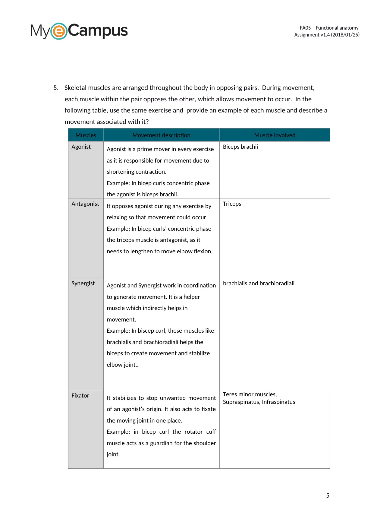
5. Skeletal muscles are arranged throughout the body in opposing pairs. During movement,
each muscle within the pair opposes the other, which allows movement to occur. In the
following table, use the same exercise and provide an example of each muscle and describe a
movement associated with it?
Muscles Movement description Muscle involved
Agonist Agonist is a prime mover in every exercise
as it is responsible for movement due to
shortening contraction.
Example: In bicep curls concentric phase
the agonist is biceps brachii.
Biceps brachii
Antagonist It opposes agonist during any exercise by
relaxing so that movement could occur.
Example: In bicep curls’ concentric phase
the triceps muscle is antagonist, as it
needs to lengthen to move elbow flexion.
Triceps
Synergist Agonist and Synergist work in coordination
to generate movement. It is a helper
muscle which indirectly helps in
movement.
Example: In biscep curl, these muscles like
brachialis and brachioradiali helps the
biceps to create movement and stabilize
elbow joint..
brachialis and brachioradiali
Fixator It stabilizes to stop unwanted movement
of an agonist’s origin. It also acts to fixate
the moving joint in one place.
Example: in bicep curl the rotator cuff
muscle acts as a guardian for the shoulder
joint.
Teres minor muscles,
Supraspinatus, Infraspinatus
5
Assignment v1.4 (2018/01/25)
5. Skeletal muscles are arranged throughout the body in opposing pairs. During movement,
each muscle within the pair opposes the other, which allows movement to occur. In the
following table, use the same exercise and provide an example of each muscle and describe a
movement associated with it?
Muscles Movement description Muscle involved
Agonist Agonist is a prime mover in every exercise
as it is responsible for movement due to
shortening contraction.
Example: In bicep curls concentric phase
the agonist is biceps brachii.
Biceps brachii
Antagonist It opposes agonist during any exercise by
relaxing so that movement could occur.
Example: In bicep curls’ concentric phase
the triceps muscle is antagonist, as it
needs to lengthen to move elbow flexion.
Triceps
Synergist Agonist and Synergist work in coordination
to generate movement. It is a helper
muscle which indirectly helps in
movement.
Example: In biscep curl, these muscles like
brachialis and brachioradiali helps the
biceps to create movement and stabilize
elbow joint..
brachialis and brachioradiali
Fixator It stabilizes to stop unwanted movement
of an agonist’s origin. It also acts to fixate
the moving joint in one place.
Example: in bicep curl the rotator cuff
muscle acts as a guardian for the shoulder
joint.
Teres minor muscles,
Supraspinatus, Infraspinatus
5

FA05 – Functional anatomy
Assignment v1.4 (2018/01/25)
6
Assignment v1.4 (2018/01/25)
6
⊘ This is a preview!⊘
Do you want full access?
Subscribe today to unlock all pages.
Trusted by 1+ million students worldwide
FA05 – Functional anatomy
Assignment v1.4 (2018/01/25)
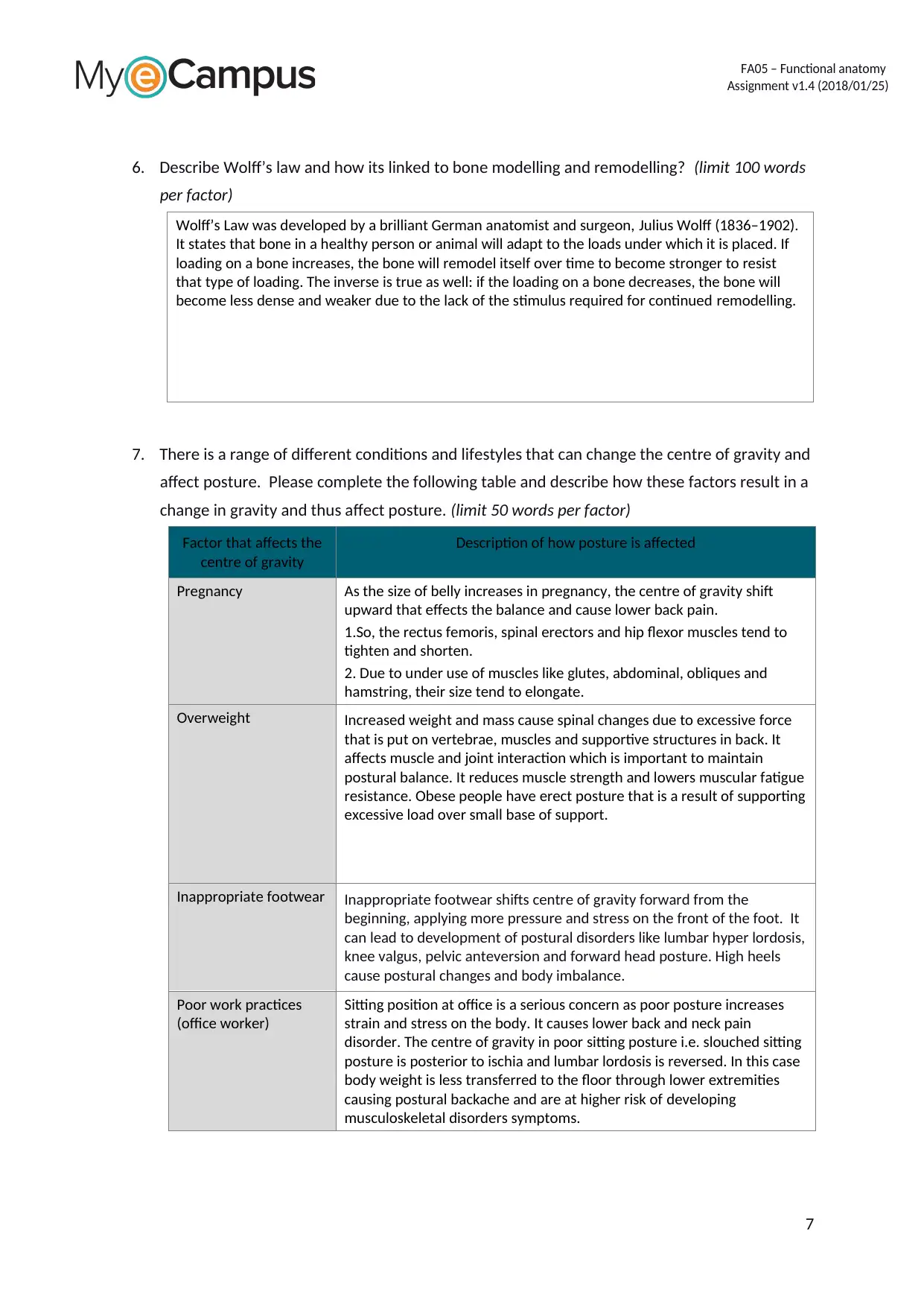
6. Describe Wolff’s law and how its linked to bone modelling and remodelling? (limit 100 words
per factor)
Wolff’s Law was developed by a brilliant German anatomist and surgeon, Julius Wolff (1836–1902).
It states that bone in a healthy person or animal will adapt to the loads under which it is placed. If
loading on a bone increases, the bone will remodel itself over time to become stronger to resist
that type of loading. The inverse is true as well: if the loading on a bone decreases, the bone will
become less dense and weaker due to the lack of the stimulus required for continued remodelling.
7. There is a range of different conditions and lifestyles that can change the centre of gravity and
affect posture. Please complete the following table and describe how these factors result in a
change in gravity and thus affect posture. (limit 50 words per factor)
Factor that affects the
centre of gravity
Description of how posture is affected
Pregnancy As the size of belly increases in pregnancy, the centre of gravity shift
upward that effects the balance and cause lower back pain.
1.So, the rectus femoris, spinal erectors and hip flexor muscles tend to
tighten and shorten.
2. Due to under use of muscles like glutes, abdominal, obliques and
hamstring, their size tend to elongate.
Overweight Increased weight and mass cause spinal changes due to excessive force
that is put on vertebrae, muscles and supportive structures in back. It
affects muscle and joint interaction which is important to maintain
postural balance. It reduces muscle strength and lowers muscular fatigue
resistance. Obese people have erect posture that is a result of supporting
excessive load over small base of support.
Inappropriate footwear Inappropriate footwear shifts centre of gravity forward from the
beginning, applying more pressure and stress on the front of the foot. It
can lead to development of postural disorders like lumbar hyper lordosis,
knee valgus, pelvic anteversion and forward head posture. High heels
cause postural changes and body imbalance.
Poor work practices
(office worker)
Sitting position at office is a serious concern as poor posture increases
strain and stress on the body. It causes lower back and neck pain
disorder. The centre of gravity in poor sitting posture i.e. slouched sitting
posture is posterior to ischia and lumbar lordosis is reversed. In this case
body weight is less transferred to the floor through lower extremities
causing postural backache and are at higher risk of developing
musculoskeletal disorders symptoms.
7
Assignment v1.4 (2018/01/25)
6. Describe Wolff’s law and how its linked to bone modelling and remodelling? (limit 100 words
per factor)
Wolff’s Law was developed by a brilliant German anatomist and surgeon, Julius Wolff (1836–1902).
It states that bone in a healthy person or animal will adapt to the loads under which it is placed. If
loading on a bone increases, the bone will remodel itself over time to become stronger to resist
that type of loading. The inverse is true as well: if the loading on a bone decreases, the bone will
become less dense and weaker due to the lack of the stimulus required for continued remodelling.
7. There is a range of different conditions and lifestyles that can change the centre of gravity and
affect posture. Please complete the following table and describe how these factors result in a
change in gravity and thus affect posture. (limit 50 words per factor)
Factor that affects the
centre of gravity
Description of how posture is affected
Pregnancy As the size of belly increases in pregnancy, the centre of gravity shift
upward that effects the balance and cause lower back pain.
1.So, the rectus femoris, spinal erectors and hip flexor muscles tend to
tighten and shorten.
2. Due to under use of muscles like glutes, abdominal, obliques and
hamstring, their size tend to elongate.
Overweight Increased weight and mass cause spinal changes due to excessive force
that is put on vertebrae, muscles and supportive structures in back. It
affects muscle and joint interaction which is important to maintain
postural balance. It reduces muscle strength and lowers muscular fatigue
resistance. Obese people have erect posture that is a result of supporting
excessive load over small base of support.
Inappropriate footwear Inappropriate footwear shifts centre of gravity forward from the
beginning, applying more pressure and stress on the front of the foot. It
can lead to development of postural disorders like lumbar hyper lordosis,
knee valgus, pelvic anteversion and forward head posture. High heels
cause postural changes and body imbalance.
Poor work practices
(office worker)
Sitting position at office is a serious concern as poor posture increases
strain and stress on the body. It causes lower back and neck pain
disorder. The centre of gravity in poor sitting posture i.e. slouched sitting
posture is posterior to ischia and lumbar lordosis is reversed. In this case
body weight is less transferred to the floor through lower extremities
causing postural backache and are at higher risk of developing
musculoskeletal disorders symptoms.
7
Paraphrase This Document
Need a fresh take? Get an instant paraphrase of this document with our AI Paraphraser
FA05 – Functional anatomy
Assignment v1.4 (2018/01/25)
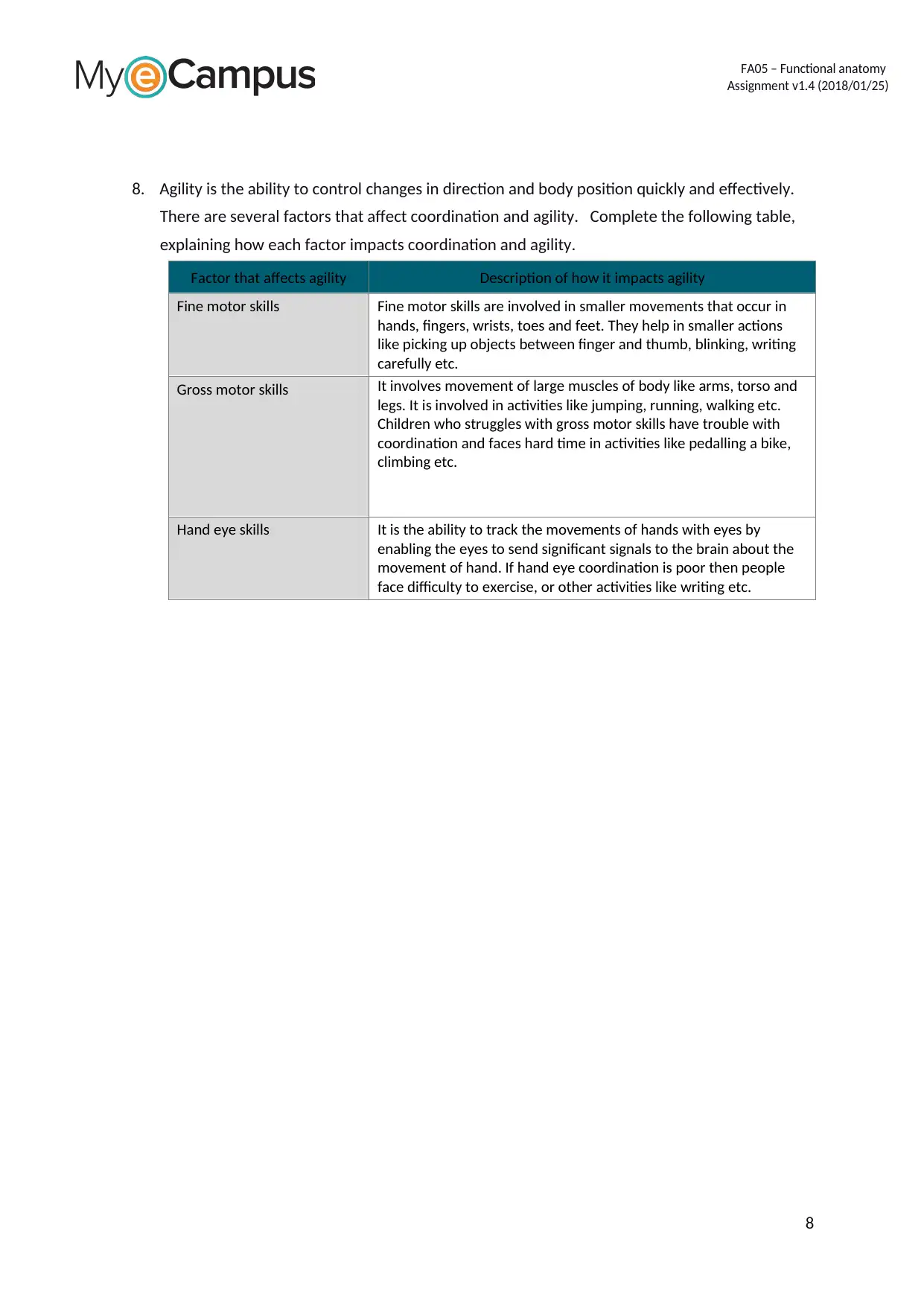
8. Agility is the ability to control changes in direction and body position quickly and effectively.
There are several factors that affect coordination and agility. Complete the following table,
explaining how each factor impacts coordination and agility.
Factor that affects agility Description of how it impacts agility
Fine motor skills Fine motor skills are involved in smaller movements that occur in
hands, fingers, wrists, toes and feet. They help in smaller actions
like picking up objects between finger and thumb, blinking, writing
carefully etc.
Gross motor skills It involves movement of large muscles of body like arms, torso and
legs. It is involved in activities like jumping, running, walking etc.
Children who struggles with gross motor skills have trouble with
coordination and faces hard time in activities like pedalling a bike,
climbing etc.
Hand eye skills It is the ability to track the movements of hands with eyes by
enabling the eyes to send significant signals to the brain about the
movement of hand. If hand eye coordination is poor then people
face difficulty to exercise, or other activities like writing etc.
8
Assignment v1.4 (2018/01/25)
8. Agility is the ability to control changes in direction and body position quickly and effectively.
There are several factors that affect coordination and agility. Complete the following table,
explaining how each factor impacts coordination and agility.
Factor that affects agility Description of how it impacts agility
Fine motor skills Fine motor skills are involved in smaller movements that occur in
hands, fingers, wrists, toes and feet. They help in smaller actions
like picking up objects between finger and thumb, blinking, writing
carefully etc.
Gross motor skills It involves movement of large muscles of body like arms, torso and
legs. It is involved in activities like jumping, running, walking etc.
Children who struggles with gross motor skills have trouble with
coordination and faces hard time in activities like pedalling a bike,
climbing etc.
Hand eye skills It is the ability to track the movements of hands with eyes by
enabling the eyes to send significant signals to the brain about the
movement of hand. If hand eye coordination is poor then people
face difficulty to exercise, or other activities like writing etc.
8
FA05 – Functional anatomy
Assignment v1.4 (2018/01/25)
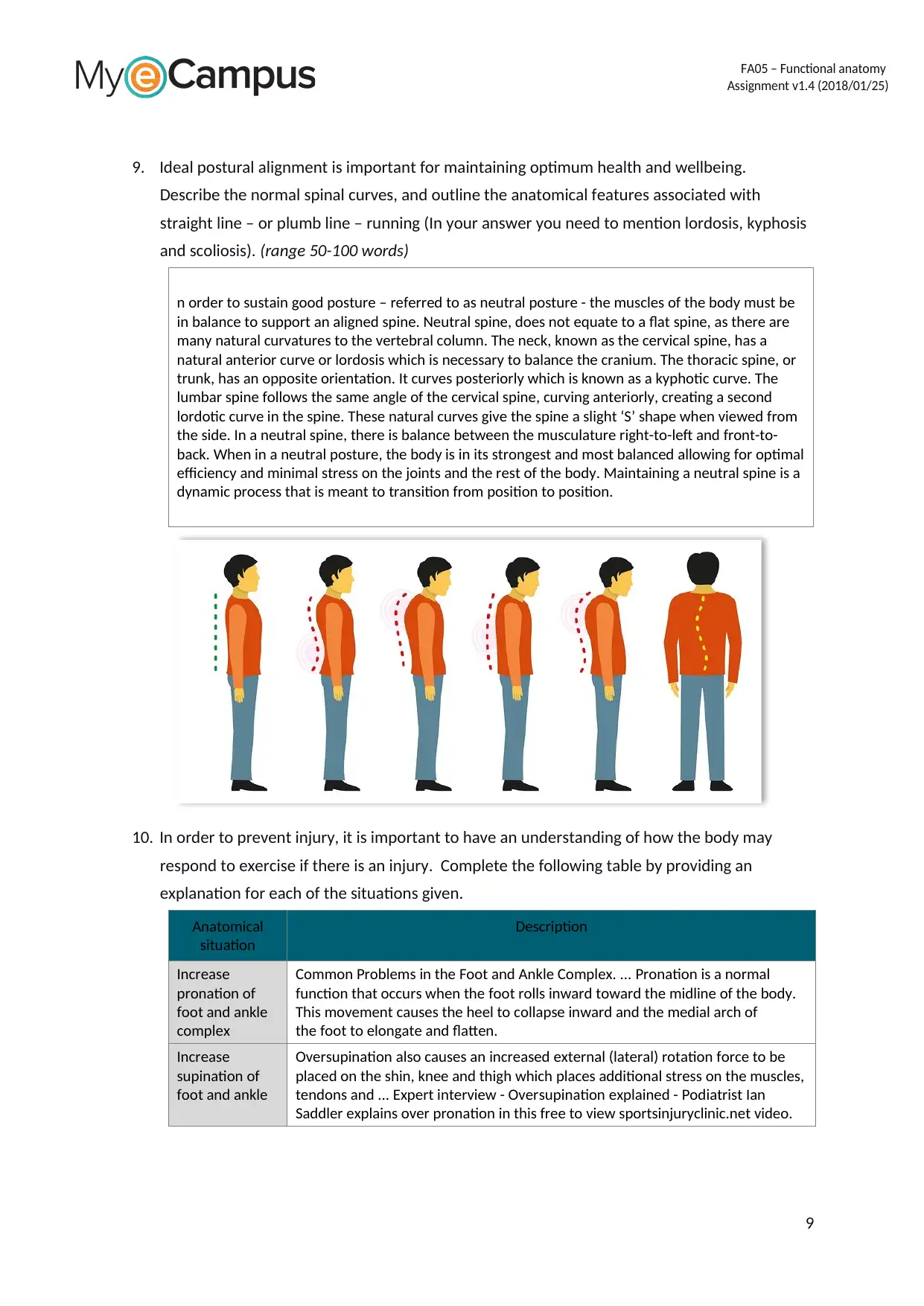
9. Ideal postural alignment is important for maintaining optimum health and wellbeing.
Describe the normal spinal curves, and outline the anatomical features associated with
straight line – or plumb line – running (In your answer you need to mention lordosis, kyphosis
and scoliosis). (range 50-100 words)
n order to sustain good posture – referred to as neutral posture - the muscles of the body must be
in balance to support an aligned spine. Neutral spine, does not equate to a flat spine, as there are
many natural curvatures to the vertebral column. The neck, known as the cervical spine, has a
natural anterior curve or lordosis which is necessary to balance the cranium. The thoracic spine, or
trunk, has an opposite orientation. It curves posteriorly which is known as a kyphotic curve. The
lumbar spine follows the same angle of the cervical spine, curving anteriorly, creating a second
lordotic curve in the spine. These natural curves give the spine a slight ‘S’ shape when viewed from
the side. In a neutral spine, there is balance between the musculature right-to-left and front-to-
back. When in a neutral posture, the body is in its strongest and most balanced allowing for optimal
efficiency and minimal stress on the joints and the rest of the body. Maintaining a neutral spine is a
dynamic process that is meant to transition from position to position.
10. In order to prevent injury, it is important to have an understanding of how the body may
respond to exercise if there is an injury. Complete the following table by providing an
explanation for each of the situations given.
Anatomical
situation
Description
Increase
pronation of
foot and ankle
complex
Common Problems in the Foot and Ankle Complex. ... Pronation is a normal
function that occurs when the foot rolls inward toward the midline of the body.
This movement causes the heel to collapse inward and the medial arch of
the foot to elongate and flatten.
Increase
supination of
foot and ankle
Oversupination also causes an increased external (lateral) rotation force to be
placed on the shin, knee and thigh which places additional stress on the muscles,
tendons and ... Expert interview - Oversupination explained - Podiatrist Ian
Saddler explains over pronation in this free to view sportsinjuryclinic.net video.
9
Assignment v1.4 (2018/01/25)
9. Ideal postural alignment is important for maintaining optimum health and wellbeing.
Describe the normal spinal curves, and outline the anatomical features associated with
straight line – or plumb line – running (In your answer you need to mention lordosis, kyphosis
and scoliosis). (range 50-100 words)
n order to sustain good posture – referred to as neutral posture - the muscles of the body must be
in balance to support an aligned spine. Neutral spine, does not equate to a flat spine, as there are
many natural curvatures to the vertebral column. The neck, known as the cervical spine, has a
natural anterior curve or lordosis which is necessary to balance the cranium. The thoracic spine, or
trunk, has an opposite orientation. It curves posteriorly which is known as a kyphotic curve. The
lumbar spine follows the same angle of the cervical spine, curving anteriorly, creating a second
lordotic curve in the spine. These natural curves give the spine a slight ‘S’ shape when viewed from
the side. In a neutral spine, there is balance between the musculature right-to-left and front-to-
back. When in a neutral posture, the body is in its strongest and most balanced allowing for optimal
efficiency and minimal stress on the joints and the rest of the body. Maintaining a neutral spine is a
dynamic process that is meant to transition from position to position.
10. In order to prevent injury, it is important to have an understanding of how the body may
respond to exercise if there is an injury. Complete the following table by providing an
explanation for each of the situations given.
Anatomical
situation
Description
Increase
pronation of
foot and ankle
complex
Common Problems in the Foot and Ankle Complex. ... Pronation is a normal
function that occurs when the foot rolls inward toward the midline of the body.
This movement causes the heel to collapse inward and the medial arch of
the foot to elongate and flatten.
Increase
supination of
foot and ankle
Oversupination also causes an increased external (lateral) rotation force to be
placed on the shin, knee and thigh which places additional stress on the muscles,
tendons and ... Expert interview - Oversupination explained - Podiatrist Ian
Saddler explains over pronation in this free to view sportsinjuryclinic.net video.
9
⊘ This is a preview!⊘
Do you want full access?
Subscribe today to unlock all pages.
Trusted by 1+ million students worldwide
FA05 – Functional anatomy
Assignment v1.4 (2018/01/25)
Anatomical
situation
Description
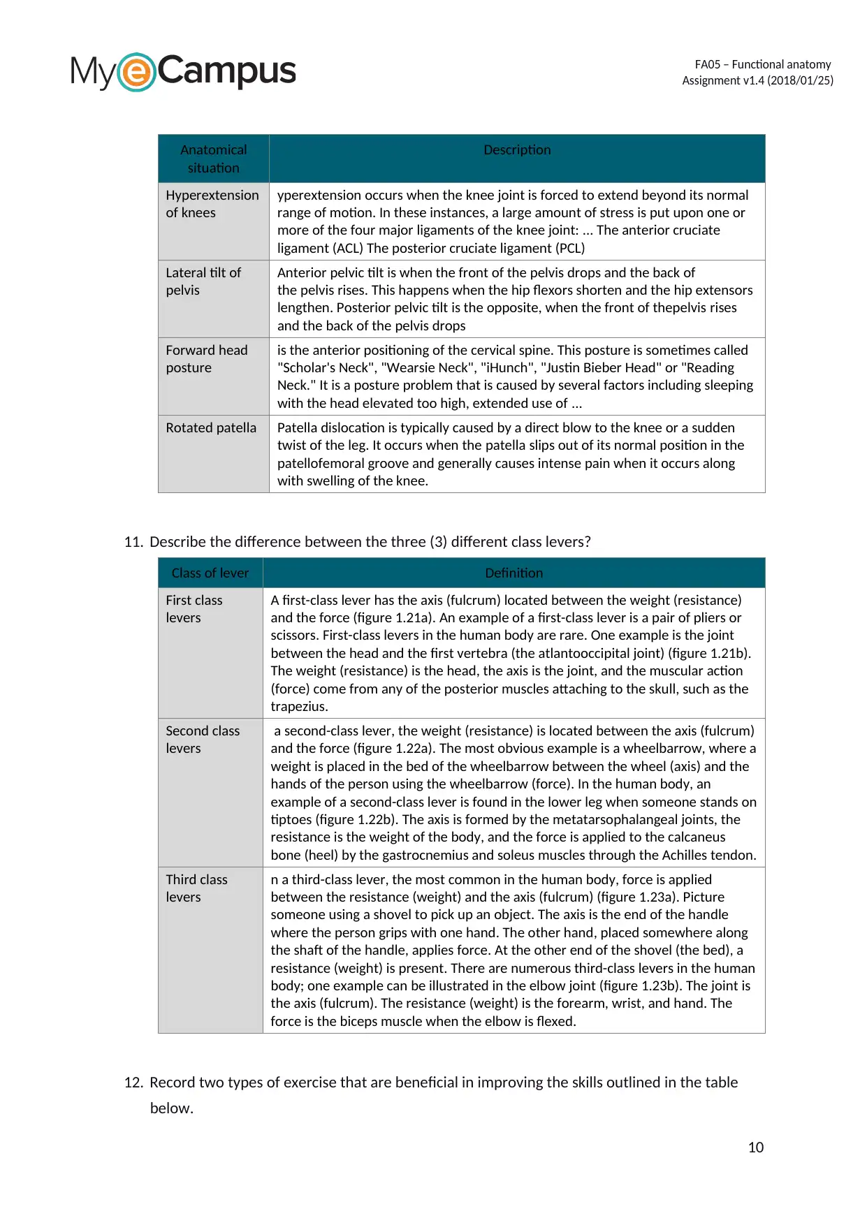
Hyperextension
of knees
yperextension occurs when the knee joint is forced to extend beyond its normal
range of motion. In these instances, a large amount of stress is put upon one or
more of the four major ligaments of the knee joint: ... The anterior cruciate
ligament (ACL) The posterior cruciate ligament (PCL)
Lateral tilt of
pelvis
Anterior pelvic tilt is when the front of the pelvis drops and the back of
the pelvis rises. This happens when the hip flexors shorten and the hip extensors
lengthen. Posterior pelvic tilt is the opposite, when the front of thepelvis rises
and the back of the pelvis drops
Forward head
posture
is the anterior positioning of the cervical spine. This posture is sometimes called
"Scholar's Neck", "Wearsie Neck", "iHunch", "Justin Bieber Head" or "Reading
Neck." It is a posture problem that is caused by several factors including sleeping
with the head elevated too high, extended use of ...
Rotated patella Patella dislocation is typically caused by a direct blow to the knee or a sudden
twist of the leg. It occurs when the patella slips out of its normal position in the
patellofemoral groove and generally causes intense pain when it occurs along
with swelling of the knee.
11. Describe the difference between the three (3) different class levers?
Class of lever Definition
First class
levers
A first-class lever has the axis (fulcrum) located between the weight (resistance)
and the force (figure 1.21a). An example of a first-class lever is a pair of pliers or
scissors. First-class levers in the human body are rare. One example is the joint
between the head and the first vertebra (the atlantooccipital joint) (figure 1.21b).
The weight (resistance) is the head, the axis is the joint, and the muscular action
(force) come from any of the posterior muscles attaching to the skull, such as the
trapezius.
Second class
levers
a second-class lever, the weight (resistance) is located between the axis (fulcrum)
and the force (figure 1.22a). The most obvious example is a wheelbarrow, where a
weight is placed in the bed of the wheelbarrow between the wheel (axis) and the
hands of the person using the wheelbarrow (force). In the human body, an
example of a second-class lever is found in the lower leg when someone stands on
tiptoes (figure 1.22b). The axis is formed by the metatarsophalangeal joints, the
resistance is the weight of the body, and the force is applied to the calcaneus
bone (heel) by the gastrocnemius and soleus muscles through the Achilles tendon.
Third class
levers
n a third-class lever, the most common in the human body, force is applied
between the resistance (weight) and the axis (fulcrum) (figure 1.23a). Picture
someone using a shovel to pick up an object. The axis is the end of the handle
where the person grips with one hand. The other hand, placed somewhere along
the shaft of the handle, applies force. At the other end of the shovel (the bed), a
resistance (weight) is present. There are numerous third-class levers in the human
body; one example can be illustrated in the elbow joint (figure 1.23b). The joint is
the axis (fulcrum). The resistance (weight) is the forearm, wrist, and hand. The
force is the biceps muscle when the elbow is flexed.
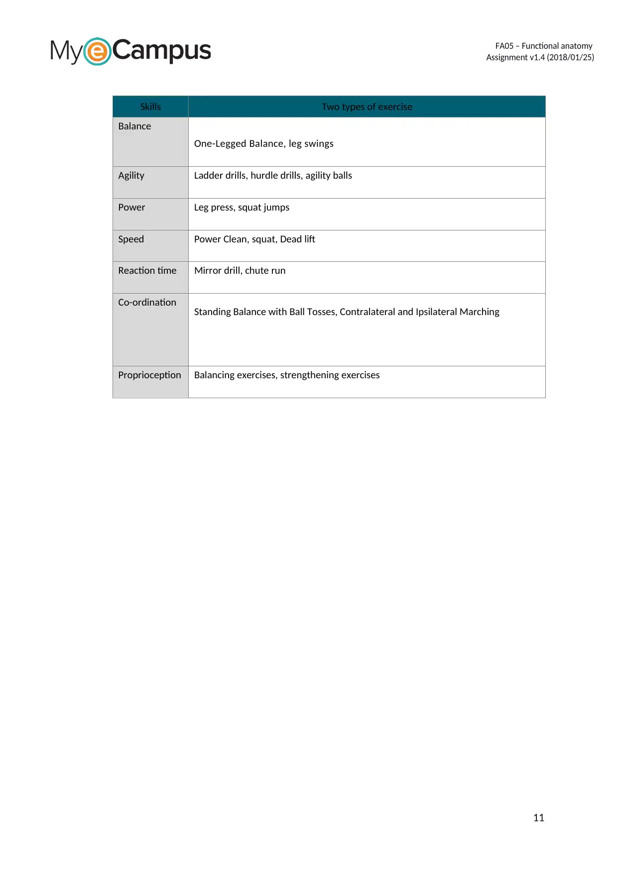
12. Record two types of exercise that are beneficial in improving the skills outlined in the table
below.
10
Assignment v1.4 (2018/01/25)
Anatomical
situation
Description
Hyperextension
of knees
yperextension occurs when the knee joint is forced to extend beyond its normal
range of motion. In these instances, a large amount of stress is put upon one or
more of the four major ligaments of the knee joint: ... The anterior cruciate
ligament (ACL) The posterior cruciate ligament (PCL)
Lateral tilt of
pelvis
Anterior pelvic tilt is when the front of the pelvis drops and the back of
the pelvis rises. This happens when the hip flexors shorten and the hip extensors
lengthen. Posterior pelvic tilt is the opposite, when the front of thepelvis rises
and the back of the pelvis drops
Forward head
posture
is the anterior positioning of the cervical spine. This posture is sometimes called
"Scholar's Neck", "Wearsie Neck", "iHunch", "Justin Bieber Head" or "Reading
Neck." It is a posture problem that is caused by several factors including sleeping
with the head elevated too high, extended use of ...
Rotated patella Patella dislocation is typically caused by a direct blow to the knee or a sudden
twist of the leg. It occurs when the patella slips out of its normal position in the
patellofemoral groove and generally causes intense pain when it occurs along
with swelling of the knee.
11. Describe the difference between the three (3) different class levers?
Class of lever Definition
First class
levers
A first-class lever has the axis (fulcrum) located between the weight (resistance)
and the force (figure 1.21a). An example of a first-class lever is a pair of pliers or
scissors. First-class levers in the human body are rare. One example is the joint
between the head and the first vertebra (the atlantooccipital joint) (figure 1.21b).
The weight (resistance) is the head, the axis is the joint, and the muscular action
(force) come from any of the posterior muscles attaching to the skull, such as the
trapezius.
Second class
levers
a second-class lever, the weight (resistance) is located between the axis (fulcrum)
and the force (figure 1.22a). The most obvious example is a wheelbarrow, where a
weight is placed in the bed of the wheelbarrow between the wheel (axis) and the
hands of the person using the wheelbarrow (force). In the human body, an
example of a second-class lever is found in the lower leg when someone stands on
tiptoes (figure 1.22b). The axis is formed by the metatarsophalangeal joints, the
resistance is the weight of the body, and the force is applied to the calcaneus
bone (heel) by the gastrocnemius and soleus muscles through the Achilles tendon.
Third class
levers
n a third-class lever, the most common in the human body, force is applied
between the resistance (weight) and the axis (fulcrum) (figure 1.23a). Picture
someone using a shovel to pick up an object. The axis is the end of the handle
where the person grips with one hand. The other hand, placed somewhere along
the shaft of the handle, applies force. At the other end of the shovel (the bed), a
resistance (weight) is present. There are numerous third-class levers in the human
body; one example can be illustrated in the elbow joint (figure 1.23b). The joint is
the axis (fulcrum). The resistance (weight) is the forearm, wrist, and hand. The
force is the biceps muscle when the elbow is flexed.
12. Record two types of exercise that are beneficial in improving the skills outlined in the table
below.
10
Paraphrase This Document
Need a fresh take? Get an instant paraphrase of this document with our AI Paraphraser
FA05 – Functional anatomy
Assignment v1.4 (2018/01/25)
Skills Two types of exercise
Balance
One-Legged Balance, leg swings
Agility Ladder drills, hurdle drills, agility balls
Power Leg press, squat jumps
Speed Power Clean, squat, Dead lift
Reaction time Mirror drill, chute run
Co-ordination Standing Balance with Ball Tosses, Contralateral and Ipsilateral Marching
Proprioception Balancing exercises, strengthening exercises
11
FA05 – Functional anatomy
Assignment v1.4 (2018/01/25)
13. Changes in musculoskeletal anatomy and physiology are fundamental to fitness
improvements. In the following table, record the changes in each anatomical and
physiological structure of the musculoskeletal system.
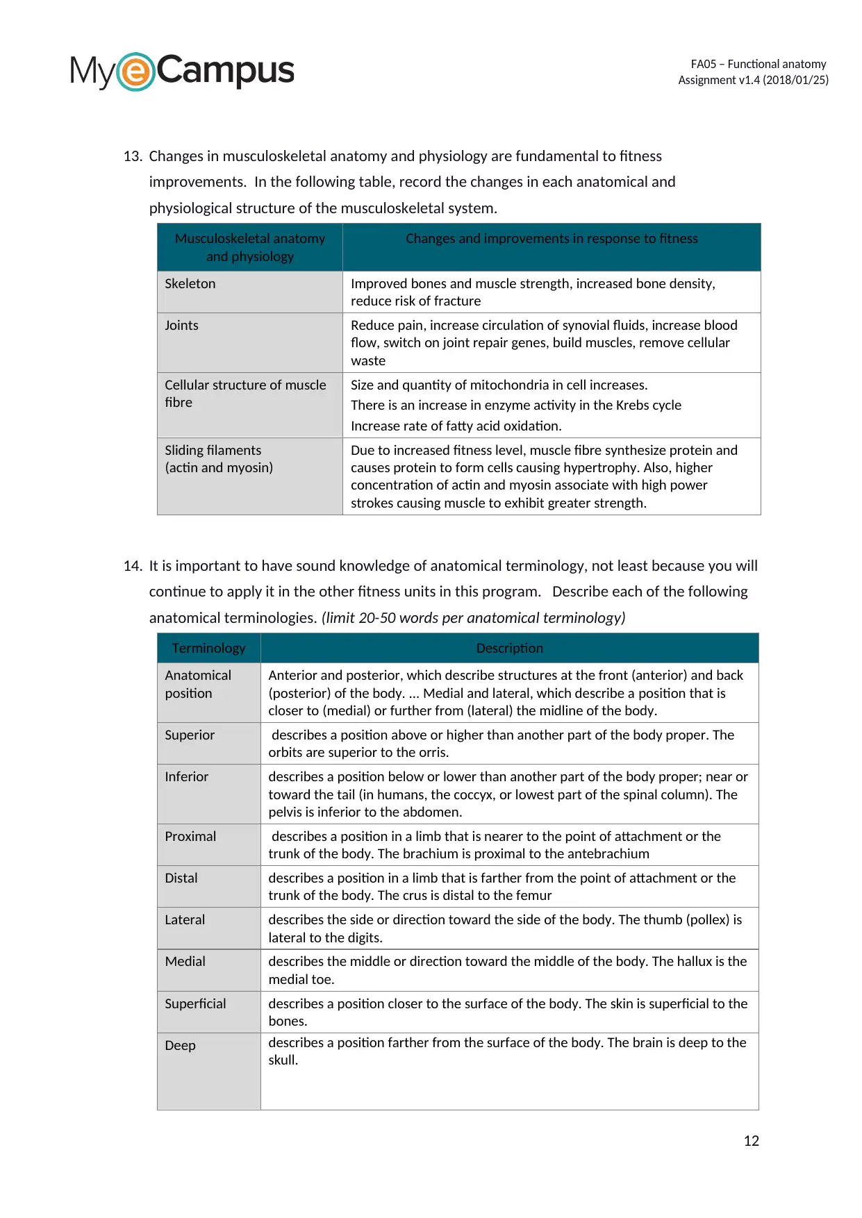
Musculoskeletal anatomy
and physiology
Changes and improvements in response to fitness
Skeleton Improved bones and muscle strength, increased bone density,
reduce risk of fracture
Joints Reduce pain, increase circulation of synovial fluids, increase blood
flow, switch on joint repair genes, build muscles, remove cellular
waste
Cellular structure of muscle
fibre
Size and quantity of mitochondria in cell increases.
There is an increase in enzyme activity in the Krebs cycle
Increase rate of fatty acid oxidation.
Sliding filaments
(actin and myosin)
Due to increased fitness level, muscle fibre synthesize protein and
causes protein to form cells causing hypertrophy. Also, higher
concentration of actin and myosin associate with high power
strokes causing muscle to exhibit greater strength.
14. It is important to have sound knowledge of anatomical terminology, not least because you will
continue to apply it in the other fitness units in this program. Describe each of the following
anatomical terminologies. (limit 20-50 words per anatomical terminology)
Terminology Description
Anatomical
position
Anterior and posterior, which describe structures at the front (anterior) and back
(posterior) of the body. ... Medial and lateral, which describe a position that is
closer to (medial) or further from (lateral) the midline of the body.
Superior describes a position above or higher than another part of the body proper. The
orbits are superior to the orris.
Inferior describes a position below or lower than another part of the body proper; near or
toward the tail (in humans, the coccyx, or lowest part of the spinal column). The
pelvis is inferior to the abdomen.
Proximal describes a position in a limb that is nearer to the point of attachment or the
trunk of the body. The brachium is proximal to the antebrachium
Distal describes a position in a limb that is farther from the point of attachment or the
trunk of the body. The crus is distal to the femur
Lateral describes the side or direction toward the side of the body. The thumb (pollex) is
lateral to the digits.
Medial describes the middle or direction toward the middle of the body. The hallux is the
medial toe.
Superficial describes a position closer to the surface of the body. The skin is superficial to the
bones.
Deep describes a position farther from the surface of the body. The brain is deep to the
skull.
12
Assignment v1.4 (2018/01/25)
13. Changes in musculoskeletal anatomy and physiology are fundamental to fitness
improvements. In the following table, record the changes in each anatomical and
physiological structure of the musculoskeletal system.
Musculoskeletal anatomy
and physiology
Changes and improvements in response to fitness
Skeleton Improved bones and muscle strength, increased bone density,
reduce risk of fracture
Joints Reduce pain, increase circulation of synovial fluids, increase blood
flow, switch on joint repair genes, build muscles, remove cellular
waste
Cellular structure of muscle
fibre
Size and quantity of mitochondria in cell increases.
There is an increase in enzyme activity in the Krebs cycle
Increase rate of fatty acid oxidation.
Sliding filaments
(actin and myosin)
Due to increased fitness level, muscle fibre synthesize protein and
causes protein to form cells causing hypertrophy. Also, higher
concentration of actin and myosin associate with high power
strokes causing muscle to exhibit greater strength.
14. It is important to have sound knowledge of anatomical terminology, not least because you will
continue to apply it in the other fitness units in this program. Describe each of the following
anatomical terminologies. (limit 20-50 words per anatomical terminology)
Terminology Description
Anatomical
position
Anterior and posterior, which describe structures at the front (anterior) and back
(posterior) of the body. ... Medial and lateral, which describe a position that is
closer to (medial) or further from (lateral) the midline of the body.
Superior describes a position above or higher than another part of the body proper. The
orbits are superior to the orris.
Inferior describes a position below or lower than another part of the body proper; near or
toward the tail (in humans, the coccyx, or lowest part of the spinal column). The
pelvis is inferior to the abdomen.
Proximal describes a position in a limb that is nearer to the point of attachment or the
trunk of the body. The brachium is proximal to the antebrachium
Distal describes a position in a limb that is farther from the point of attachment or the
trunk of the body. The crus is distal to the femur
Lateral describes the side or direction toward the side of the body. The thumb (pollex) is
lateral to the digits.
Medial describes the middle or direction toward the middle of the body. The hallux is the
medial toe.
Superficial describes a position closer to the surface of the body. The skin is superficial to the
bones.
Deep describes a position farther from the surface of the body. The brain is deep to the
skull.
12
⊘ This is a preview!⊘
Do you want full access?
Subscribe today to unlock all pages.
Trusted by 1+ million students worldwide
1 out of 28
Related Documents


Your All-in-One AI-Powered Toolkit for Academic Success.
+13062052269
info@desklib.com
Available 24*7 on WhatsApp / Email
![[object Object]](/_next/static/media/star-bottom.7253800d.svg)
Unlock your academic potential
Copyright © 2020–2026 A2Z Services. All Rights Reserved. Developed and managed by ZUCOL.