AIPT - FA05 Functional Anatomy: Posture Analysis, Injuries & Movement
VerifiedAdded on 2023/06/12
|20
|7445
|378
Homework Assignment
AI Summary
This assignment focuses on functional anatomy, specifically addressing posture analysis, common injuries resulting from poor posture, and the relationship between posture, injury risk, and muscular deficits. It covers identifying tight and weak muscles associated with postural abnormalities like lordosis, kyphosis, and winged scapula, along with suggesting corrective exercises. The assignment also explores conditions like sciatica, neck pain, patellofemoral knee pain, lower back pain, and shoulder impingement, detailing their impact on posture and the muscles involved. Furthermore, it delves into how poor posture exacerbates diminished muscle strength, limited flexibility, increased muscle tension, and limited function. The assignment includes an analysis of joint complexes, planes of movement, movement types, and range of motion, as well as a discussion of agonist, antagonist, synergist, and fixator muscles. Finally, it examines Wolff's law, factors affecting the center of gravity (pregnancy, overweight, inappropriate footwear, poor work practices), and factors impacting agility (fine motor skills, gross motor skills, hand-eye coordination).
FA05 Functional anatomy
Assignment v1.4 (2018/01/25)
FA05 Functional anatomy
Name
Email address
Assessment
© 2017 Australian Institute of Personal Trainers Pty Ltd and its licensors (AIPT) Commonwealth of Australia Copyright
Regulations 1969
Warning - This material has been reproduced and communicated to you by or on behalf of AIPT, pursuant to Part VB
of the Copyright Act 1968 (the Act).
The material in this communication may be subject to copyright under the Act. Any further reproduction or
communication of this material by you may be the subject of copyright protection under the Act.
All rights are reserved and you must obtain the prior written permission of AIPT for the republication or redistribution
of any content. Do not remove this notice.
Assignment – Short answer
Assignments may include a variety of questions, this can include short or longer answer questions.
These questions are designed to test how you apply your knowledge into a real-world situation.
All assignments are completed as a Microsoft Word document and must be submitted through My
eCampus for grading. Your assessor is looking for how you apply your knowledge and how you
think critically about the topic area.
1. It is important to have a sound knowledge of posture and the involvement of musculoskeletal
anatomy and appropriate corrective actions. In the following table, you will need to identify
the postural abnormality, identify the tight and weak muscles, and provide suggestive
corrective actions. (limit 50-100words per postural abnormality)
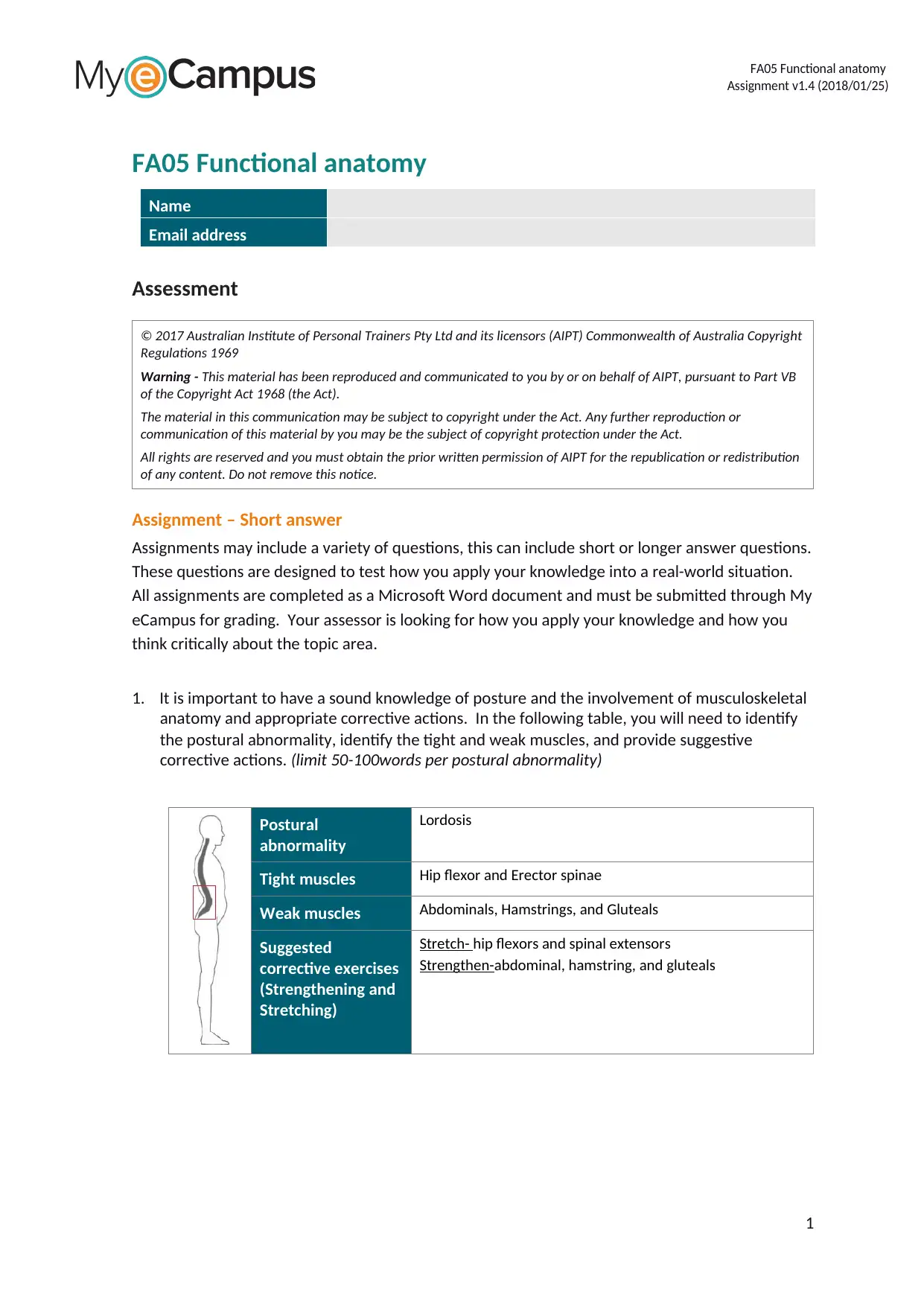
Postural
abnormality
Lordosis
Tight muscles Hip flexor and Erector spinae
Weak muscles Abdominals, Hamstrings, and Gluteals
Suggested
corrective exercises
(Strengthening and
Stretching)
Stretch- hip flexors and spinal extensors
Strengthen-abdominal, hamstring, and gluteals
1
Assignment v1.4 (2018/01/25)
FA05 Functional anatomy
Name
Email address
Assessment
© 2017 Australian Institute of Personal Trainers Pty Ltd and its licensors (AIPT) Commonwealth of Australia Copyright
Regulations 1969
Warning - This material has been reproduced and communicated to you by or on behalf of AIPT, pursuant to Part VB
of the Copyright Act 1968 (the Act).
The material in this communication may be subject to copyright under the Act. Any further reproduction or
communication of this material by you may be the subject of copyright protection under the Act.
All rights are reserved and you must obtain the prior written permission of AIPT for the republication or redistribution
of any content. Do not remove this notice.
Assignment – Short answer
Assignments may include a variety of questions, this can include short or longer answer questions.
These questions are designed to test how you apply your knowledge into a real-world situation.
All assignments are completed as a Microsoft Word document and must be submitted through My
eCampus for grading. Your assessor is looking for how you apply your knowledge and how you
think critically about the topic area.
1. It is important to have a sound knowledge of posture and the involvement of musculoskeletal
anatomy and appropriate corrective actions. In the following table, you will need to identify
the postural abnormality, identify the tight and weak muscles, and provide suggestive
corrective actions. (limit 50-100words per postural abnormality)
Postural
abnormality
Lordosis
Tight muscles Hip flexor and Erector spinae
Weak muscles Abdominals, Hamstrings, and Gluteals
Suggested
corrective exercises
(Strengthening and
Stretching)
Stretch- hip flexors and spinal extensors
Strengthen-abdominal, hamstring, and gluteals
1
Paraphrase This Document
Need a fresh take? Get an instant paraphrase of this document with our AI Paraphraser
FA05 Functional anatomy
Assignment v1.4 (2018/01/25)
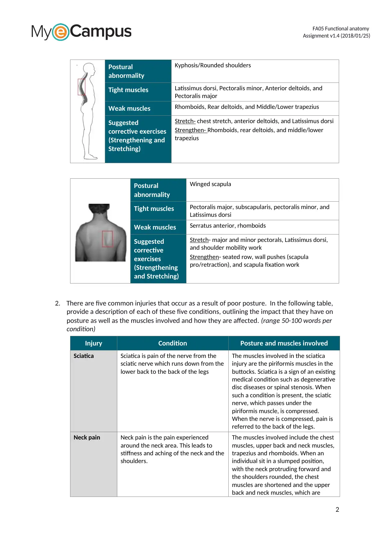
Postural
abnormality
Kyphosis/Rounded shoulders
Tight muscles Latissimus dorsi, Pectoralis minor, Anterior deltoids, and
Pectoralis major
Weak muscles Rhomboids, Rear deltoids, and Middle/Lower trapezius
Suggested
corrective exercises
(Strengthening and
Stretching)
Stretch- chest stretch, anterior deltoids, and Latissimus dorsi
Strengthen- Rhomboids, rear deltoids, and middle/lower
trapezius
Postural
abnormality
Winged scapula
Tight muscles Pectoralis major, subscapularis, pectoralis minor, and
Latissimus dorsi
Weak muscles Serratus anterior, rhomboids
Suggested
corrective
exercises
(Strengthening
and Stretching)
Stretch- major and minor pectorals, Latissimus dorsi,
and shoulder mobility work
Strengthen- seated row, wall pushes (scapula
pro/retraction), and scapula fixation work
2. There are five common injuries that occur as a result of poor posture. In the following table,
provide a description of each of these five conditions, outlining the impact that they have on
posture as well as the muscles involved and how they are affected. (range 50-100 words per
condition)
Injury Condition Posture and muscles involved
Sciatica Sciatica is pain of the nerve from the
sciatic nerve which runs down from the
lower back to the back of the legs
The muscles involved in the sciatica
injury are the piriformis muscles in the
buttocks. Sciatica is a sign of an existing
medical condition such as degenerative
disc diseases or spinal stenosis. When
such a condition is present, the sciatic
nerve, which passes under the
piriformis muscle, is compressed.
When the nerve is compressed, pain is
referred to the back of the legs.
Neck pain Neck pain is the pain experienced
around the neck area. This leads to
stiffness and aching of the neck and the
shoulders.
The muscles involved include the chest
muscles, upper back and neck muscles,
trapezius and rhomboids. When an
individual sit in a slumped position,
with the neck protruding forward and
the shoulders rounded, the chest
muscles are shortened and the upper
back and neck muscles, which are
2
Assignment v1.4 (2018/01/25)
Postural
abnormality
Kyphosis/Rounded shoulders
Tight muscles Latissimus dorsi, Pectoralis minor, Anterior deltoids, and
Pectoralis major
Weak muscles Rhomboids, Rear deltoids, and Middle/Lower trapezius
Suggested
corrective exercises
(Strengthening and
Stretching)
Stretch- chest stretch, anterior deltoids, and Latissimus dorsi
Strengthen- Rhomboids, rear deltoids, and middle/lower
trapezius
Postural
abnormality
Winged scapula
Tight muscles Pectoralis major, subscapularis, pectoralis minor, and
Latissimus dorsi
Weak muscles Serratus anterior, rhomboids
Suggested
corrective
exercises
(Strengthening
and Stretching)
Stretch- major and minor pectorals, Latissimus dorsi,
and shoulder mobility work
Strengthen- seated row, wall pushes (scapula
pro/retraction), and scapula fixation work
2. There are five common injuries that occur as a result of poor posture. In the following table,
provide a description of each of these five conditions, outlining the impact that they have on
posture as well as the muscles involved and how they are affected. (range 50-100 words per
condition)
Injury Condition Posture and muscles involved
Sciatica Sciatica is pain of the nerve from the
sciatic nerve which runs down from the
lower back to the back of the legs
The muscles involved in the sciatica
injury are the piriformis muscles in the
buttocks. Sciatica is a sign of an existing
medical condition such as degenerative
disc diseases or spinal stenosis. When
such a condition is present, the sciatic
nerve, which passes under the
piriformis muscle, is compressed.
When the nerve is compressed, pain is
referred to the back of the legs.
Neck pain Neck pain is the pain experienced
around the neck area. This leads to
stiffness and aching of the neck and the
shoulders.
The muscles involved include the chest
muscles, upper back and neck muscles,
trapezius and rhomboids. When an
individual sit in a slumped position,
with the neck protruding forward and
the shoulders rounded, the chest
muscles are shortened and the upper
back and neck muscles, which are
2
FA05 Functional anatomy
Assignment v1.4 (2018/01/25)
Injury Condition Posture and muscles involved
responsible for pulling the shoulders
back are weakened. The rhomboids
and the trapezius are therefore forced
to work harder becoming achy and
tight as they try to keep the shoulders
back.
Patellofemora
l knee pain
This is pain that is felt at the front of
the knee.
The muscles involved are the
quadriceps muscles. The pain is
heightened by activities such as walking
down the hill or upstairs or standing up
after long hours of sitting. When there
is an imbalance between the
quadriceps muscles, the patella will
track laterally.
Lower back
pain
Lower back pain is the pain that is
experienced at the lower back due to
several factors such as trauma, poor
posture or overuse
The muscles that are involved include
the quadratus lumborum, and the
spinal erectors. When extra strain is
put on these muscles due to poor
posture, certain muscles such as the
flexor and extensor muscles are caused
to overwork leading to muscle spasms
and strains.
Shoulder
impingement
This injury is caused when shoulder
joint space is decreased leading to
pinching of any tendons that pass
through the space. The space is often
reduced due to poor posture
The muscles involved are the pectoralis
major, the pectoralis minor, the lower
trapezius, and the serratus anterior.
When a person sits for long periods
slouched at a desk, the chest muscles
become tight while the upper back
muscles become weak causing the
shoulder joint to lean forward.
3
⊘ This is a preview!⊘
Do you want full access?
Subscribe today to unlock all pages.
Trusted by 1+ million students worldwide
FA05 Functional anatomy
Assignment v1.4 (2018/01/25)
3. There is a definitive relationship between poor posture, increased risk of injury, and muscular
deficit. Please describe how poor posture exacerbates each of the following.
1. Diminished muscle
strength and
endurance
Poor posture requires help from phasic fibres causing the wasting
away of supporting muscles because of disuse. The frail unused
muscles then become tight and shorten the length of the muscle.
This can compress the vertebrae and worsen posture.
2. Limited flexibility
The balance between the initial length of a muscle at rest and the
contraction of a muscle creates the correct tension to allow
movements of joints. This affects the range of motion of the joint.
Poor posture can cause the muscle length at rest to be too long or
too short thereby limiting the flexibility of the muscle to contract.
3. Increased muscle
tension and tone
Increased muscle tension and tone occurs when the muscle is
overworked or tensed due to poor posture. This means the muscle
is not fully relaxed and so the length at rest is altered. This leads to
limited full range of motion of the joint.
4. Limited function
The functions of usually limited by poor posture. Poor posture
overtime leads to permanently stretched or shortened muscles.
Muscles that have been stretched or shortened leads to limited
function as they no longer function as expected.
4. In the following table, several joint complexes are listed. You will need to record the main
planes of movement that the joint can work within, the movement types and the range of
motion for each movement type.
Joint complex Planes of movement Movement types
Range of motion in
degrees for each
movement type
Shoulder joint
(example)
1. Sagittal Plane
2. Frontal Plane
3. Horizontal Plane
1. Flexion and extension
2. Adduction and
abduction
3. Horizontal Flexion and
horizontal extension
1. 0-90 degrees
2. 0-90 degrees
3. 0-180 degrees
Elbow joint 1.Sagittal plane 1.Flexion
2.extension
2.Pronation and
supination
1.0-45 degrees
2. 0
3.0-90 degrees
Hip joint 1.Sagittal Plane
2.Frontal Plane
3. Horizontal Plane
4. All planes
1.Flexion
2. Extension
3.Adduction
4. abduction
5.Horizontal Flexion
6.Horizontal extension
1. 0-125-145 degrees
2. 0-10-20 degrees
3. 0- 20-30 degrees
4.0-40-45 degrees
5.0-35-45 degrees
6.0-45-60 degrees
Knee joint 1.Sagittal Plane 1.Flexion
2.Extension
1.0- 125-145 degrees
2.0-10 degrees
Ankle joint 1.Sagittal Plane 1.Plantar flexion
2.Dorsiflexion
3. Inversion and eversion
1.0-45-50 degrees
2.0-20 degrees
3.0-35 degrees
4
Paraphrase This Document
Need a fresh take? Get an instant paraphrase of this document with our AI Paraphraser
FA05 Functional anatomy
Assignment v1.4 (2018/01/25)
Wrist joint 1.Sagittal Plane
2.Frontal Plane
1.Flexion
2.Extension
3.Adduction
4.abduction
1.0-80-90 degrees
2.0-70 degrees
3.0-45 degrees
4.0-45 degrees
5. Skeletal muscles are arranged throughout the body in opposing pairs. During movement,
each muscle within the pair opposes the other, which allows movement to occur. In the
following table, use the same exercise and provide an example of each muscle and describe a
movement associated with it?
Muscles Movement description Muscle involved
Agonist It causes movement because of its
shortening contraction. The muscles cause
movements to occur
Biceps branchii
Antagonist It relaxes and lengthen by opposing the
agonist for movement to take place
Triceps muscle
Synergist The muscles coordinates with the agonist
to cause movement to take place
Brachioradialis and brachialis
Fixator It works by fixing the moving joint in place Subscapularis, Supraspinatus, and
Infraspinatus
6. Describe Wolff’s law and how its linked to bone modelling and remodelling? (limit 100 words
per factor)
Wolf law states that the bone of a healthy person or an animal adapts to the loads under which it
is placed. Over some period of time, a bone will remodel itself if the load placed under it increases.
Bones constantly remodel in the course of life. If the stress is increased, the bone becomes stronger
by remodeling to resist that type of load. Similarly, if the load becomes lighter, the bone will
become weaker and less dense because of the absence of inducement needed for continual
remodeling.
7. There is a range of different conditions and lifestyles that can change the centre of gravity and
affect posture. Please complete the following table and describe how these factors result in a
change in gravity and thus affect posture. (limit 50 words per factor)
Factor that affects
the centre of gravity Description of how posture is affected
Pregnancy The centre of gravity shifts upwards during pregnancy. As the belly
enlarges, the pelvic begins to roll forward naturally into an anterior pelvic
tilt. The shift of the centre of gravity affects body balance. Muscles such
as the hip flexors and the spinal erectors tend to tighten and shorten
while some muscles such as the glutes, the hamstrings and the
abdominals tend to lengthen causing body causing some discomfort or
pain in the lower back.
Overweight Obesity changes the body movements by leading to changes in
anthropometry. Obesity decreases the fatigue resistance of muscles and
reduces the relative strength of the muscles. The two limitations cause a
delay in the motors and inadequate remedial torque. These lead to an
incapability to affect a suitable response to perturbation that allows for
5
FA05 Functional anatomy
Assignment v1.4 (2018/01/25)
the posture control maintenance.
Inappropriate
footwear
With appropriate foot ware, the centre of gravity moves from the heels
and become distributed evenly along the foot. With inappropriate shoes
such as high heels or poorly fitting shoes, the centre of gravity shifts.
With high heels, the centre of gravity shifts forwards from the very start
and puts a lot of pressure and stress toward the front of the foot.
Poor work practices
(office worker)
In a slouched sitting position in the office, the lumbar lordosis is in a
reversed position, the centre of gravity is posterior to the ischia, and far
less weight of the body is transferred to the floor through the lower
extremities. In a normal erect sitting position, the centre of gravity is
forward to the ischia, the lumbar lordosis is flattened slightly and 25% of
body weight is transferred to the floor through the lower extremities.
8. Agility is the ability to control changes in direction and body position quickly and effectively.
There are several factors that affect coordination and agility. Complete the following table,
explaining how each factor impacts coordination and agility.
Factor that affects agility Description of how it impacts agility
Fine motor skills Fine motor skills are the skills that require the usage of small
muscles that controls the fingers, hand, thumb, and hand eye
coordination. These include skills such as grasping, picking objects
and moving objects
Gross motor skills Gross motor skills are the skills that that involve usage of large
muscles in the legs, torso, and arm. These include skills such as
walking, jumping, and running.
Hand eye skills Hand eye coordination is the ability of the eye to track the
movements of the hands. Poor hand eye coordination can affect the
ability to do exercise and similarly affect the everyday activities such
as writing.
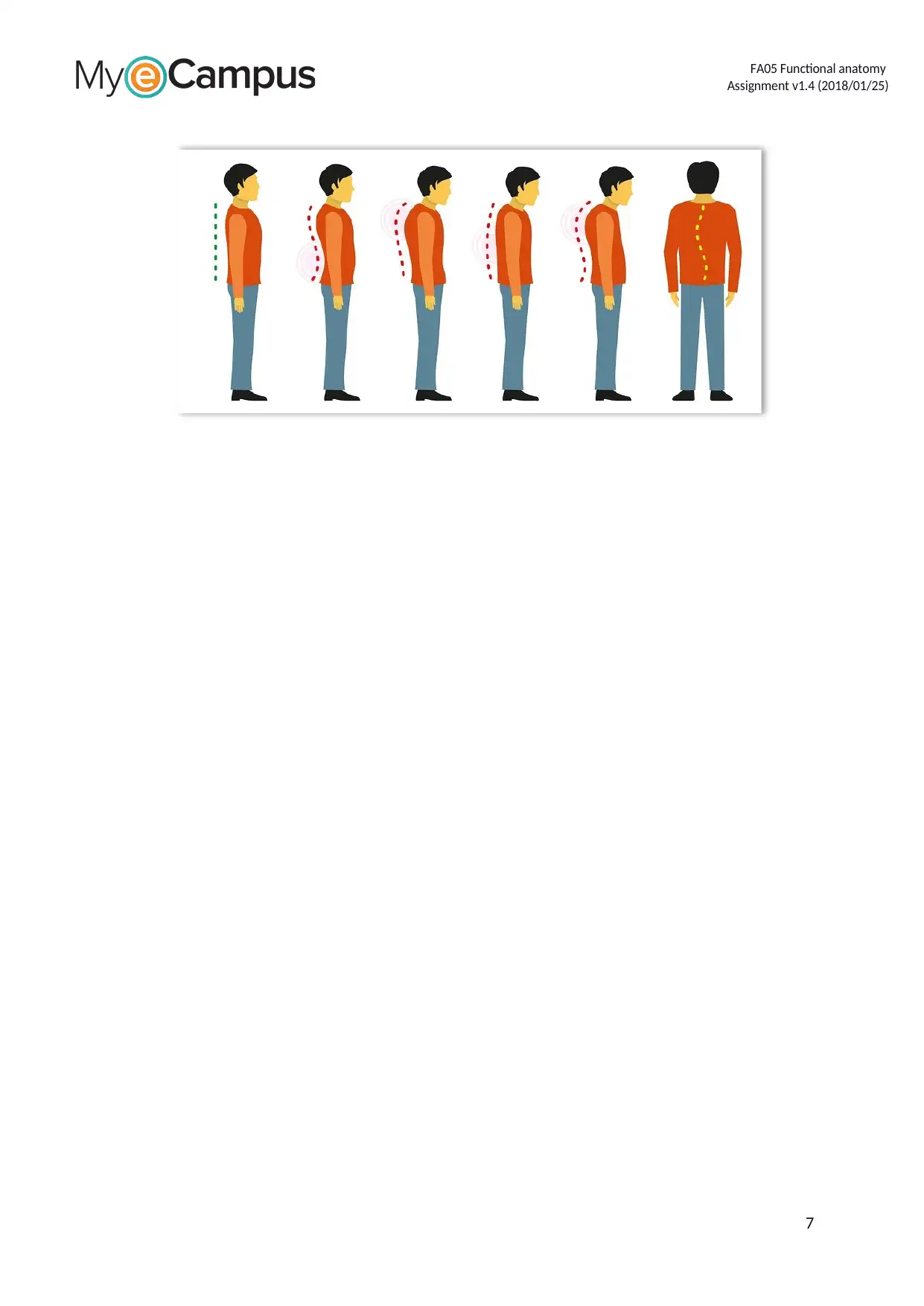
9. Ideal postural alignment is important for maintaining optimum health and wellbeing.
Describe the normal spinal curves, and outline the anatomical features associated with
straight line – or plumb line – running (In your answer you need to mention lordosis, kyphosis
and scoliosis). (range 50-100 words)
The skeleton a human being has some natural spinal curves that may decrease or increase in size
depending on factors like lifestyle and genetics. The lumbar and the cervical spines both have a
normal spine curve known as lordosis. Lordosis is an increased curvature of the spine. The thoracic
spine has a more complex normal curve known as the kyphosis. Kyphosis is an increased curvature
at the posterior of the thorax. Scoliosis is also a normal curve of the spine. Scoliosis is a vertebra
with a lateral curve manifesting with a rotation of the vertebrae and lateral flexion. The anatomical
features associated with the plumbline include: slightly posterior to the coronal suture, through the
axis of the odontoid process, through the external auditory meatus, midway through acromion
process, slightly posterior to the hip joint, through the bodies of the lumbar vertebrae, slightly
anterior to the axis of the knee joint, through the calcaneocuboid, and slightly anterior to the
lateral malleolus.
6
Assignment v1.4 (2018/01/25)
the posture control maintenance.
Inappropriate
footwear
With appropriate foot ware, the centre of gravity moves from the heels
and become distributed evenly along the foot. With inappropriate shoes
such as high heels or poorly fitting shoes, the centre of gravity shifts.
With high heels, the centre of gravity shifts forwards from the very start
and puts a lot of pressure and stress toward the front of the foot.
Poor work practices
(office worker)
In a slouched sitting position in the office, the lumbar lordosis is in a
reversed position, the centre of gravity is posterior to the ischia, and far
less weight of the body is transferred to the floor through the lower
extremities. In a normal erect sitting position, the centre of gravity is
forward to the ischia, the lumbar lordosis is flattened slightly and 25% of
body weight is transferred to the floor through the lower extremities.
8. Agility is the ability to control changes in direction and body position quickly and effectively.
There are several factors that affect coordination and agility. Complete the following table,
explaining how each factor impacts coordination and agility.
Factor that affects agility Description of how it impacts agility
Fine motor skills Fine motor skills are the skills that require the usage of small
muscles that controls the fingers, hand, thumb, and hand eye
coordination. These include skills such as grasping, picking objects
and moving objects
Gross motor skills Gross motor skills are the skills that that involve usage of large
muscles in the legs, torso, and arm. These include skills such as
walking, jumping, and running.
Hand eye skills Hand eye coordination is the ability of the eye to track the
movements of the hands. Poor hand eye coordination can affect the
ability to do exercise and similarly affect the everyday activities such
as writing.
9. Ideal postural alignment is important for maintaining optimum health and wellbeing.
Describe the normal spinal curves, and outline the anatomical features associated with
straight line – or plumb line – running (In your answer you need to mention lordosis, kyphosis
and scoliosis). (range 50-100 words)
The skeleton a human being has some natural spinal curves that may decrease or increase in size
depending on factors like lifestyle and genetics. The lumbar and the cervical spines both have a
normal spine curve known as lordosis. Lordosis is an increased curvature of the spine. The thoracic
spine has a more complex normal curve known as the kyphosis. Kyphosis is an increased curvature
at the posterior of the thorax. Scoliosis is also a normal curve of the spine. Scoliosis is a vertebra
with a lateral curve manifesting with a rotation of the vertebrae and lateral flexion. The anatomical
features associated with the plumbline include: slightly posterior to the coronal suture, through the
axis of the odontoid process, through the external auditory meatus, midway through acromion
process, slightly posterior to the hip joint, through the bodies of the lumbar vertebrae, slightly
anterior to the axis of the knee joint, through the calcaneocuboid, and slightly anterior to the
lateral malleolus.
6
⊘ This is a preview!⊘
Do you want full access?
Subscribe today to unlock all pages.
Trusted by 1+ million students worldwide
FA05 Functional anatomy
Assignment v1.4 (2018/01/25)
7
Assignment v1.4 (2018/01/25)
7
Paraphrase This Document
Need a fresh take? Get an instant paraphrase of this document with our AI Paraphraser
FA05 Functional anatomy
Assignment v1.4 (2018/01/25)
10. In order to prevent injury, it is important to have an understanding of how the body may
respond to exercise if there is an injury. Complete the following table by providing an
explanation for each of the situations given.
Anatomical situation Description
Increase pronation of
foot and ankle complex
Increased pronation is when there is too much pronation or the bones
of the hindfoot are in a more pronated position than they as supposed
to be either when standing of walking. Increased pronation is due to
instability of the ankle bone on the heel bone.
Increase supination of
foot and ankle
At the state of increased supination, the foot is not able or less able to
absorb shock. Increase supination occurs mostly in runners. The foot
that over supinates, under pronates hence passing the absorbed stress
to the lower limb.
Hyperextension of
knees
Hyperextension of the knees usually have symptoms such as reduced
motion range, swelling, instability of the affected leg, and sharp
localized pain.
Lateral tilt of pelvis Lateral tilt of the pelvis is when one side of the hip appears higher or
lower than the normal hip position. These two conditions are medically
referred to as hip hiking and hip dropping respectively.
Forward head posture Forward head posture is a posture problem which leads to the anterior
positioning of the cervical spine. It is caused by several factors such as
prolonged use of computers or improper development of the strength
of the back muscle.
Rotated patella Rotated patella is when the patella is slips from its natural position or
place in the patellofemoral groove. Rotated patella, when it occurs a
long with a swollen knee, leads to intense pain. Rotated patella is
caused when there is an unexpected twist of the leg or a direct blow to
the knee
11. Describe the difference between the three (3) different class levers?
Class of lever Definition
First class levers The fulcrum is in the middle, the effort is applied on one side of the fulcrum
while the resistance is positioned on the other side of the fulcrum.
Second class levers The resistance is located in the middle, the fulcrum is located on one side of
the resistance while the effort is applied on the other side of the resistance.
Third class levers The effort is in the middle, the fulcrum is located on one side of the effort
ahile the resistance is on the other side of the effort.
12. Record two types of exercise that are beneficial in improving the skills outlined in the table
below.
Skills Two types of exercise
Balance One legged balance and leg swings
Agility Ladder drills and hurdle drills
Power Squat jumps and medicine ball overhead throws
Speed Sled drags and standing triple jumps
8
FA05 Functional anatomy
Assignment v1.4 (2018/01/25)
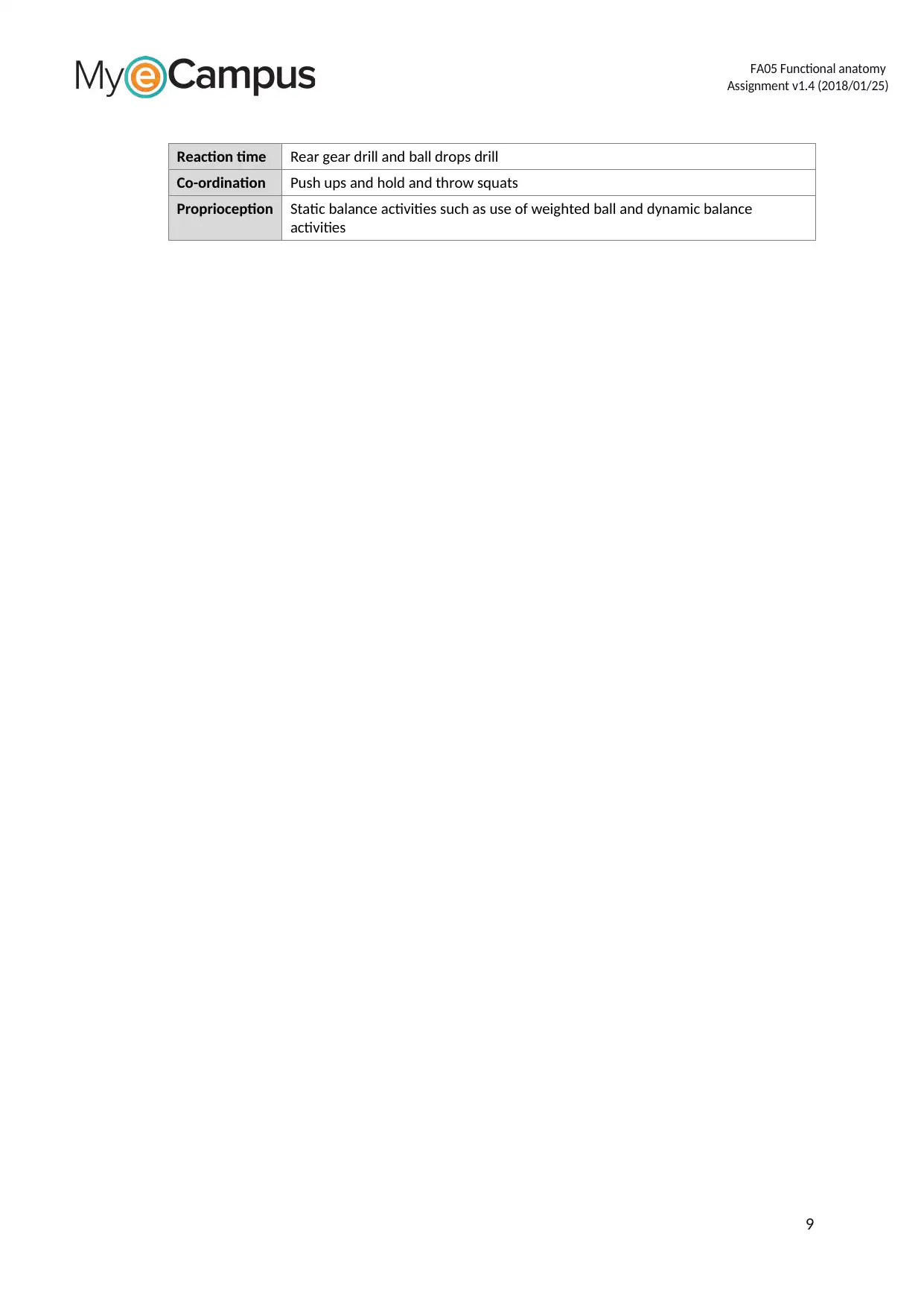
Reaction time Rear gear drill and ball drops drill
Co-ordination Push ups and hold and throw squats
Proprioception Static balance activities such as use of weighted ball and dynamic balance
activities
9
Assignment v1.4 (2018/01/25)
Reaction time Rear gear drill and ball drops drill
Co-ordination Push ups and hold and throw squats
Proprioception Static balance activities such as use of weighted ball and dynamic balance
activities
9
⊘ This is a preview!⊘
Do you want full access?
Subscribe today to unlock all pages.
Trusted by 1+ million students worldwide
FA05 Functional anatomy
Assignment v1.4 (2018/01/25)
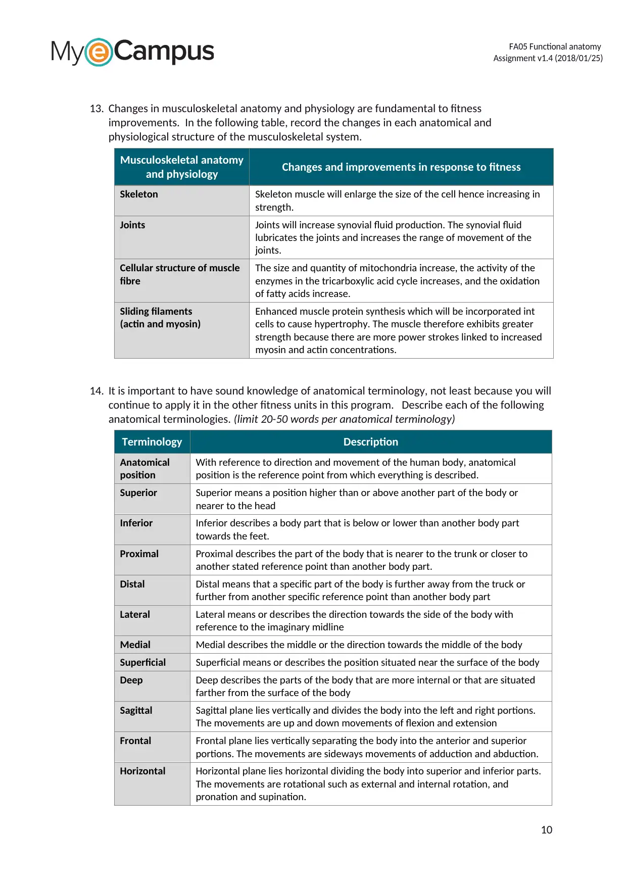
13. Changes in musculoskeletal anatomy and physiology are fundamental to fitness
improvements. In the following table, record the changes in each anatomical and
physiological structure of the musculoskeletal system.
Musculoskeletal anatomy
and physiology Changes and improvements in response to fitness
Skeleton Skeleton muscle will enlarge the size of the cell hence increasing in
strength.
Joints Joints will increase synovial fluid production. The synovial fluid
lubricates the joints and increases the range of movement of the
joints.
Cellular structure of muscle
fibre
The size and quantity of mitochondria increase, the activity of the
enzymes in the tricarboxylic acid cycle increases, and the oxidation
of fatty acids increase.
Sliding filaments
(actin and myosin)
Enhanced muscle protein synthesis which will be incorporated int
cells to cause hypertrophy. The muscle therefore exhibits greater
strength because there are more power strokes linked to increased
myosin and actin concentrations.
14. It is important to have sound knowledge of anatomical terminology, not least because you will
continue to apply it in the other fitness units in this program. Describe each of the following
anatomical terminologies. (limit 20-50 words per anatomical terminology)
Terminology Description
Anatomical
position
With reference to direction and movement of the human body, anatomical
position is the reference point from which everything is described.
Superior Superior means a position higher than or above another part of the body or
nearer to the head
Inferior Inferior describes a body part that is below or lower than another body part
towards the feet.
Proximal Proximal describes the part of the body that is nearer to the trunk or closer to
another stated reference point than another body part.
Distal Distal means that a specific part of the body is further away from the truck or
further from another specific reference point than another body part
Lateral Lateral means or describes the direction towards the side of the body with
reference to the imaginary midline
Medial Medial describes the middle or the direction towards the middle of the body
Superficial Superficial means or describes the position situated near the surface of the body
Deep Deep describes the parts of the body that are more internal or that are situated
farther from the surface of the body
Sagittal Sagittal plane lies vertically and divides the body into the left and right portions.
The movements are up and down movements of flexion and extension
Frontal Frontal plane lies vertically separating the body into the anterior and superior
portions. The movements are sideways movements of adduction and abduction.
Horizontal Horizontal plane lies horizontal dividing the body into superior and inferior parts.
The movements are rotational such as external and internal rotation, and
pronation and supination.
10
Assignment v1.4 (2018/01/25)
13. Changes in musculoskeletal anatomy and physiology are fundamental to fitness
improvements. In the following table, record the changes in each anatomical and
physiological structure of the musculoskeletal system.
Musculoskeletal anatomy
and physiology Changes and improvements in response to fitness
Skeleton Skeleton muscle will enlarge the size of the cell hence increasing in
strength.
Joints Joints will increase synovial fluid production. The synovial fluid
lubricates the joints and increases the range of movement of the
joints.
Cellular structure of muscle
fibre
The size and quantity of mitochondria increase, the activity of the
enzymes in the tricarboxylic acid cycle increases, and the oxidation
of fatty acids increase.
Sliding filaments
(actin and myosin)
Enhanced muscle protein synthesis which will be incorporated int
cells to cause hypertrophy. The muscle therefore exhibits greater
strength because there are more power strokes linked to increased
myosin and actin concentrations.
14. It is important to have sound knowledge of anatomical terminology, not least because you will
continue to apply it in the other fitness units in this program. Describe each of the following
anatomical terminologies. (limit 20-50 words per anatomical terminology)
Terminology Description
Anatomical
position
With reference to direction and movement of the human body, anatomical
position is the reference point from which everything is described.
Superior Superior means a position higher than or above another part of the body or
nearer to the head
Inferior Inferior describes a body part that is below or lower than another body part
towards the feet.
Proximal Proximal describes the part of the body that is nearer to the trunk or closer to
another stated reference point than another body part.
Distal Distal means that a specific part of the body is further away from the truck or
further from another specific reference point than another body part
Lateral Lateral means or describes the direction towards the side of the body with
reference to the imaginary midline
Medial Medial describes the middle or the direction towards the middle of the body
Superficial Superficial means or describes the position situated near the surface of the body
Deep Deep describes the parts of the body that are more internal or that are situated
farther from the surface of the body
Sagittal Sagittal plane lies vertically and divides the body into the left and right portions.
The movements are up and down movements of flexion and extension
Frontal Frontal plane lies vertically separating the body into the anterior and superior
portions. The movements are sideways movements of adduction and abduction.
Horizontal Horizontal plane lies horizontal dividing the body into superior and inferior parts.
The movements are rotational such as external and internal rotation, and
pronation and supination.
10
Paraphrase This Document
Need a fresh take? Get an instant paraphrase of this document with our AI Paraphraser
FA05 Functional anatomy
Assignment v1.4 (2018/01/25)
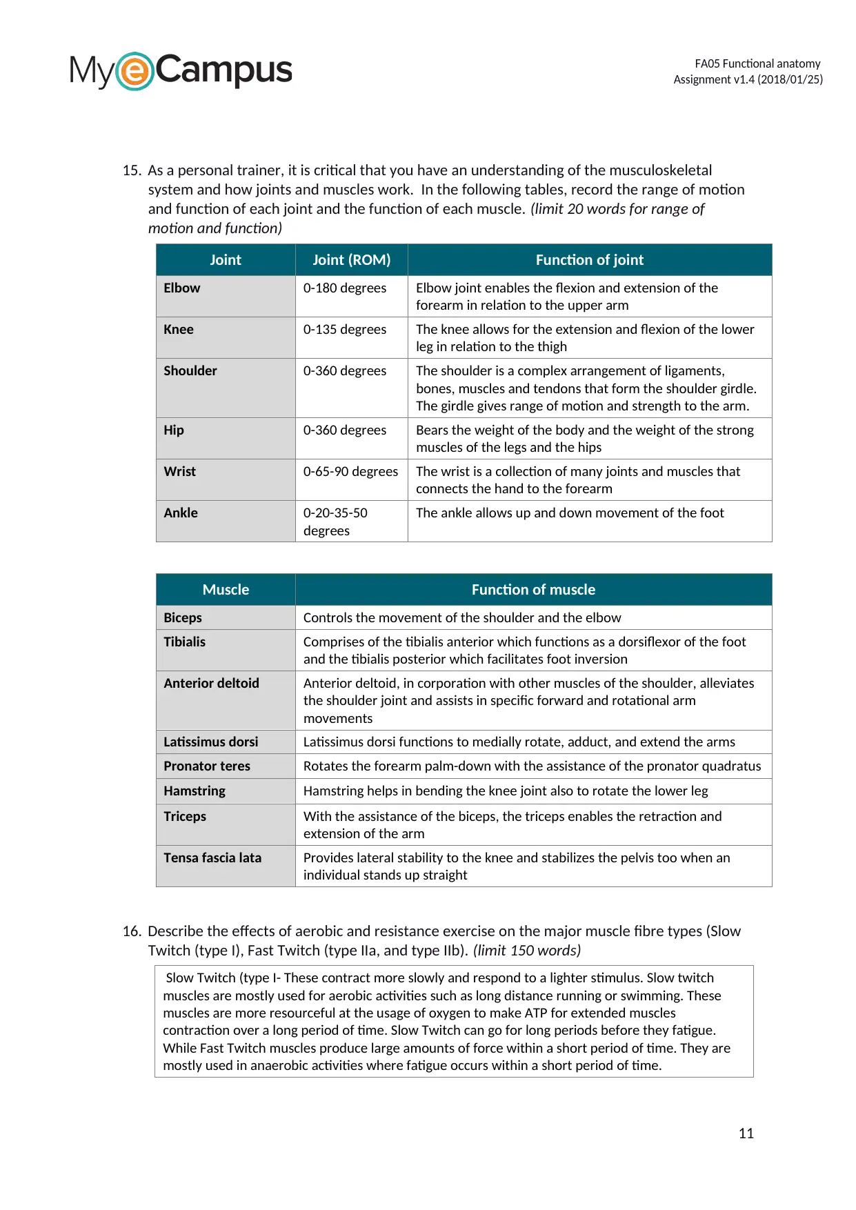
15. As a personal trainer, it is critical that you have an understanding of the musculoskeletal
system and how joints and muscles work. In the following tables, record the range of motion
and function of each joint and the function of each muscle. (limit 20 words for range of
motion and function)
Joint Joint (ROM) Function of joint
Elbow 0-180 degrees Elbow joint enables the flexion and extension of the
forearm in relation to the upper arm
Knee 0-135 degrees The knee allows for the extension and flexion of the lower
leg in relation to the thigh
Shoulder 0-360 degrees The shoulder is a complex arrangement of ligaments,
bones, muscles and tendons that form the shoulder girdle.
The girdle gives range of motion and strength to the arm.
Hip 0-360 degrees Bears the weight of the body and the weight of the strong
muscles of the legs and the hips
Wrist 0-65-90 degrees The wrist is a collection of many joints and muscles that
connects the hand to the forearm
Ankle 0-20-35-50
degrees
The ankle allows up and down movement of the foot
Muscle Function of muscle
Biceps Controls the movement of the shoulder and the elbow
Tibialis Comprises of the tibialis anterior which functions as a dorsiflexor of the foot
and the tibialis posterior which facilitates foot inversion
Anterior deltoid Anterior deltoid, in corporation with other muscles of the shoulder, alleviates
the shoulder joint and assists in specific forward and rotational arm
movements
Latissimus dorsi Latissimus dorsi functions to medially rotate, adduct, and extend the arms
Pronator teres Rotates the forearm palm-down with the assistance of the pronator quadratus
Hamstring Hamstring helps in bending the knee joint also to rotate the lower leg
Triceps With the assistance of the biceps, the triceps enables the retraction and
extension of the arm
Tensa fascia lata Provides lateral stability to the knee and stabilizes the pelvis too when an
individual stands up straight
16. Describe the effects of aerobic and resistance exercise on the major muscle fibre types (Slow
Twitch (type I), Fast Twitch (type IIa, and type IIb). (limit 150 words)
Slow Twitch (type I- These contract more slowly and respond to a lighter stimulus. Slow twitch
muscles are mostly used for aerobic activities such as long distance running or swimming. These
muscles are more resourceful at the usage of oxygen to make ATP for extended muscles
contraction over a long period of time. Slow Twitch can go for long periods before they fatigue.
While Fast Twitch muscles produce large amounts of force within a short period of time. They are
mostly used in anaerobic activities where fatigue occurs within a short period of time.
11
Assignment v1.4 (2018/01/25)
15. As a personal trainer, it is critical that you have an understanding of the musculoskeletal
system and how joints and muscles work. In the following tables, record the range of motion
and function of each joint and the function of each muscle. (limit 20 words for range of
motion and function)
Joint Joint (ROM) Function of joint
Elbow 0-180 degrees Elbow joint enables the flexion and extension of the
forearm in relation to the upper arm
Knee 0-135 degrees The knee allows for the extension and flexion of the lower
leg in relation to the thigh
Shoulder 0-360 degrees The shoulder is a complex arrangement of ligaments,
bones, muscles and tendons that form the shoulder girdle.
The girdle gives range of motion and strength to the arm.
Hip 0-360 degrees Bears the weight of the body and the weight of the strong
muscles of the legs and the hips
Wrist 0-65-90 degrees The wrist is a collection of many joints and muscles that
connects the hand to the forearm
Ankle 0-20-35-50
degrees
The ankle allows up and down movement of the foot
Muscle Function of muscle
Biceps Controls the movement of the shoulder and the elbow
Tibialis Comprises of the tibialis anterior which functions as a dorsiflexor of the foot
and the tibialis posterior which facilitates foot inversion
Anterior deltoid Anterior deltoid, in corporation with other muscles of the shoulder, alleviates
the shoulder joint and assists in specific forward and rotational arm
movements
Latissimus dorsi Latissimus dorsi functions to medially rotate, adduct, and extend the arms
Pronator teres Rotates the forearm palm-down with the assistance of the pronator quadratus
Hamstring Hamstring helps in bending the knee joint also to rotate the lower leg
Triceps With the assistance of the biceps, the triceps enables the retraction and
extension of the arm
Tensa fascia lata Provides lateral stability to the knee and stabilizes the pelvis too when an
individual stands up straight
16. Describe the effects of aerobic and resistance exercise on the major muscle fibre types (Slow
Twitch (type I), Fast Twitch (type IIa, and type IIb). (limit 150 words)
Slow Twitch (type I- These contract more slowly and respond to a lighter stimulus. Slow twitch
muscles are mostly used for aerobic activities such as long distance running or swimming. These
muscles are more resourceful at the usage of oxygen to make ATP for extended muscles
contraction over a long period of time. Slow Twitch can go for long periods before they fatigue.
While Fast Twitch muscles produce large amounts of force within a short period of time. They are
mostly used in anaerobic activities where fatigue occurs within a short period of time.
11
FA05 Functional anatomy
Assignment v1.4 (2018/01/25)
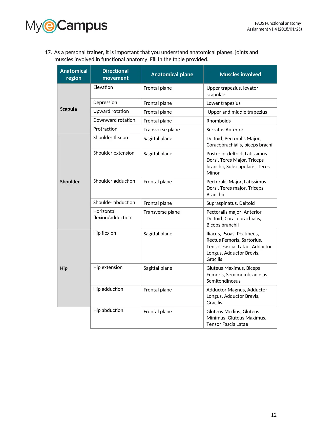
17. As a personal trainer, it is important that you understand anatomical planes, joints and
muscles involved in functional anatomy. Fill in the table provided.
Anatomical
region
Directional
movement Anatomical plane Muscles involved
Scapula
Elevation Frontal plane Upper trapezius, levator
scapulae
Depression Frontal plane Lower trapezius
Upward rotation Frontal plane Upper and middle trapezius
Downward rotation Frontal plane Rhomboids
Protraction Transverse plane Serratus Anterior
Shoulder
Shoulder flexion Sagittal plane Deltoid, Pectoralis Major,
Coracobrachialis, biceps brachii
Shoulder extension Sagittal plane Posterior deltoid, Latissimus
Dorsi, Teres Major, Triceps
branchii, Subscapularis, Teres
Minor
Shoulder adduction Frontal plane Pectoralis Major, Latissimus
Dorsi, Teres major, Triceps
Branchii
Shoulder abduction Frontal plane Supraspinatus, Deltoid
Horizontal
flexion/adduction
Transverse plane Pectoralis major, Anterior
Deltoid, Coracobrachialis,
Biceps branchii
Hip
Hip flexion Sagittal plane Iliacus, Psoas, Pectineus,
Rectus Femoris, Sartorius,
Tensor Fascia, Latae, Adductor
Longus, Adductor Brevis,
Gracilis
Hip extension Sagittal plane Gluteus Maximus, Biceps
Femoris, Semimembranosus,
Semitendinosus
Hip adduction Frontal plane Adductor Magnus, Adductor
Longus, Adductor Brevis,
Gracilis
Hip abduction Frontal plane Gluteus Medius, Gluteus
Minimus, Gluteus Maximus,
Tensor Fascia Latae
12
Assignment v1.4 (2018/01/25)
17. As a personal trainer, it is important that you understand anatomical planes, joints and
muscles involved in functional anatomy. Fill in the table provided.
Anatomical
region
Directional
movement Anatomical plane Muscles involved
Scapula
Elevation Frontal plane Upper trapezius, levator
scapulae
Depression Frontal plane Lower trapezius
Upward rotation Frontal plane Upper and middle trapezius
Downward rotation Frontal plane Rhomboids
Protraction Transverse plane Serratus Anterior
Shoulder
Shoulder flexion Sagittal plane Deltoid, Pectoralis Major,
Coracobrachialis, biceps brachii
Shoulder extension Sagittal plane Posterior deltoid, Latissimus
Dorsi, Teres Major, Triceps
branchii, Subscapularis, Teres
Minor
Shoulder adduction Frontal plane Pectoralis Major, Latissimus
Dorsi, Teres major, Triceps
Branchii
Shoulder abduction Frontal plane Supraspinatus, Deltoid
Horizontal
flexion/adduction
Transverse plane Pectoralis major, Anterior
Deltoid, Coracobrachialis,
Biceps branchii
Hip
Hip flexion Sagittal plane Iliacus, Psoas, Pectineus,
Rectus Femoris, Sartorius,
Tensor Fascia, Latae, Adductor
Longus, Adductor Brevis,
Gracilis
Hip extension Sagittal plane Gluteus Maximus, Biceps
Femoris, Semimembranosus,
Semitendinosus
Hip adduction Frontal plane Adductor Magnus, Adductor
Longus, Adductor Brevis,
Gracilis
Hip abduction Frontal plane Gluteus Medius, Gluteus
Minimus, Gluteus Maximus,
Tensor Fascia Latae
12
⊘ This is a preview!⊘
Do you want full access?
Subscribe today to unlock all pages.
Trusted by 1+ million students worldwide
1 out of 20


Your All-in-One AI-Powered Toolkit for Academic Success.
+13062052269
info@desklib.com
Available 24*7 on WhatsApp / Email
![[object Object]](/_next/static/media/star-bottom.7253800d.svg)
Unlock your academic potential
Copyright © 2020–2026 A2Z Services. All Rights Reserved. Developed and managed by ZUCOL.