Biology Homework: Genetics Concepts, Cell Cycle, and Cancer Analysis

Verified

Added on  2022/05/25

|6
|1817
|41
Homework Assignment
AI Summary
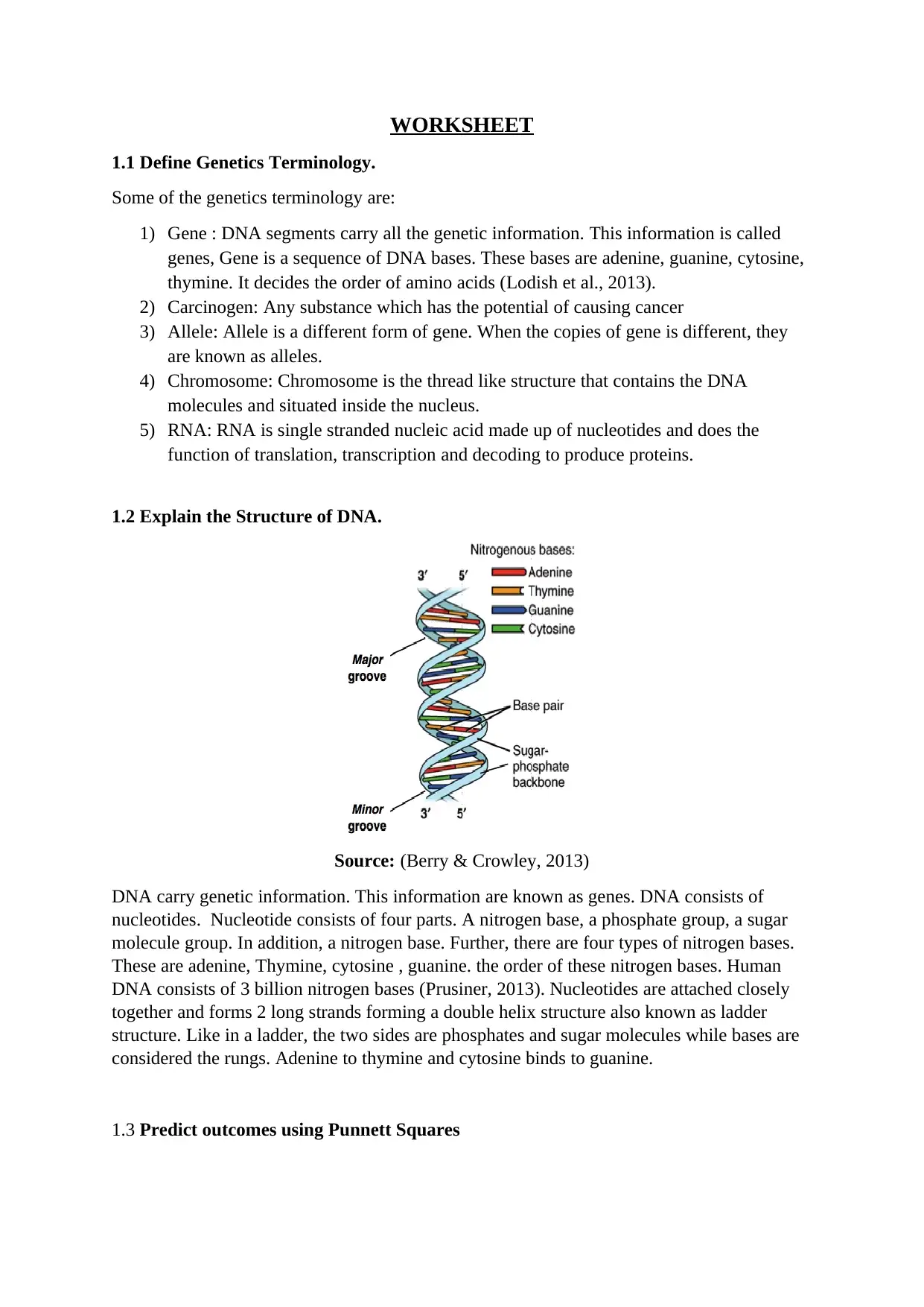
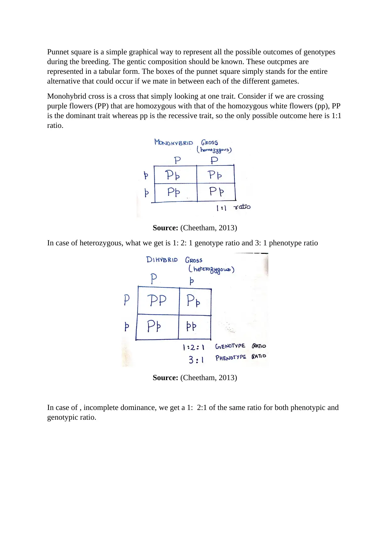
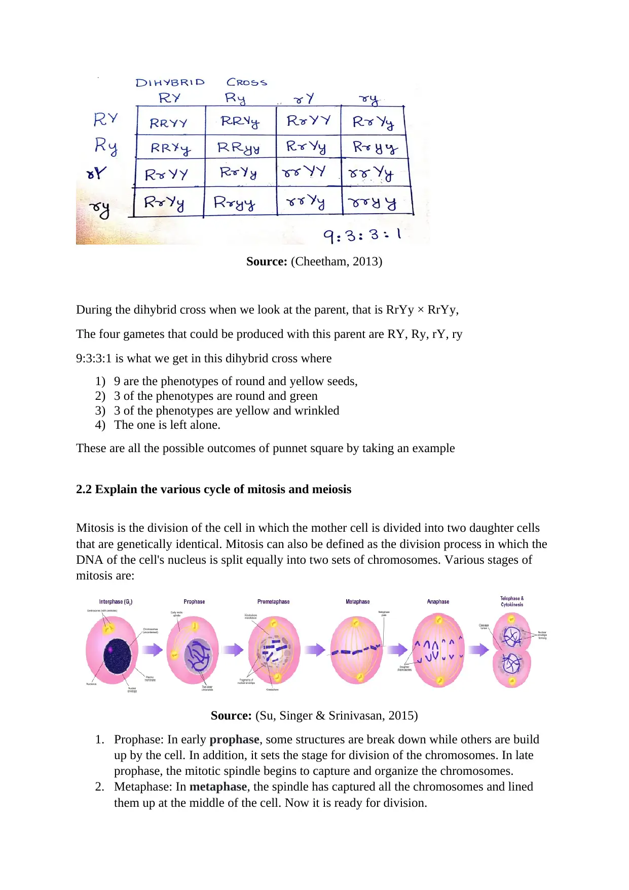
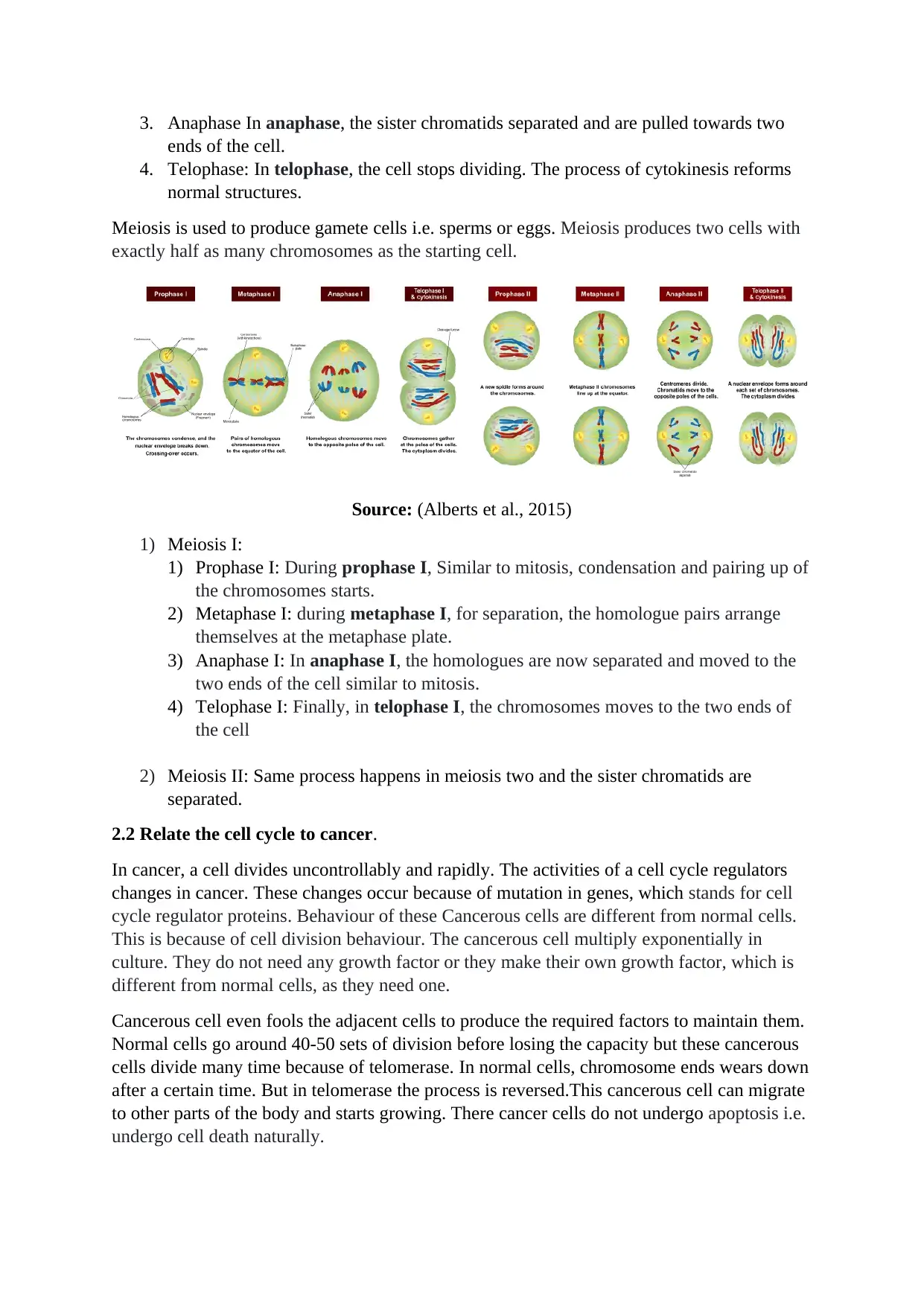
This document is a comprehensive genetics worksheet covering a range of topics within biology. It begins by defining key genetics terminology such as genes, alleles, chromosomes, and RNA. The worksheet then explains the structure of DNA, including its components and the arrangement of nitrogenous bases. It further delves into the use of Punnett squares to predict genetic outcomes in monohybrid and dihybrid crosses, including examples of complete dominance, incomplete dominance, and heterozygous crosses. The document also explains the processes of mitosis and meiosis, detailing the various stages of each cell division cycle. It then explores the relationship between the cell cycle and cancer, highlighting how disruptions in cell cycle regulators can lead to uncontrolled cell division and tumor formation. Finally, the worksheet presents cancer facts, including its global impact, risk factors, and the different stages of the cell cycle.
Document Page
WORKSHEET
1.1 Define Genetics Terminology.
Some of the genetics terminology are:
1) Gene : DNA segments carry all the genetic information. This information is called
genes, Gene is a sequence of DNA bases. These bases are adenine, guanine, cytosine,
thymine. It decides the order of amino acids (Lodish et al., 2013).
2) Carcinogen: Any substance which has the potential of causing cancer
3) Allele: Allele is a different form of gene. When the copies of gene is different, they
are known as alleles.
4) Chromosome: Chromosome is the thread like structure that contains the DNA
molecules and situated inside the nucleus.
5) RNA: RNA is single stranded nucleic acid made up of nucleotides and does the
function of translation, transcription and decoding to produce proteins.
1.2 Explain the Structure of DNA.
Source: (Berry & Crowley, 2013)
DNA carry genetic information. This information are known as genes. DNA consists of
nucleotides. Nucleotide consists of four parts. A nitrogen base, a phosphate group, a sugar
molecule group. In addition, a nitrogen base. Further, there are four types of nitrogen bases.
These are adenine, Thymine, cytosine , guanine. the order of these nitrogen bases. Human
DNA consists of 3 billion nitrogen bases (Prusiner, 2013). Nucleotides are attached closely
together and forms 2 long strands forming a double helix structure also known as ladder
structure. Like in a ladder, the two sides are phosphates and sugar molecules while bases are
considered the rungs. Adenine to thymine and cytosine binds to guanine.
1.3 Predict outcomes using Punnett Squares
tabler-icon-diamond-filled.svg

Secure Best Marks with AI Grader

Need help grading? Try our AI Grader for instant feedback on your assignments.
Document Page
Punnet square is a simple graphical way to represent all the possible outcomes of genotypes
during the breeding. The gentic composition should be known. These outcpmes are
represented in a tabular form. The boxes of the punnet square simply stands for the entire
alternative that could occur if we mate in between each of the different gametes.
Monohybrid cross is a cross that simply looking at one trait. Consider if we are crossing
purple flowers (PP) that are homozygous with that of the homozygous white flowers (pp), PP
is the dominant trait whereas pp is the recessive trait, so the only possible outcome here is 1:1
ratio.
Source: (Cheetham, 2013)
In case of heterozygous, what we get is 1: 2: 1 genotype ratio and 3: 1 phenotype ratio
Source: (Cheetham, 2013)
In case of , incomplete dominance, we get a 1: 2:1 of the same ratio for both phenotypic and
genotypic ratio.
Document Page
Source: (Cheetham, 2013)
During the dihybrid cross when we look at the parent, that is RrYy × RrYy,
The four gametes that could be produced with this parent are RY, Ry, rY, ry
9:3:3:1 is what we get in this dihybrid cross where
1) 9 are the phenotypes of round and yellow seeds,
2) 3 of the phenotypes are round and green
3) 3 of the phenotypes are yellow and wrinkled
4) The one is left alone.
These are all the possible outcomes of punnet square by taking an example
2.2 Explain the various cycle of mitosis and meiosis
Mitosis is the division of the cell in which the mother cell is divided into two daughter cells
that are genetically identical. Mitosis can also be defined as the division process in which the
DNA of the cell's nucleus is split equally into two sets of chromosomes. Various stages of
mitosis are:
Source: (Su, Singer & Srinivasan, 2015)
1. Prophase: In early prophase, some structures are break down while others are build
up by the cell. In addition, it sets the stage for division of the chromosomes. In late
prophase, the mitotic spindle begins to capture and organize the chromosomes.
2. Metaphase: In metaphase, the spindle has captured all the chromosomes and lined
them up at the middle of the cell. Now it is ready for division.
Document Page
3. Anaphase In anaphase, the sister chromatids separated and are pulled towards two
ends of the cell.
4. Telophase: In telophase, the cell stops dividing. The process of cytokinesis reforms
normal structures.
Meiosis is used to produce gamete cells i.e. sperms or eggs. Meiosis produces two cells with
exactly half as many chromosomes as the starting cell.
Source: (Alberts et al., 2015)
1) Meiosis I:
1) Prophase I: During prophase I, Similar to mitosis, condensation and pairing up of
the chromosomes starts.
2) Metaphase I: during metaphase I, for separation, the homologue pairs arrange
themselves at the metaphase plate.
3) Anaphase I: In anaphase I, the homologues are now separated and moved to the
two ends of the cell similar to mitosis.
4) Telophase I: Finally, in telophase I, the chromosomes moves to the two ends of
the cell
2) Meiosis II: Same process happens in meiosis two and the sister chromatids are
separated.
2.2 Relate the cell cycle to cancer.
In cancer, a cell divides uncontrollably and rapidly. The activities of a cell cycle regulators
changes in cancer. These changes occur because of mutation in genes, which stands for cell
cycle regulator proteins. Behaviour of these Cancerous cells are different from normal cells.
This is because of cell division behaviour. The cancerous cell multiply exponentially in
culture. They do not need any growth factor or they make their own growth factor, which is
different from normal cells, as they need one.
Cancerous cell even fools the adjacent cells to produce the required factors to maintain them.
Normal cells go around 40-50 sets of division before losing the capacity but these cancerous
cells divide many time because of telomerase. In normal cells, chromosome ends wears down
after a certain time. But in telomerase the process is reversed.This cancerous cell can migrate
to other parts of the body and starts growing. There cancer cells do not undergo apoptosis i.e.
undergo cell death naturally.
tabler-icon-diamond-filled.svg

Secure Best Marks with AI Grader

Need help grading? Try our AI Grader for instant feedback on your assignments.
Document Page
8.1 Identify cancer facts.
Young, old, rich, men, women or children, cancer can affect anyone around us. Cancer is
considered as one of the leading causes of death in the world. Cancer cells are like normal
cells but they grow rapidly and uncontrollably. Some of the cancer related facts are:
1) Cancer is a type of disease, which occurs due uncontrolled and rapid growth of the
cells, which may be due to genetic factor or external factor. There are 100 types of
cancer. Any part of the body can be affected.
2) Cancer is the major cause of death in the world. According to the stats, around 10
million death is caused due to cancer in 2018 (Saleem & Kobayashi, 2016).
3) The major risk factor for cancer is the excessive consumption of tobacco.
4) About 70 percentage of death from cancer occur from countries of Asia, Africa,
Central and South America
5) In 2018 , The most common type of cancer for men were lung, liver ,stomach,
Collateral, and prostate cancer
6) In 2018, the most common type of cancer for men were breast, lung, colorectal,
cervical and stomach cancer (Alver, Chadha & Blow, 2014).
8.2 Identify the sages of cell cycle.
The cell cycle is made up of two phases. These are
1) Interphase
2) Mitotic phase
During the interphase process, cells makes a copy of the DNA and starts growing. The
cell grows larger, copies organelles and makes the molecular building blocks during the G1
phase. The cell synthesizes a complete copy of the DNA in its nucleus during the S phase. At
G2 phase for the preparation of mitosis, the cell grows further, makes proteins and organelles
recognises its contents.
Now, during mitotic phase, DNA is divided into two sets by the cell and divides its
cytoplasm. Two new cells are formed.
The chromosomes are pulled apart by the mitotic spindle. The four different stages of mitosis
are Prophase, metaphase, telophase and anaphase.
As discussed, the process of cytokinesis forms two new cells after the division of cytoplasm
of the cell. At the end of mitosis, the cytokinesis begins with a little overlap.
Document Page
REFERENCES:
Alberts, B., Bray, D., Hopkin, K., Johnson, A. D., Lewis, J., Raff, M., ... & Walter, P.
(2015). Essential cell biology. Garland Science.
Alver, R. C., Chadha, G. S., & Blow, J. J. (2014). The contribution of dormant origins to
genome stability: from cell biology to human genetics. DNA repair, 19, 182-189.
Berry, D. P., & Crowley, J. J. (2013). Cell biology symposium: genetics of feed efficiency in
dairy and beef cattle. Journal of Animal Science, 91(4), 1594-1613.
Cheetham, S. W., Gruhl, F., Mattick, J. S., & Dinger, M. E. (2013). Long noncoding RNAs
and the genetics of cancer. British journal of cancer, 108(12), 2419.
Lodish, H., Berk, A., Zipursky, S. L., Matsudaira, P., Baltimore, D., & Darnell, J.
(2013). Molecular cell biology (Vol. 7). New York: WH freeman.
Prusiner, S. B. (2013). Biology and genetics of prions causing neurodegeneration. Annual
review of genetics, 47, 601-623.
Saleem, M. A., & Kobayashi, Y. (2016). Cell biology and genetics of minimal change
disease. F1000Research, 5.
Su, D., Singer, E. A., & Srinivasan, R. (2015). Molecular pathways in renal cell carcinoma:
recent advances in genetics and molecular biology. Current opinion in
oncology, 27(3), 217-223.
chevron_up_icon
1 out of 6
circle_padding
hide_on_mobile
zoom_out_icon
logo.png

Your All-in-One AI-Powered Toolkit for Academic Success.

Available 24*7 on WhatsApp / Email

[object Object]