Report on Hashimoto Disease: Pathophysiology and Diagnosis
VerifiedAdded on 2022/10/17
|8
|1600
|5
Report
AI Summary
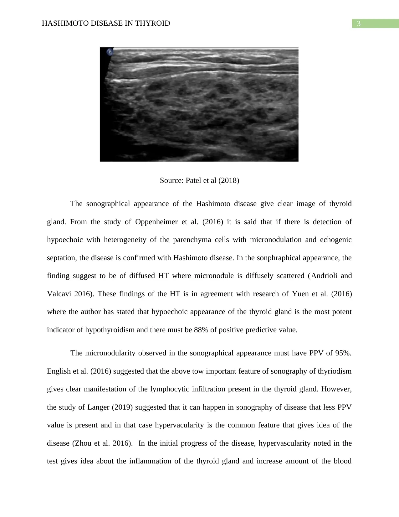
This report provides a comprehensive overview of Hashimoto's disease, a form of hypothyroidism characterized by low T3 and T4 hormone secretion and high TSH levels. It delves into the pathophysiology, explaining the autoimmune processes involving CD4 and T helper cells, the role of autoantibodies, and the destruction of thyroid gland parenchyma. The report also examines the sonographical appearance of Hashimoto's disease, detailing the significance of echogenicity, heterogeneity, micronodularity, and hypervascularity in diagnosis. It references key studies and findings related to these features, highlighting their importance in identifying the disease and understanding its progression. The report emphasizes the value of sonography in diagnosing Hashimoto's disease and the correlation between sonographical findings and the underlying lymphocytic infiltration of the thyroid gland.

Running head: HASHIMOTO DISEASE IN THYROID
HASHIMOTO DISEASE IN THYROID
Name of Student:
Name of University:
Author’s Note:
HASHIMOTO DISEASE IN THYROID
Name of Student:
Name of University:
Author’s Note:
Paraphrase This Document
Need a fresh take? Get an instant paraphrase of this document with our AI Paraphraser

1HASHIMOTO DISEASE IN THYROID
Part A pathophysiology of Hashimoto disease in thyroid
Hashimoto disease is the one of the form of hypothyroidism which is caused when there
is low secretion of T3 and T4 in the body. As the consequence of this, there is high production of
TSH hormones. Hashimoto disease is also known as autoimmune thyroiditis (D'Aurizio et al.
2015). Formation of Goitre is important in this form of hypothyroidism which occur because of
inflammation. Hashimoto disease is caused by the activation of CD4 and T helper cells which is
explicit to the thyroid. Such activation is caused by the presence of virus and bacteria. From the
study of Trohman et al. (2018) it was known that Hashimoto disease is caused by the intake of
iodine, high smoking and hepatitis.
In normal functioning of hormonal regulation, one of the function of hypothalamus
secrete TSH releasing hormone which is known as TRH. It is responsible for the further
activation of pituitary gland to release thyroid stimulating hormone TSH, which in turn also
activate the thyroid gland to produce TH particularly triiodothyronine, T3 and T4 (Kahaly et al.
2016). The regulation of the TRH and TSH is done through negative feedback mechanism. In the
situation of Hashimoto disease, there is low production of TH which is mainly caused by the loss
of the function of the thyroid gland. Due to such dysfunction of thyroid gland, there is low
secretion of TH and high release of TRH and TSH (Page et al. 2015).
The pathophysiology of the Hashimoto disease, can be further attributed by looking into
the autoimmunity disorder. Hashimoto disease is the chronic lymphocytic thyroiditis in which
antibodies is produced in against the thyroid gland which cause chronic inflammation of it.
Abnormal functioning of the thyroid gland resulted in activation of helper T cell and makes
CD4+. This in turn causes B cells to makes thyroid specific autoantibodies which is against the

2HASHIMOTO DISEASE IN THYROID
thyroid gland itself. From the study of Mincer and Jialal (2018) it was seen that there are four
different type of identified antibodies in Hashimoto thyroiditis which includes antimicrosomal
antibody, antithyroglobulin antibody, antibody to thyroid stimulating hormone receptor and
antibody to iodine transporters. These autoantibodies leads to antibody dependent cytotoxicity. It
can is summarized by the fact that activated helper T cell induce the production of CD8+ T cells,
which is responsible of thyroid cell cytotoxicity and Fas-FasL mediated apoptosis of thyroid
follicular cells. The all the above mentioned activity of the autoantibodies and CD8+ cells
causes destruction of the parenchyma of thyroid glands and infiltration of the lymphoid cells. In
response to all such activity happening in the body, antibody is produced that attack the thyroid
gland (Berghi 2017). These lymphoid cells starts to makes antibodies in the thyroid gland and
autoimmune process gets initiated. These all result in Hashimoto thyroiditis and because of it,
thyroid gland fails to produce required amount of thyroid glands to function in the body and
results in development of hypothyroidism.
Part B sonographical appearance of the Hashimoto disease
The sonographical test of the Hashimoto disease reveals the risk of the disease. The
examination is mainly done to detect five major appearance of the disease like presence and
absence of echogenicity, heterogeneity of parenchyma, septation of the gland, micronodularity
and hypervascularity of the disease (Durfee et al. 2015).
⊘ This is a preview!⊘
Do you want full access?
Subscribe today to unlock all pages.

Trusted by 1+ million students worldwide

3HASHIMOTO DISEASE IN THYROID
Source: Patel et al (2018)
The sonographical appearance of the Hashimoto disease give clear image of thyroid
gland. From the study of Oppenheimer et al. (2016) it is said that if there is detection of
hypoechoic with heterogeneity of the parenchyma cells with micronodulation and echogenic
septation, the disease is confirmed with Hashimoto disease. In the sonphraphical appearance, the
finding suggest to be of diffused HT where micronodule is diffusely scattered (Andrioli and
Valcavi 2016). These findings of the HT is in agreement with research of Yuen et al. (2016)
where the author has stated that hypoechoic appearance of the thyroid gland is the most potent
indicator of hypothyroidism and there must be 88% of positive predictive value.
The micronodularity observed in the sonographical appearance must have PPV of 95%.
English et al. (2016) suggested that the above tow important feature of sonography of thyriodism
gives clear manifestation of the lymphocytic infiltration present in the thyroid gland. However,
the study of Langer (2019) suggested that it can happen in sonography of disease that less PPV
value is present and in that case hypervacularity is the common feature that gives idea of the
disease (Zhou et al. 2016). In the initial progress of the disease, hypervascularity noted in the
test gives idea about the inflammation of the thyroid gland and increase amount of the blood
Paraphrase This Document
Need a fresh take? Get an instant paraphrase of this document with our AI Paraphraser

4HASHIMOTO DISEASE IN THYROID
flow can be interpreted (Andrioli and Valcavi 2016). Overall, it can be understood that various
feature of sonograhy of HT improvise the sensitivity in diagnosis.
flow can be interpreted (Andrioli and Valcavi 2016). Overall, it can be understood that various
feature of sonograhy of HT improvise the sensitivity in diagnosis.

5HASHIMOTO DISEASE IN THYROID
Reference
Andrioli, M. and Valcavi, R., 2016. Sonography of normal and abnormal thyroid and parathyroid
glands. In Imaging in Endocrine Disorders (Vol. 45, pp. 1-15). Karger Publishers.
Berghi, N.O., 2017. Immunological mechanisms implicated in the pathogenesis of chronic
Urticaria and Hashimoto thyroiditis. Iranian Journal of Allergy, Asthma and Immunology, 16(4),
pp.358-366.
D'Aurizio, F., Villalta, D., Metus, P., Doretto, P. and Tozzoli, R., 2015. Is vitamin D a player or
not in the pathophysiology of autoimmune thyroid diseases?. Autoimmunity reviews, 14(5),
pp.363-369.
Dighe, M., Barr, R., Bojunga, J., Cantisani, V., Chammas, M.C., Cosgrove, D., Cui, X.W., Dong,
Y., Fenner, F., Radzina, M. and Vinayak, S., 2017. Thyroid ultrasound: state of the art part 1–
thyroid ultrasound reporting and diffuse thyroid diseases. Medical ultrasonography, 19(1),
pp.79-93.
Durfee, S.M., Benson, C.B., Arthaud, D.M., Alexander, E.K. and Frates, M.C., 2015.
Sonographic appearance of thyroid cancer in patients with Hashimoto thyroiditis. Journal of
Ultrasound in Medicine, 34(4), pp.697-704.
English, C., Casey, R., Bell, M., Bergin, D. and Murphy, J., 2016. The Sonographic Features of
the Thyroid Gland After Treatment with Radioiodine Therapy in Patients with Graves'
Disease. Ultrasound in medicine & biology, 42(1), pp.60-67.
⊘ This is a preview!⊘
Do you want full access?
Subscribe today to unlock all pages.

Trusted by 1+ million students worldwide

6HASHIMOTO DISEASE IN THYROID
Kahaly, G.J., Diana, T., Glang, J., Kanitz, M., Pitz, S. and König, J., 2016. Thyroid stimulating
antibodies are highly prevalent in Hashimoto's thyroiditis and associated Orbitopathy. The
Journal of Clinical Endocrinology & Metabolism, 101(5), pp.1998-2004.
Langer, J.E., 2019. Sonography of the Thyroid. Radiologic Clinics, 57(3), pp.469-483.
Mincer, D.L. and Jialal, I., 2018. Hashimoto thyroiditis. In StatPearls [Internet]. StatPearls
Publishing.
Oppenheimer, D.C., Giampoli, E., Montoya, S., Patel, S. and Dogra, V., 2016. Sonographic
features of nodular hashimoto thyroiditis. Ultrasound quarterly, 32(3), pp.271-276.
Page, C., Duverlie, G., Sevestre, H. and Desailloud, R., 2015. Erythrovirus B19 and autoimmune
thyroid diseases. Review of the literature and pathophysiological hypotheses. Journal of medical
virology, 87(1), pp.162-169.
Patel, S., Giampoli, E., Oppenheimer, D., Montoya, S., Rupasov, A. and Dogra, V., 2018.
Sonographic features of diffuse Hashimoto thyroiditis: determining sensitivity of features and
predictors of malignancy. Am J Sonogr, 1(6), pp.1-7.
Trohman, R.G., Sharma, P.S., McAninch, E.A. and Bianco, A.C., 2018. Amiodarone and the
thyroid physiology, pathophysiology, diagnosis and management. Trends in cardiovascular
medicine.
Yuen, H.Y., Wong, K.T. and Ahuja, A.T., 2016. Sonography of diffuse thyroid
disease. Australasian Journal of Ultrasound in Medicine, 19(1), pp.13-29.
Paraphrase This Document
Need a fresh take? Get an instant paraphrase of this document with our AI Paraphraser

7HASHIMOTO DISEASE IN THYROID
Zhou, H., Yue, W.W., Du, L.Y., Xu, J.M., Liu, B.J., Li, X.L., Wang, D., Zhou, X.L. and Xu,
H.X., 2016. A modified Thyroid Imaging Reporting and Data System (mTI-RADS) for thyroid
nodules in coexisting Hashimoto’s Thyroiditis. Scientific reports, 6, p.26410.
Zhou, H., Yue, W.W., Du, L.Y., Xu, J.M., Liu, B.J., Li, X.L., Wang, D., Zhou, X.L. and Xu,
H.X., 2016. A modified Thyroid Imaging Reporting and Data System (mTI-RADS) for thyroid
nodules in coexisting Hashimoto’s Thyroiditis. Scientific reports, 6, p.26410.
1 out of 8
Related Documents

Your All-in-One AI-Powered Toolkit for Academic Success.
+13062052269
info@desklib.com
Available 24*7 on WhatsApp / Email
Unlock your academic potential
Copyright © 2020–2025 A2Z Services. All Rights Reserved. Developed and managed by ZUCOL.