Case Study: HCC Diagnosis, Treatment, and Patient Outcome Analysis

Verified

Added on  2022/11/19

|1
|1085
|72
Case Study
AI Summary
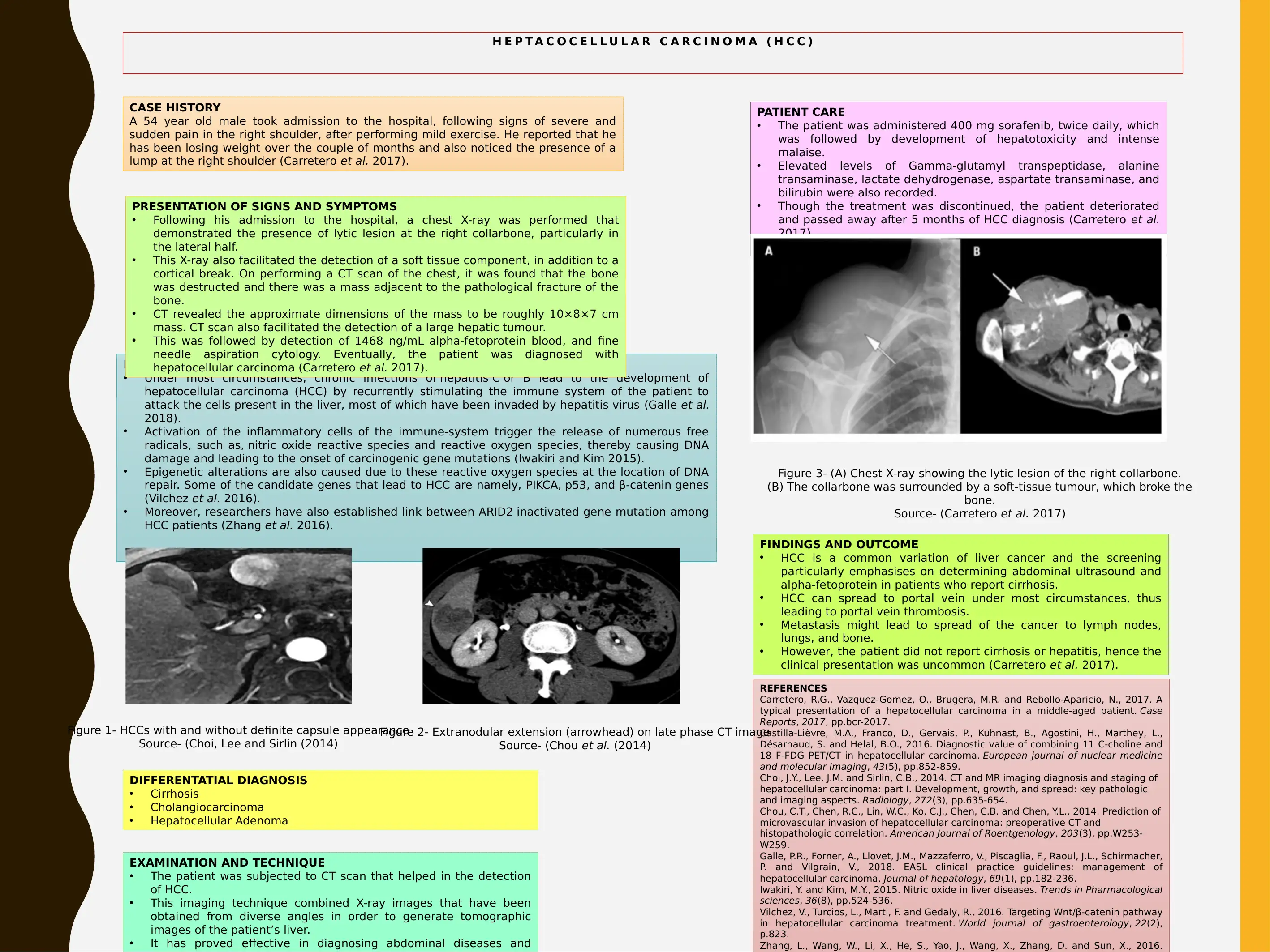
This case study details the diagnosis and treatment of a 54-year-old male patient diagnosed with hepatocellular carcinoma (HCC). The patient presented with right shoulder pain and weight loss, leading to the detection of a lump. Differential diagnoses included cirrhosis, cholangiocarcinoma, and hepatocellular adenoma. The study outlines the pathophysiology, emphasizing the role of chronic hepatitis infections and the resulting DNA damage. The patient's care involved sorafenib treatment, which was discontinued due to hepatotoxicity. Diagnostic methods included chest X-rays, CT scans, and alpha-fetoprotein blood tests. The findings revealed a lytic lesion in the right collarbone and a large hepatic tumor. The outcome was fatal, with the patient passing away five months after diagnosis. The case highlights the importance of imaging techniques like CT scans in diagnosing HCC and the challenges in treating advanced stages of the disease. References to relevant research papers are also included.
Document Page
H E P T A C O C E L L U L A R C A R C I N O M A ( H C C )
CASE HISTORY
A 54 year old male took admission to the hospital, following signs of severe and
sudden pain in the right shoulder, after performing mild exercise. He reported that he
has been losing weight over the couple of months and also noticed the presence of a
lump at the right shoulder (Carretero et al. 2017).
DIFFERENTATIAL DIAGNOSIS
Cirrhosis
Cholangiocarcinoma
Hepatocellular Adenoma
PATHOPHYSIOLOGY
Under most circumstances, chronic infections of hepatitis C or B lead to the development of
hepatocellular carcinoma (HCC) by recurrently stimulating the immune system of the patient to
attack the cells present in the liver, most of which have been invaded by hepatitis virus (Galle et al.
2018).
Activation of the inflammatory cells of the immune-system trigger the release of numerous free
radicals, such as, nitric oxide reactive species and reactive oxygen species, thereby causing DNA
damage and leading to the onset of carcinogenic gene mutations (Iwakiri and Kim 2015).
Epigenetic alterations are also caused due to these reactive oxygen species at the location of DNA
repair. Some of the candidate genes that lead to HCC are namely, PIKCA, p53, and β-catenin genes
(Vilchez et al. 2016).
Moreover, researchers have also established link between ARID2 inactivated gene mutation among
HCC patients (Zhang et al. 2016).
PATIENT CARE
The patient was administered 400 mg sorafenib, twice daily, which
was followed by development of hepatotoxicity and intense
malaise.
Elevated levels of Gamma-glutamyl transpeptidase, alanine
transaminase, lactate dehydrogenase, aspartate transaminase, and
bilirubin were also recorded.
Though the treatment was discontinued, the patient deteriorated
and passed away after 5 months of HCC diagnosis (Carretero et al.
2017).
REFERENCES
Carretero, R.G., Vazquez-Gomez, O., Brugera, M.R. and Rebollo-Aparicio, N., 2017. A
typical presentation of a hepatocellular carcinoma in a middle-aged patient. Case
Reports, 2017, pp.bcr-2017.
Castilla-Lièvre, M.A., Franco, D., Gervais, P., Kuhnast, B., Agostini, H., Marthey, L.,
Désarnaud, S. and Helal, B.O., 2016. Diagnostic value of combining 11 C-choline and
18 F-FDG PET/CT in hepatocellular carcinoma. European journal of nuclear medicine
and molecular imaging, 43(5), pp.852-859.
Choi, J.Y., Lee, J.M. and Sirlin, C.B., 2014. CT and MR imaging diagnosis and staging of
hepatocellular carcinoma: part I. Development, growth, and spread: key pathologic
and imaging aspects. Radiology, 272(3), pp.635-654.
Chou, C.T., Chen, R.C., Lin, W.C., Ko, C.J., Chen, C.B. and Chen, Y.L., 2014. Prediction of
microvascular invasion of hepatocellular carcinoma: preoperative CT and
histopathologic correlation. American Journal of Roentgenology, 203(3), pp.W253-
W259.
Galle, P.R., Forner, A., Llovet, J.M., Mazzaferro, V., Piscaglia, F., Raoul, J.L., Schirmacher,
P. and Vilgrain, V., 2018. EASL clinical practice guidelines: management of
hepatocellular carcinoma. Journal of hepatology, 69(1), pp.182-236.
Iwakiri, Y. and Kim, M.Y., 2015. Nitric oxide in liver diseases. Trends in Pharmacological
sciences, 36(8), pp.524-536.
Vilchez, V., Turcios, L., Marti, F. and Gedaly, R., 2016. Targeting Wnt/β-catenin pathway
in hepatocellular carcinoma treatment. World journal of gastroenterology, 22(2),
p.823.
Zhang, L., Wang, W., Li, X., He, S., Yao, J., Wang, X., Zhang, D. and Sun, X., 2016.
Figure 3- (A) Chest X-ray showing the lytic lesion of the right collarbone.
(B) The collarbone was surrounded by a soft-tissue tumour, which broke the
bone.
Source- (Carretero et al. 2017)
PRESENTATION OF SIGNS AND SYMPTOMS
Following his admission to the hospital, a chest X-ray was performed that
demonstrated the presence of lytic lesion at the right collarbone, particularly in
the lateral half.
This X-ray also facilitated the detection of a soft tissue component, in addition to a
cortical break. On performing a CT scan of the chest, it was found that the bone
was destructed and there was a mass adjacent to the pathological fracture of the
bone.
CT revealed the approximate dimensions of the mass to be roughly 10×8×7 cm
mass. CT scan also facilitated the detection of a large hepatic tumour.
This was followed by detection of 1468 ng/mL alpha-fetoprotein blood, and fine
needle aspiration cytology. Eventually, the patient was diagnosed with
hepatocellular carcinoma (Carretero et al. 2017).
EXAMINATION AND TECHNIQUE
The patient was subjected to CT scan that helped in the detection
of HCC.
This imaging technique combined X-ray images that have been
obtained from diverse angles in order to generate tomographic
images of the patient’s liver.
It has proved effective in diagnosing abdominal diseases and
FINDINGS AND OUTCOME
HCC is a common variation of liver cancer and the screening
particularly emphasises on determining abdominal ultrasound and
alpha-fetoprotein in patients who report cirrhosis.
HCC can spread to portal vein under most circumstances, thus
leading to portal vein thrombosis.
Metastasis might lead to spread of the cancer to lymph nodes,
lungs, and bone.
However, the patient did not report cirrhosis or hepatitis, hence the
clinical presentation was uncommon (Carretero et al. 2017).
Figure 1- HCCs with and without definite capsule appearance
Source- (Choi, Lee and Sirlin (2014)
Figure 2- Extranodular extension (arrowhead) on late phase CT image
Source- (Chou et al. (2014)
tabler-icon-diamond-filled.svg

Secure Best Marks with AI Grader

Need help grading? Try our AI Grader for instant feedback on your assignments.
chevron_up_icon
1 out of 1
circle_padding
hide_on_mobile
zoom_out_icon
logo.png

Your All-in-One AI-Powered Toolkit for Academic Success.

Available 24*7 on WhatsApp / Email

[object Object]