Advanced Health Assessment: Head and Neck Presentation (NURS 511)
VerifiedAdded on 2022/08/13
|12
|1468
|40
Presentation
AI Summary
This presentation provides a comprehensive overview of the anatomical and physiological structures of the head and neck, including the eyes, nose, ears, and throat. It explores the health history of these organs, highlighting potential abnormalities and diagnostic tests such as urinalysis, CT scans, and eye exams. The presentation also covers the normal and abnormal findings associated with various conditions and emphasizes the role of nurses in addressing these issues. Furthermore, it delves into specific diagnostic procedures for eye, ear, nose, and neck problems, along with relevant nursing interventions. The presentation concludes with a discussion on the overall assessment and management of head and neck-related health concerns, supported by evidence-based research and practical applications.
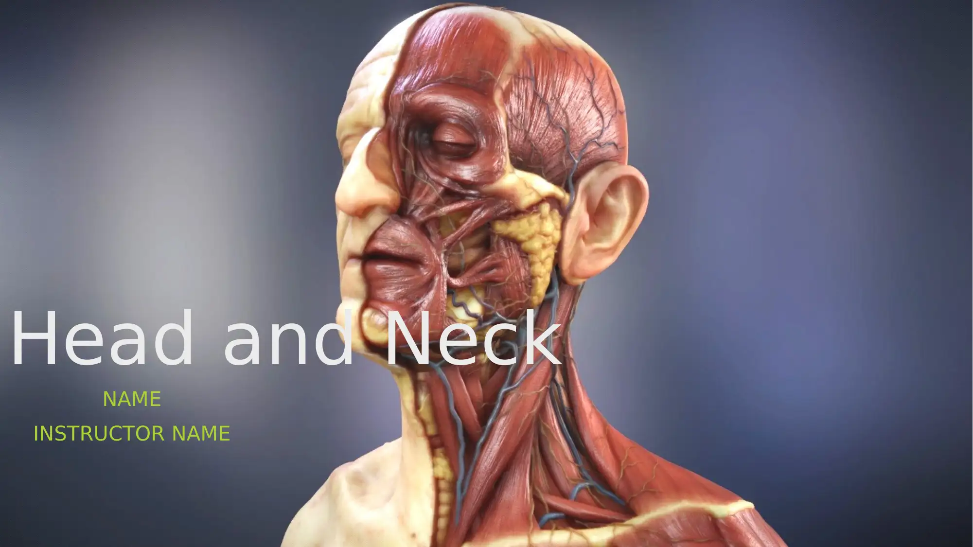
Head and Neck
NAME
INSTRUCTOR NAME
NAME
INSTRUCTOR NAME
Paraphrase This Document
Need a fresh take? Get an instant paraphrase of this document with our AI Paraphraser
Outline and Objective of the
Presentation
The objective of this presentation is to shed light on the anatomical and
physiological structure of head and neck.
Head and neck comprises of significant structures of the whole body. The
critical structures that can be found within the head and the neck are the Eyes,
the Nose, the Ears and the Throat. The major senses and the main controller of
the central nervous system lies in this region. The previous health issues
related to these organs will be highlighted.
A general study on the recent techniques to detect the risk related to these
organs will be assessed.
A physical and diagnostic tests for assessing the onset of risk will be
mentioned.
The normal range and abnormal range during the disease will be evaluated.
The approach of the nurses and their role to deal with such abnormalities will
be studied.
Introduction
Head is the most significant part of the system. The head and
neck portion deals with the most critical function of the body
which includes brain, muscles, blood vessels, mouth, nose,
tongue, throat, teeth, ears, eyes and glands. The anatomical and
their physiological structures are complex and has a history of
numerous health issues from the past. The diagnosis and
abnormal findings of the diseases related to head and neck has
been determined
⊘ This is a preview!⊘
Do you want full access?
Subscribe today to unlock all pages.
Trusted by 1+ million students worldwide
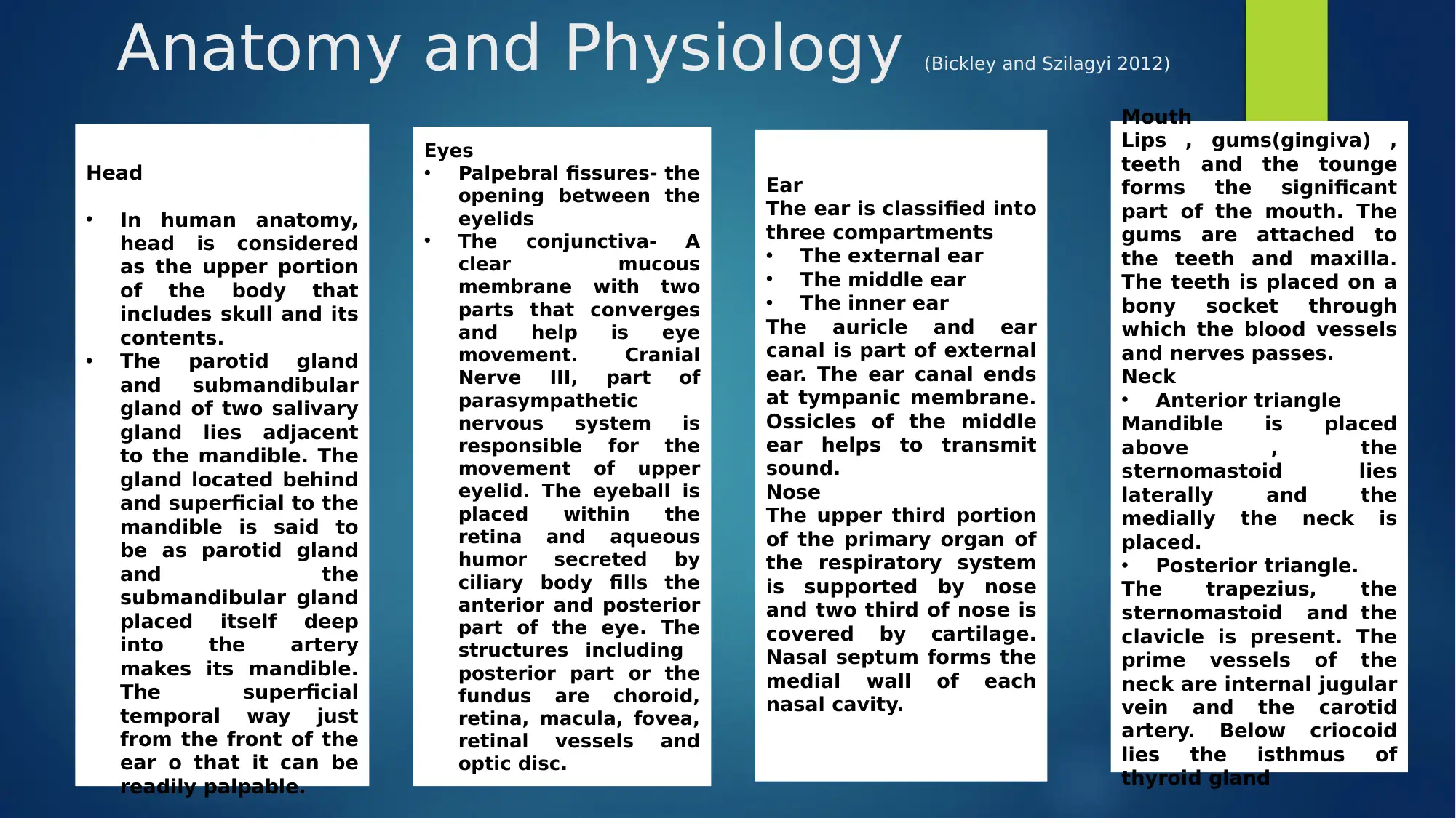
Anatomy and Physiology (Bickley and Szilagyi 2012)
Head
• In human anatomy,
head is considered
as the upper portion
of the body that
includes skull and its
contents.
• The parotid gland
and submandibular
gland of two salivary
gland lies adjacent
to the mandible. The
gland located behind
and superficial to the
mandible is said to
be as parotid gland
and the
submandibular gland
placed itself deep
into the artery
makes its mandible.
The superficial
temporal way just
from the front of the
ear o that it can be
readily palpable.
Eyes
• Palpebral fissures- the
opening between the
eyelids
• The conjunctiva- A
clear mucous
membrane with two
parts that converges
and help is eye
movement. Cranial
Nerve III, part of
parasympathetic
nervous system is
responsible for the
movement of upper
eyelid. The eyeball is
placed within the
retina and aqueous
humor secreted by
ciliary body fills the
anterior and posterior
part of the eye. The
structures including
posterior part or the
fundus are choroid,
retina, macula, fovea,
retinal vessels and
optic disc.
Ear
The ear is classified into
three compartments
• The external ear
• The middle ear
• The inner ear
The auricle and ear
canal is part of external
ear. The ear canal ends
at tympanic membrane.
Ossicles of the middle
ear helps to transmit
sound.
Nose
The upper third portion
of the primary organ of
the respiratory system
is supported by nose
and two third of nose is
covered by cartilage.
Nasal septum forms the
medial wall of each
nasal cavity.
Mouth
Lips , gums(gingiva) ,
teeth and the tounge
forms the significant
part of the mouth. The
gums are attached to
the teeth and maxilla.
The teeth is placed on a
bony socket through
which the blood vessels
and nerves passes.
Neck
• Anterior triangle
Mandible is placed
above , the
sternomastoid lies
laterally and the
medially the neck is
placed.
• Posterior triangle.
The trapezius, the
sternomastoid and the
clavicle is present. The
prime vessels of the
neck are internal jugular
vein and the carotid
artery. Below criocoid
lies the isthmus of
thyroid gland
Paraphrase This Document
Need a fresh take? Get an instant paraphrase of this document with our AI Paraphraser
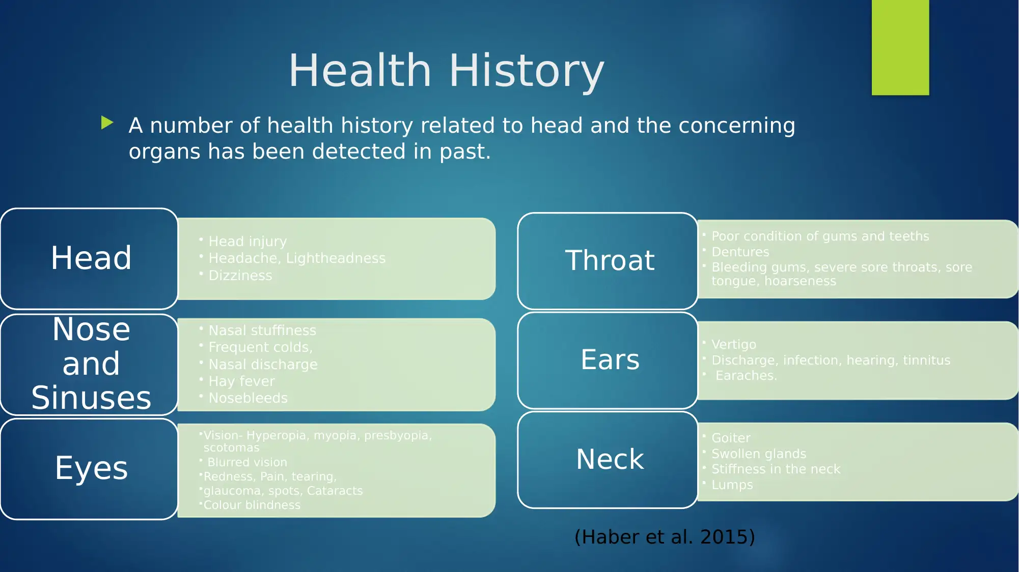
Health History
A number of health history related to head and the concerning
organs has been detected in past.
• Head injury
• Headache, Lightheadness
• Dizziness
Head
• Nasal stuffiness
• Frequent colds,
• Nasal discharge
• Hay fever
• Nosebleeds
Nose
and
Sinuses •Vision- Hyperopia, myopia, presbyopia,
scotomas
• Blurred vision
•Redness, Pain, tearing,
•glaucoma, spots, Cataracts
•Colour blindness
Eyes
• Poor condition of gums and teeths
• Dentures
• Bleeding gums, severe sore throats, sore
tongue, hoarseness
Throat
• Vertigo
• Discharge, infection, hearing, tinnitus
• Earaches.
Ears
• Goiter
• Swollen glands
• Stiffness in the neck
• Lumps
Neck
(Haber et al. 2015)
Physical examination
Head
• Hair
• Scalp
• Skull
• Face
• Skin
EYES •Screening of
temporal field.
•Position of the
eyes
•Abnormalities
in eyelids
•Conjunctiva
and Sclera
•Lens and
cornea-
inspection of
cornea can be
done by
passing
oblique light.
•Pupils
•Convergence
test
Ear • Ear canal
and drum
• Webers
test-
lateralizati
on test
• Compariso
n of bone
onduction
and air
conduction
.
Nose • Pressing
each ala
nasi and
make the
patient
breathe
• Inspecting
the interior
portion of
the nose
with an
otoscope
Mouth • Inspectio
n of the
color of
the gums
Neck • Preauricula
r, posterior
auricular,
occipital,
tonsillar,
submandib
ular, sub
mental,
superficial
cervical,
posterior
cervical,
deep
cervical
chain,
supraclavi
cular.
(Elisseou, Puranam and Nandi 2018)
⊘ This is a preview!⊘
Do you want full access?
Subscribe today to unlock all pages.
Trusted by 1+ million students worldwide
Diagnostic tests
To detect abnormal condition of the head related problems like headaches, migraines following diagnostic test needs to
be done (George et al. 2014)
Urinalysis, CT Scan, MRI, Sinus X-Ray, EEG, Spinal Tap, Eye test
Eye abnormalities (Baudouin et al. 2014. Diagnosing the severity of dry eye: a clear and practical algorithm. British
Journal of Ophthalmology, 98(9), pp.1168-1176.
Color Vision test- the color vision is tested by Farnworth- Munsell 100 Hue.
Nerve Fiber Analysis and Computerized Optic Disc Imaging
Corneal Topography- This test measures the shape of the cornea.
Electro- diagnostic testing- This test provides information about the function of the retina and optic pathways to the brain.
Fluorescein angiography- It’s the test to ensure the presence of abnormal blood vessels and recognize the presence of
leaks in the retinal fluid.
Nose
Nasal endoscopy, Allergy test, Blood test, Imaging studies
Ear
General screening test, Tuning fork test, App-based hearing test , Audiometer test
Neck test
CT scan, MRI, Bone scan, Electromyograph, Discogram, Myelogram
Paraphrase This Document
Need a fresh take? Get an instant paraphrase of this document with our AI Paraphraser
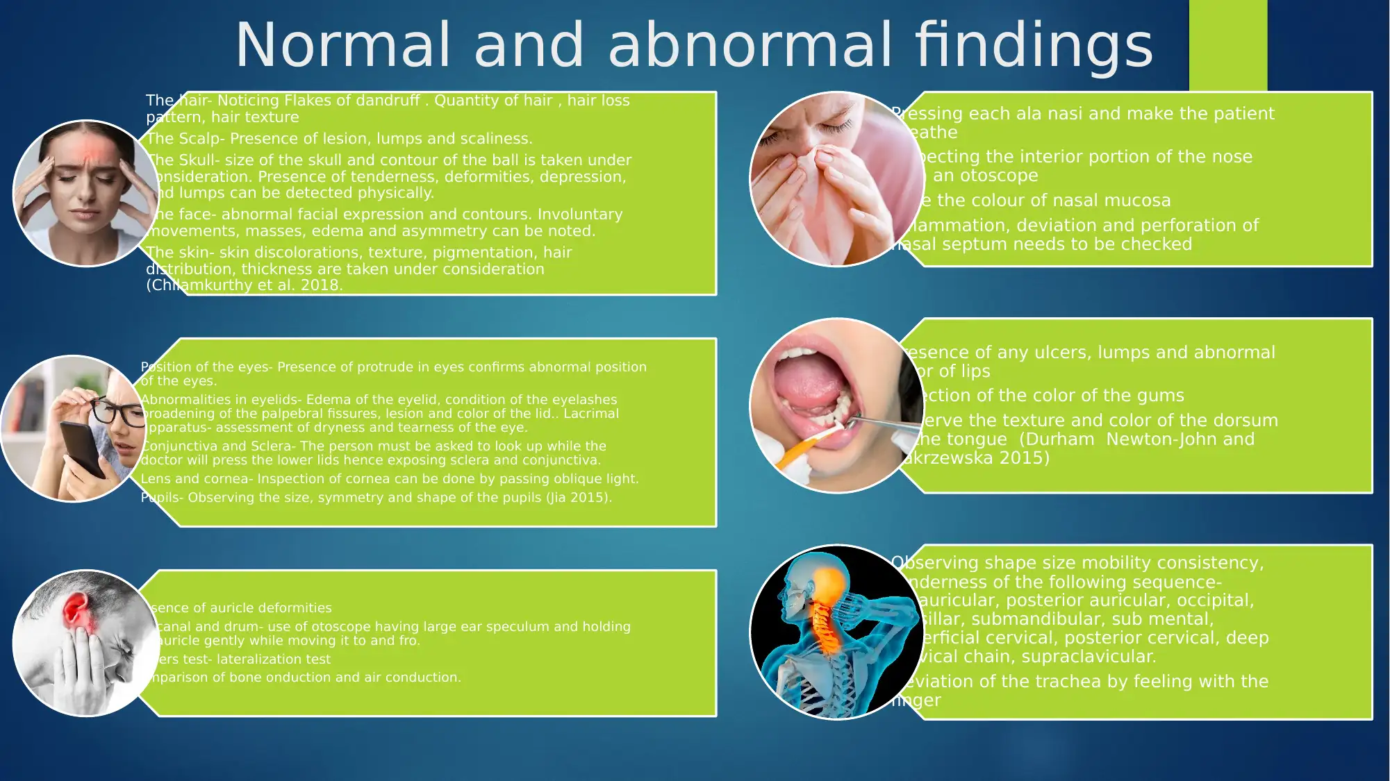
Normal and abnormal findings
The hair- Noticing Flakes of dandruff . Quantity of hair , hair loss
pattern, hair texture
The Scalp- Presence of lesion, lumps and scaliness.
The Skull- size of the skull and contour of the ball is taken under
consideration. Presence of tenderness, deformities, depression,
and lumps can be detected physically.
The face- abnormal facial expression and contours. Involuntary
movements, masses, edema and asymmetry can be noted.
The skin- skin discolorations, texture, pigmentation, hair
distribution, thickness are taken under consideration
(Chilamkurthy et al. 2018.
Position of the eyes- Presence of protrude in eyes confirms abnormal position
of the eyes.
Abnormalities in eyelids- Edema of the eyelid, condition of the eyelashes
broadening of the palpebral fissures, lesion and color of the lid.. Lacrimal
apparatus- assessment of dryness and tearness of the eye.
Conjunctiva and Sclera- The person must be asked to look up while the
doctor will press the lower lids hence exposing sclera and conjunctiva.
Lens and cornea- Inspection of cornea can be done by passing oblique light.
Pupils- Observing the size, symmetry and shape of the pupils (Jia 2015).
Presence of auricle deformities
Ear canal and drum- use of otoscope having large ear speculum and holding
the auricle gently while moving it to and fro.
Webers test- lateralization test
Comparison of bone onduction and air conduction.
Pressing each ala nasi and make the patient
breathe
Inspecting the interior portion of the nose
with an otoscope
Note the colour of nasal mucosa
Inflammation, deviation and perforation of
nasal septum needs to be checked
Presence of any ulcers, lumps and abnormal
color of lips
Inpection of the color of the gums
Observe the texture and color of the dorsum
of the tongue (Durham Newton-John and
Zakrzewska 2015)
Observing shape size mobility consistency,
tenderness of the following sequence-
Preauricular, posterior auricular, occipital,
tonsillar, submandibular, sub mental,
superficial cervical, posterior cervical, deep
cervical chain, supraclavicular.
Deviation of the trachea by feeling with the
finger
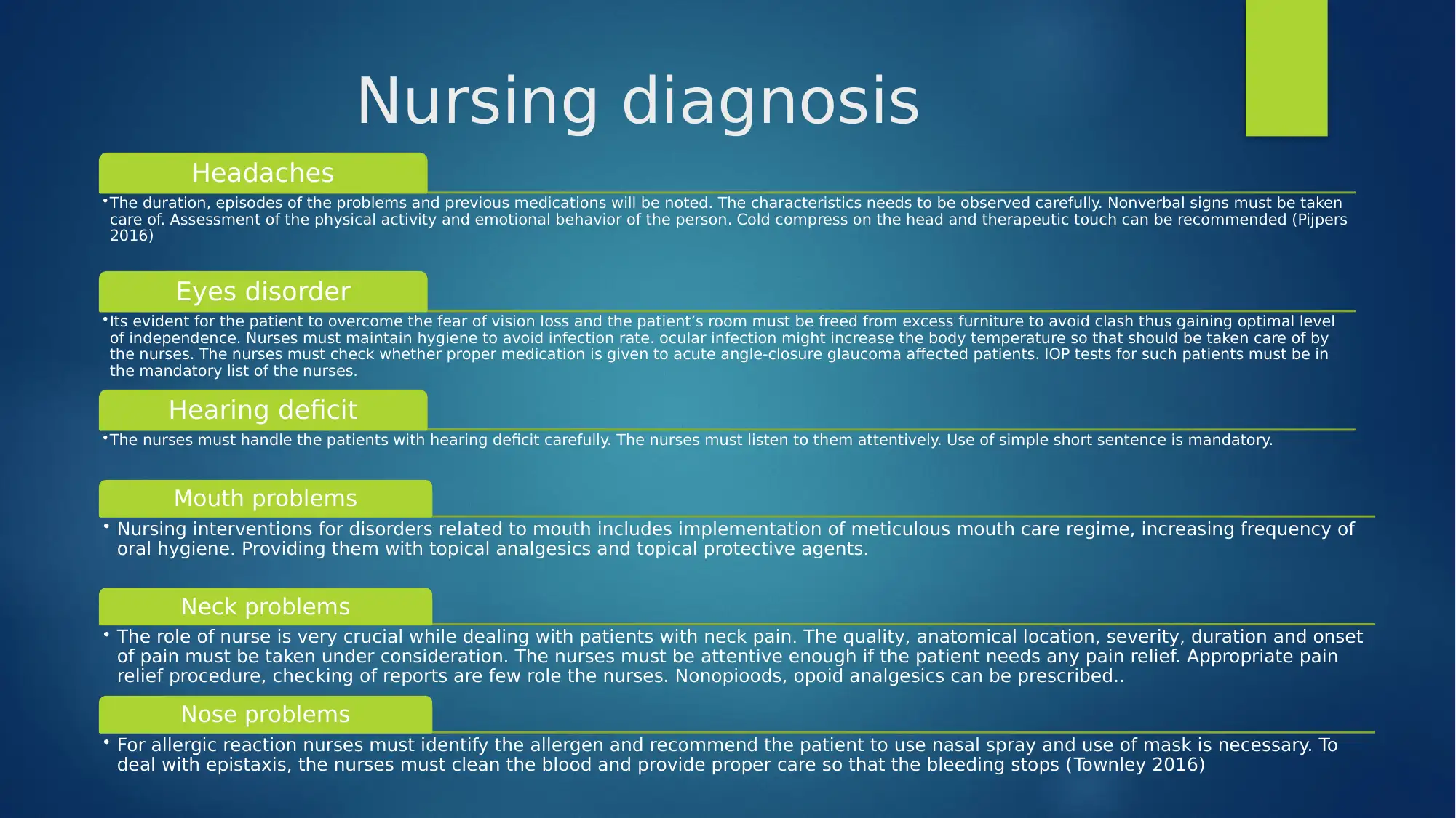
Nursing diagnosis
Headaches
•The duration, episodes of the problems and previous medications will be noted. The characteristics needs to be observed carefully. Nonverbal signs must be taken
care of. Assessment of the physical activity and emotional behavior of the person. Cold compress on the head and therapeutic touch can be recommended (Pijpers
2016)
Eyes disorder
•Its evident for the patient to overcome the fear of vision loss and the patient’s room must be freed from excess furniture to avoid clash thus gaining optimal level
of independence. Nurses must maintain hygiene to avoid infection rate. ocular infection might increase the body temperature so that should be taken care of by
the nurses. The nurses must check whether proper medication is given to acute angle-closure glaucoma affected patients. IOP tests for such patients must be in
the mandatory list of the nurses.
Hearing deficit
•The nurses must handle the patients with hearing deficit carefully. The nurses must listen to them attentively. Use of simple short sentence is mandatory.
Mouth problems
• Nursing interventions for disorders related to mouth includes implementation of meticulous mouth care regime, increasing frequency of
oral hygiene. Providing them with topical analgesics and topical protective agents.
Neck problems
• The role of nurse is very crucial while dealing with patients with neck pain. The quality, anatomical location, severity, duration and onset
of pain must be taken under consideration. The nurses must be attentive enough if the patient needs any pain relief. Appropriate pain
relief procedure, checking of reports are few role the nurses. Nonopioods, opoid analgesics can be prescribed..
Nose problems
• For allergic reaction nurses must identify the allergen and recommend the patient to use nasal spray and use of mask is necessary. To
deal with epistaxis, the nurses must clean the blood and provide proper care so that the bleeding stops (Townley 2016)
⊘ This is a preview!⊘
Do you want full access?
Subscribe today to unlock all pages.
Trusted by 1+ million students worldwide
Summary
This presentation concludes the anatomical and physiological
structures of all the organs involved in head and neck. Their
health history has been discussed along with which their
abnormal physical appearance is highlighted. The presentation
includes the diagnostic test and nursing interventions of the head
and neck related problems.
Paraphrase This Document
Need a fresh take? Get an instant paraphrase of this document with our AI Paraphraser
Reference
Baudouin, C., Aragona, P., Van Setten, G., Rolando, M., Irkeç, M., del Castillo, J.B., Geerling, G., Labetoulle, M. and Bonini, S.,
2014. Diagnosing the severity of dry eye: a clear and practical algorithm. British Journal of Ophthalmology, 98(9), pp.1168-
1176.
Bickley, L. and Szilagyi, P.G., 2012. Bates' guide to physical examination and history-taking. Lippincott Williams & Wilkins.
Chilamkurthy, S., Ghosh, R., Tanamala, S., Biviji, M., Campeau, N.G., Venugopal, V.K., Mahajan, V., Rao, P. and Warier, P., 2018.
Deep learning algorithms for detection of critical findings in head CT scans: a retrospective study. The Lancet, 392(10162),
pp.2388-2396.
Durham, J., Newton-John, T.R. and Zakrzewska, J.M., 2015. Temporomandibular disorders. bmj, 350, p.h1154.
Elisseou, S., Puranam, S. and Nandi, M., 2018. A novel, trauma-informed physical examination curriculum. Med Educ, 52(5),
pp.555-556.
George, R.T., Mehra, V.C., Chen, M.Y., Kitagawa, K., Arbab-Zadeh, A., Miller, J.M., Matheson, M.B., Vavere, A.L., Kofoed, K.F.,
Rochitte, C.E. and Dewey, M., 2014. Myocardial CT perfusion imaging and SPECT for the diagnosis of coronary artery
disease: a head-to-head comparison from the CORE320 multicenter diagnostic performance study. Radiology, 272(2),
pp.407-416.
Haber, J., Hartnett, E., Allen, K., Hallas, D., Dorsen, C., Lange-Kessler, J., Lloyd, M., Thomas, E. and Wholihan, D., 2015. Putting
the mouth back in the head: HEENT to HEENOT. American journal of public health, 105(3), pp.437-441.
Jia, Y., Bailey, S.T., Hwang, T.S., McClintic, S.M., Gao, S.S., Pennesi, M.E., Flaxel, C.J., Lauer, A.K., Wilson, D.J., Hornegger, J. and
Fujimoto, J.G., 2015. Quantitative optical coherence tomography angiography of vascular abnormalities in the living human
eye. Proceedings of the National Academy of Sciences, 112(18), pp.E2395-E2402.
Pijpers, J.A., Louter, M.A., De Bruin, M.E., Van Zwet, E.W., Zitman, F.G., Ferrari, M.D. and Terwindt, G.M., 2016. Detoxification in
medication-overuse headache, a retrospective controlled follow-up study: does care by a headache nurse lead to
cure?. Cephalalgia, 36(2), pp.122-130.
Townley, D., Shields, B., Keogh, I., Zhan, M.Q. and Farrell, C., National University of Ireland Galway (NUI Galway), 2016. Devices
for therapeutic nasal neuromodulation and associated methods and systems. U.S. Patent Application 15/153,217.

⊘ This is a preview!⊘
Do you want full access?
Subscribe today to unlock all pages.
Trusted by 1+ million students worldwide
1 out of 12
Your All-in-One AI-Powered Toolkit for Academic Success.
+13062052269
info@desklib.com
Available 24*7 on WhatsApp / Email
![[object Object]](/_next/static/media/star-bottom.7253800d.svg)
Unlock your academic potential
Copyright © 2020–2025 A2Z Services. All Rights Reserved. Developed and managed by ZUCOL.