Infectious Conjunctivitis Case Study: Health Variations 2 Assignment
VerifiedAdded on 2022/10/12
|6
|2125
|79
Case Study
AI Summary
This case study presents a detailed analysis of infectious conjunctivitis, addressing its background, causes, and the role of microorganisms. The assignment explores the mechanism of action and adverse reactions of gentamicin, a common treatment. It further examines the physiological basis of the signs and symptoms observed in a patient, including redness, swelling, and discharge. The study also delves into infection control issues, transmission pathways, and strategies to break the chain of infection within a healthcare setting. Finally, the assignment emphasizes proper referencing using the APA 6th edition style.
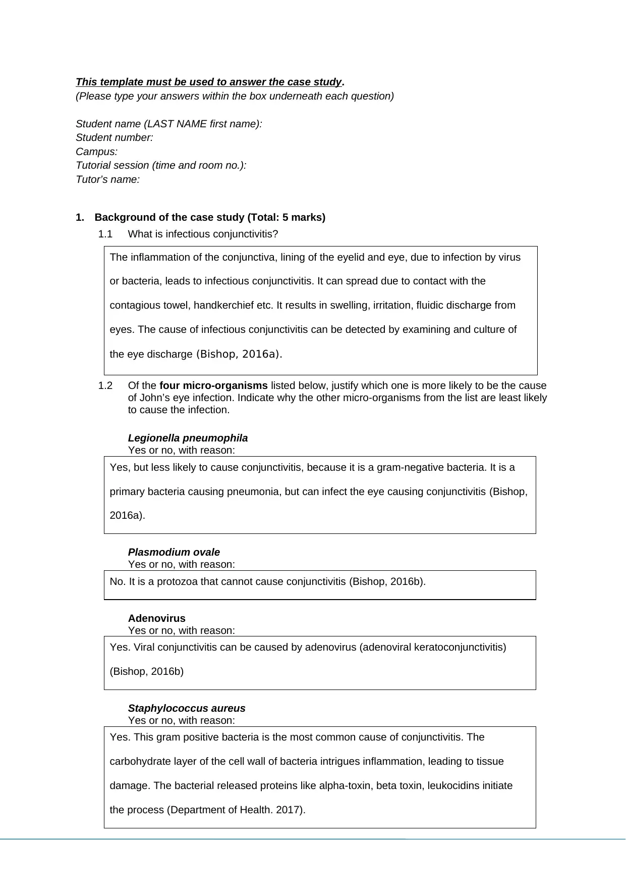
This template must be used to answer the case study.
(Please type your answers within the box underneath each question)
Student name (LAST NAME first name):
Student number:
Campus:
Tutorial session (time and room no.):
Tutor’s name:
1. Background of the case study (Total: 5 marks)
1.1 What is infectious conjunctivitis?
The inflammation of the conjunctiva, lining of the eyelid and eye, due to infection by virus
or bacteria, leads to infectious conjunctivitis. It can spread due to contact with the
contagious towel, handkerchief etc. It results in swelling, irritation, fluidic discharge from
eyes. The cause of infectious conjunctivitis can be detected by examining and culture of
the eye discharge (Bishop, 2016a).
1.2 Of the four micro-organisms listed below, justify which one is more likely to be the cause
of John’s eye infection. Indicate why the other micro-organisms from the list are least likely
to cause the infection.
Legionella pneumophila
Yes or no, with reason:
Yes, but less likely to cause conjunctivitis, because it is a gram-negative bacteria. It is a
primary bacteria causing pneumonia, but can infect the eye causing conjunctivitis (Bishop,
2016a).
Plasmodium ovale
Yes or no, with reason:
No. It is a protozoa that cannot cause conjunctivitis (Bishop, 2016b).
Adenovirus
Yes or no, with reason:
Yes. Viral conjunctivitis can be caused by adenovirus (adenoviral keratoconjunctivitis)
(Bishop, 2016b)
Staphylococcus aureus
Yes or no, with reason:
Yes. This gram positive bacteria is the most common cause of conjunctivitis. The
carbohydrate layer of the cell wall of bacteria intrigues inflammation, leading to tissue
damage. The bacterial released proteins like alpha-toxin, beta toxin, leukocidins initiate
the process (Department of Health. 2017).
Paraphrase This Document
Need a fresh take? Get an instant paraphrase of this document with our AI Paraphraser
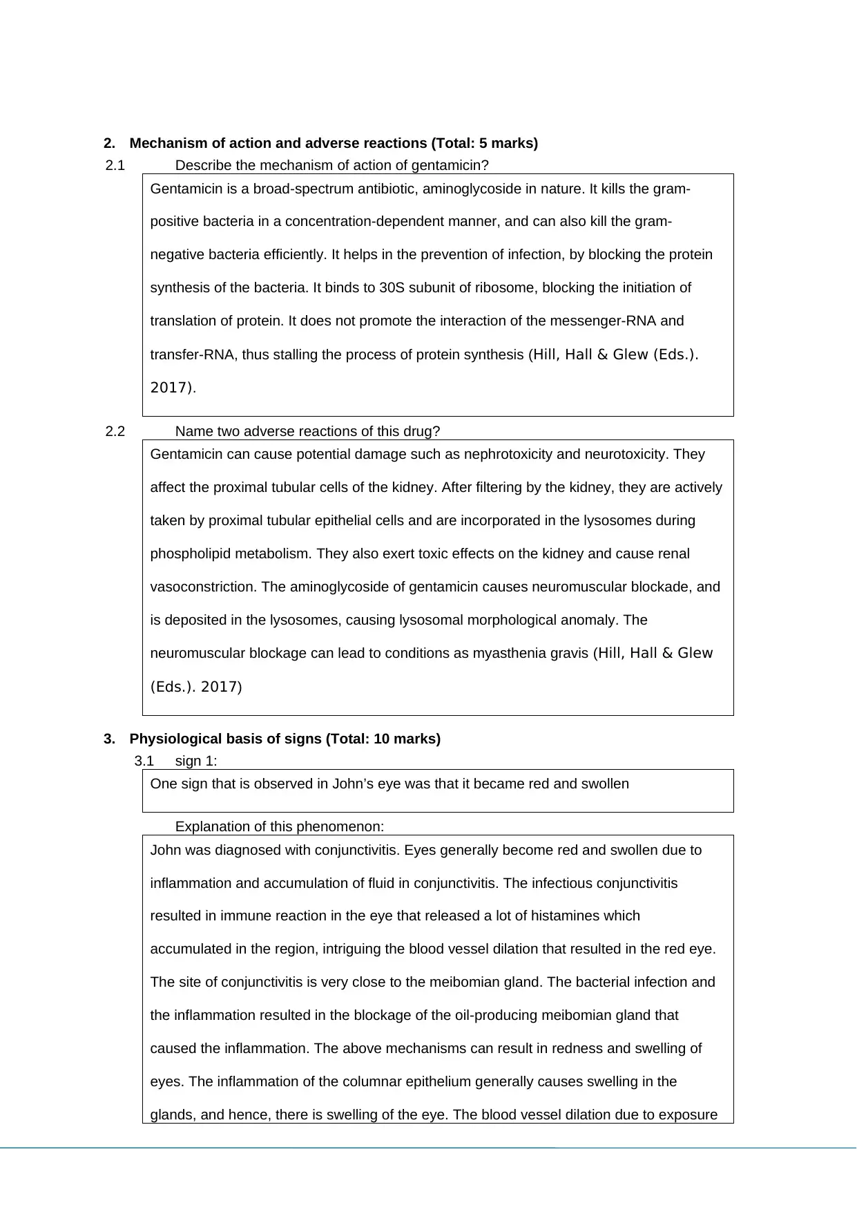
2. Mechanism of action and adverse reactions (Total: 5 marks)
2.1 Describe the mechanism of action of gentamicin?
Gentamicin is a broad-spectrum antibiotic, aminoglycoside in nature. It kills the gram-
positive bacteria in a concentration-dependent manner, and can also kill the gram-
negative bacteria efficiently. It helps in the prevention of infection, by blocking the protein
synthesis of the bacteria. It binds to 30S subunit of ribosome, blocking the initiation of
translation of protein. It does not promote the interaction of the messenger-RNA and
transfer-RNA, thus stalling the process of protein synthesis (Hill, Hall & Glew (Eds.).
2017).
2.2 Name two adverse reactions of this drug?
Gentamicin can cause potential damage such as nephrotoxicity and neurotoxicity. They
affect the proximal tubular cells of the kidney. After filtering by the kidney, they are actively
taken by proximal tubular epithelial cells and are incorporated in the lysosomes during
phospholipid metabolism. They also exert toxic effects on the kidney and cause renal
vasoconstriction. The aminoglycoside of gentamicin causes neuromuscular blockade, and
is deposited in the lysosomes, causing lysosomal morphological anomaly. The
neuromuscular blockage can lead to conditions as myasthenia gravis (Hill, Hall & Glew
(Eds.). 2017)
3. Physiological basis of signs (Total: 10 marks)
3.1 sign 1:
One sign that is observed in John’s eye was that it became red and swollen
Explanation of this phenomenon:
John was diagnosed with conjunctivitis. Eyes generally become red and swollen due to
inflammation and accumulation of fluid in conjunctivitis. The infectious conjunctivitis
resulted in immune reaction in the eye that released a lot of histamines which
accumulated in the region, intriguing the blood vessel dilation that resulted in the red eye.
The site of conjunctivitis is very close to the meibomian gland. The bacterial infection and
the inflammation resulted in the blockage of the oil-producing meibomian gland that
caused the inflammation. The above mechanisms can result in redness and swelling of
eyes. The inflammation of the columnar epithelium generally causes swelling in the
glands, and hence, there is swelling of the eye. The blood vessel dilation due to exposure
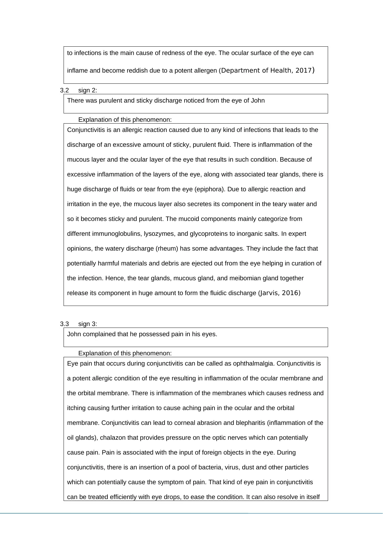
to infections is the main cause of redness of the eye. The ocular surface of the eye can
inflame and become reddish due to a potent allergen (Department of Health, 2017)
3.2 sign 2:
There was purulent and sticky discharge noticed from the eye of John
Explanation of this phenomenon:
Conjunctivitis is an allergic reaction caused due to any kind of infections that leads to the
discharge of an excessive amount of sticky, purulent fluid. There is inflammation of the
mucous layer and the ocular layer of the eye that results in such condition. Because of
excessive inflammation of the layers of the eye, along with associated tear glands, there is
huge discharge of fluids or tear from the eye (epiphora). Due to allergic reaction and
irritation in the eye, the mucous layer also secretes its component in the teary water and
so it becomes sticky and purulent. The mucoid components mainly categorize from
different immunoglobulins, lysozymes, and glycoproteins to inorganic salts. In expert
opinions, the watery discharge (rheum) has some advantages. They include the fact that
potentially harmful materials and debris are ejected out from the eye helping in curation of
the infection. Hence, the tear glands, mucous gland, and meibomian gland together
release its component in huge amount to form the fluidic discharge (Jarvis, 2016)
3.3 sign 3:
John complained that he possessed pain in his eyes.
Explanation of this phenomenon:
Eye pain that occurs during conjunctivitis can be called as ophthalmalgia. Conjunctivitis is
a potent allergic condition of the eye resulting in inflammation of the ocular membrane and
the orbital membrane. There is inflammation of the membranes which causes redness and
itching causing further irritation to cause aching pain in the ocular and the orbital
membrane. Conjunctivitis can lead to corneal abrasion and blepharitis (inflammation of the
oil glands), chalazon that provides pressure on the optic nerves which can potentially
cause pain. Pain is associated with the input of foreign objects in the eye. During
conjunctivitis, there is an insertion of a pool of bacteria, virus, dust and other particles
which can potentially cause the symptom of pain. That kind of eye pain in conjunctivitis
can be treated efficiently with eye drops, to ease the condition. It can also resolve in itself
⊘ This is a preview!⊘
Do you want full access?
Subscribe today to unlock all pages.

Trusted by 1+ million students worldwide
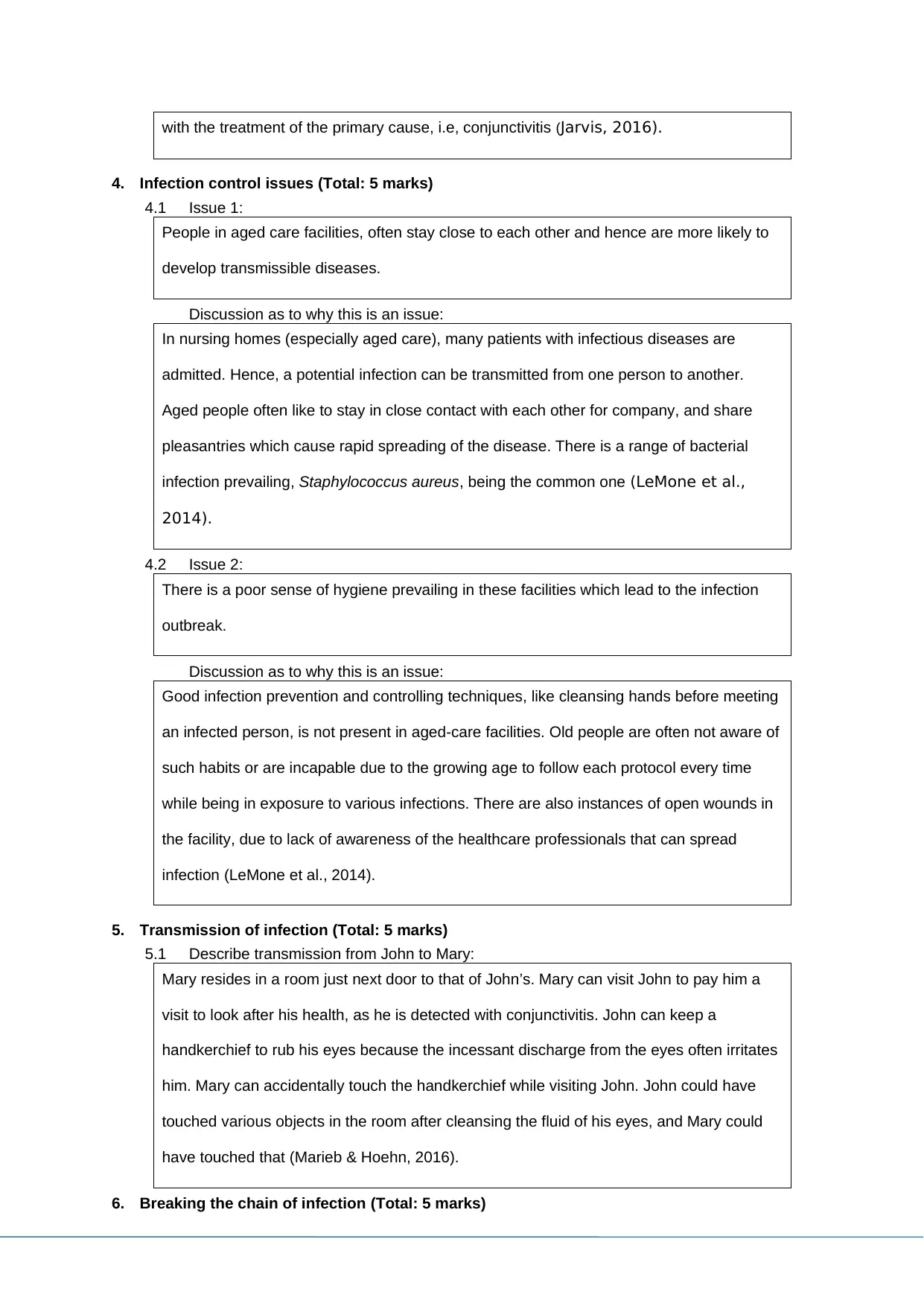
with the treatment of the primary cause, i.e, conjunctivitis (Jarvis, 2016).
4. Infection control issues (Total: 5 marks)
4.1 Issue 1:
People in aged care facilities, often stay close to each other and hence are more likely to
develop transmissible diseases.
Discussion as to why this is an issue:
In nursing homes (especially aged care), many patients with infectious diseases are
admitted. Hence, a potential infection can be transmitted from one person to another.
Aged people often like to stay in close contact with each other for company, and share
pleasantries which cause rapid spreading of the disease. There is a range of bacterial
infection prevailing, Staphylococcus aureus, being the common one (LeMone et al.,
2014).
4.2 Issue 2:
There is a poor sense of hygiene prevailing in these facilities which lead to the infection
outbreak.
Discussion as to why this is an issue:
Good infection prevention and controlling techniques, like cleansing hands before meeting
an infected person, is not present in aged-care facilities. Old people are often not aware of
such habits or are incapable due to the growing age to follow each protocol every time
while being in exposure to various infections. There are also instances of open wounds in
the facility, due to lack of awareness of the healthcare professionals that can spread
infection (LeMone et al., 2014).
5. Transmission of infection (Total: 5 marks)
5.1 Describe transmission from John to Mary:
Mary resides in a room just next door to that of John’s. Mary can visit John to pay him a
visit to look after his health, as he is detected with conjunctivitis. John can keep a
handkerchief to rub his eyes because the incessant discharge from the eyes often irritates
him. Mary can accidentally touch the handkerchief while visiting John. John could have
touched various objects in the room after cleansing the fluid of his eyes, and Mary could
have touched that (Marieb & Hoehn, 2016).
6. Breaking the chain of infection (Total: 5 marks)
Paraphrase This Document
Need a fresh take? Get an instant paraphrase of this document with our AI Paraphraser
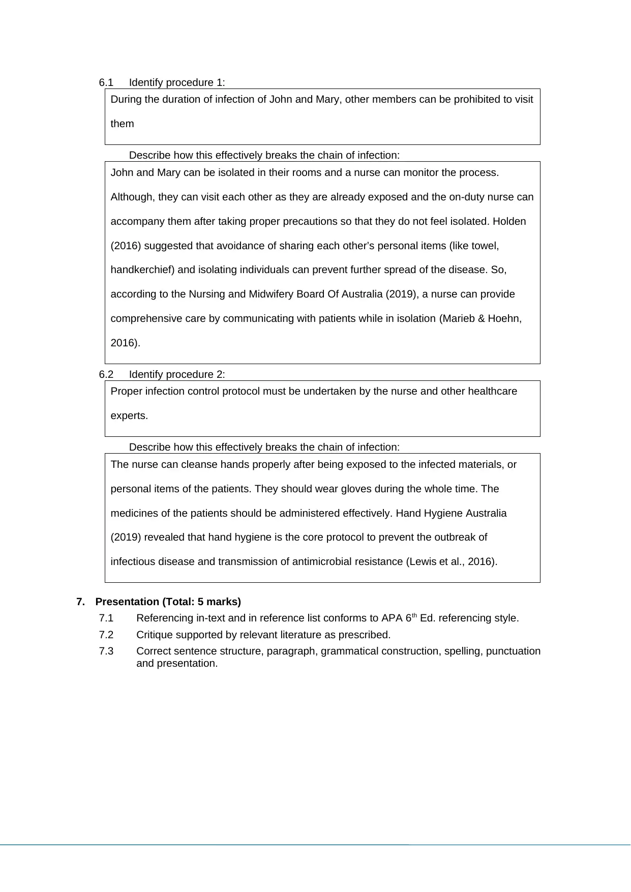
6.1 Identify procedure 1:
During the duration of infection of John and Mary, other members can be prohibited to visit
them
Describe how this effectively breaks the chain of infection:
John and Mary can be isolated in their rooms and a nurse can monitor the process.
Although, they can visit each other as they are already exposed and the on-duty nurse can
accompany them after taking proper precautions so that they do not feel isolated. Holden
(2016) suggested that avoidance of sharing each other’s personal items (like towel,
handkerchief) and isolating individuals can prevent further spread of the disease. So,
according to the Nursing and Midwifery Board Of Australia (2019), a nurse can provide
comprehensive care by communicating with patients while in isolation (Marieb & Hoehn,
2016).
6.2 Identify procedure 2:
Proper infection control protocol must be undertaken by the nurse and other healthcare
experts.
Describe how this effectively breaks the chain of infection:
The nurse can cleanse hands properly after being exposed to the infected materials, or
personal items of the patients. They should wear gloves during the whole time. The
medicines of the patients should be administered effectively. Hand Hygiene Australia
(2019) revealed that hand hygiene is the core protocol to prevent the outbreak of
infectious disease and transmission of antimicrobial resistance (Lewis et al., 2016).
7. Presentation (Total: 5 marks)
7.1 Referencing in-text and in reference list conforms to APA 6th Ed. referencing style.
7.2 Critique supported by relevant literature as prescribed.
7.3 Correct sentence structure, paragraph, grammatical construction, spelling, punctuation
and presentation.
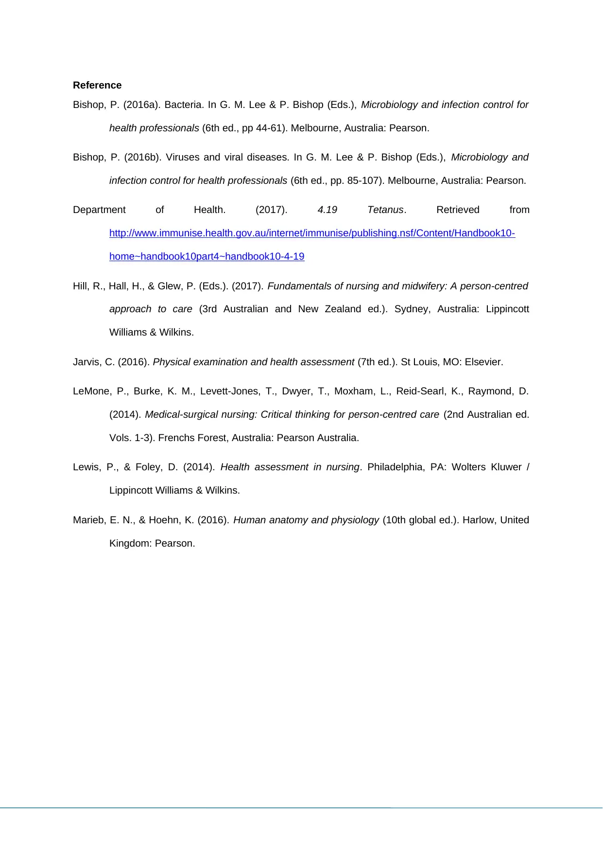
Reference
Bishop, P. (2016a). Bacteria. In G. M. Lee & P. Bishop (Eds.), Microbiology and infection control for
health professionals (6th ed., pp 44-61). Melbourne, Australia: Pearson.
Bishop, P. (2016b). Viruses and viral diseases. In G. M. Lee & P. Bishop (Eds.), Microbiology and
infection control for health professionals (6th ed., pp. 85-107). Melbourne, Australia: Pearson.
Department of Health. (2017). 4.19 Tetanus. Retrieved from
http://www.immunise.health.gov.au/internet/immunise/publishing.nsf/Content/Handbook10-
home~handbook10part4~handbook10-4-19
Hill, R., Hall, H., & Glew, P. (Eds.). (2017). Fundamentals of nursing and midwifery: A person-centred
approach to care (3rd Australian and New Zealand ed.). Sydney, Australia: Lippincott
Williams & Wilkins.
Jarvis, C. (2016). Physical examination and health assessment (7th ed.). St Louis, MO: Elsevier.
LeMone, P., Burke, K. M., Levett-Jones, T., Dwyer, T., Moxham, L., Reid-Searl, K., Raymond, D.
(2014). Medical-surgical nursing: Critical thinking for person-centred care (2nd Australian ed.
Vols. 1-3). Frenchs Forest, Australia: Pearson Australia.
Lewis, P., & Foley, D. (2014). Health assessment in nursing. Philadelphia, PA: Wolters Kluwer /
Lippincott Williams & Wilkins.
Marieb, E. N., & Hoehn, K. (2016). Human anatomy and physiology (10th global ed.). Harlow, United
Kingdom: Pearson.
⊘ This is a preview!⊘
Do you want full access?
Subscribe today to unlock all pages.

Trusted by 1+ million students worldwide
1 out of 6
Related Documents
Your All-in-One AI-Powered Toolkit for Academic Success.
+13062052269
info@desklib.com
Available 24*7 on WhatsApp / Email
![[object Object]](/_next/static/media/star-bottom.7253800d.svg)
Unlock your academic potential
Copyright © 2020–2026 A2Z Services. All Rights Reserved. Developed and managed by ZUCOL.


