The Crucial Role of Hormones in Maternal Physiology During Pregnancy
VerifiedAdded on 2023/06/15
|14
|2798
|84
Report
AI Summary
This report provides a detailed overview of the crucial role hormones play in maternal physiology during pregnancy. It discusses the functions of key hormones such as progesterone, estrogen, human chorionic gonadotropin (hCG), and human placental lactogen (hPL) at different stages of pregnancy. Progesterone is highlighted for its role in maintaining the uterine lining and preventing contractions, while estrogen supports blood vessel development and prepares the body for lactation. The report also explains how hCG maintains the corpus luteum and promotes immune tolerance, and how hPL affects insulin sensitivity. Furthermore, the report emphasizes the importance of monitoring hormone levels to prevent complications and ensure a healthy pregnancy. This resource is ideal for students looking for detailed insights into the endocrinology of pregnancy and the adaptive physiological changes in pregnant women. Desklib provides a platform for students to access similar reports and solved assignments.
Running head: PHYSIOLOGY
Physiology
Name of the student:
Name of the University:
Author’s note
Physiology
Name of the student:
Name of the University:
Author’s note
Paraphrase This Document
Need a fresh take? Get an instant paraphrase of this document with our AI Paraphraser
1PHYSIOLOGY
Introduction:
Pregnancy is associated with many physiological changes in a woman’s body and these
changes promotes healthy development of the fetus. Pregnancy related physiological changes
initiate after conception and affect every organ systems. Pregnant women are more likely to
experience symptoms of nausea and vomiting, which are adaptive mechanisms during
pregnancy, to prevent women from consuming teratogenic substances. Such etiology of
pregnancy is also linked to many pregnancy associated hormones such as human chorionic
gonadotropin (hCG) hormone and oestrogen (Soma-Pillay et al. 2016). The main purpose of this
report is to discuss the role of hormones during pregnancy and provide a detailed insight into the
function of different hormones during different stages of pregnancy.
Endocrinology of pregnancy:
The endocrinology of human pregnancy is an important factor that helps to maintain a
healthy pregnancy. The endocrine changes occur because of physiological alterations in the feto-
placental unit (FPU). The FPU is the boundary site between fetus and mother and it is the site
where hormonal production and secretion takes places. Hormonal signals originating from FPU
during pregnancy are linked to endocrine and metabolic changes in pregnant women. Both
neuronal and hormonal factors maintain the pregnancy and its associated changes. Proper timing
between neuro-endocrine events facilitates fetal growth. Progesterone, estrogen and hCG are
some key hormones involved during pregnancy and the function and role of each hormone in
2PHYSIOLOGY
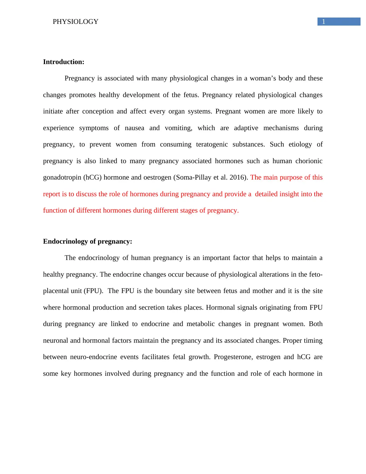
pregnancy physiology is explained in detail in the next section (Bazer 2012).
.
Figure 1: FPU, the boundary between fetus and mother and a major site for protein and steroid
hormone production and secretion. Source: (Bazer 2012).
Pregnancy associated hormones and their role in different stages of pregnancy:
Progesterone:
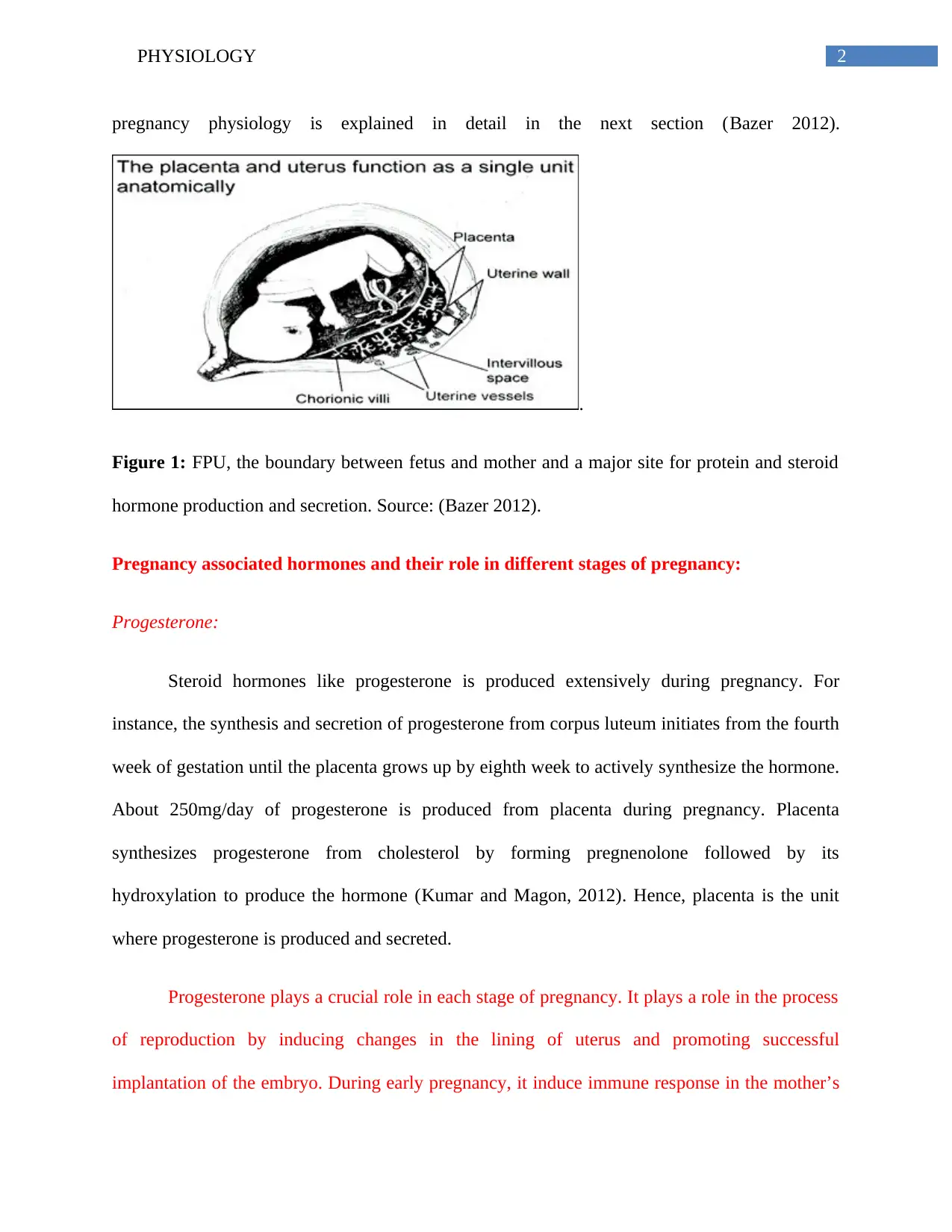
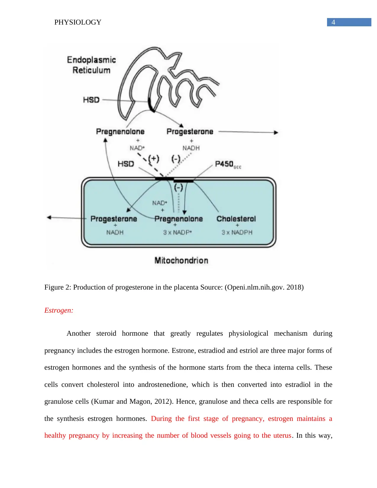
Steroid hormones like progesterone is produced extensively during pregnancy. For
instance, the synthesis and secretion of progesterone from corpus luteum initiates from the fourth
week of gestation until the placenta grows up by eighth week to actively synthesize the hormone.
About 250mg/day of progesterone is produced from placenta during pregnancy. Placenta
synthesizes progesterone from cholesterol by forming pregnenolone followed by its
hydroxylation to produce the hormone (Kumar and Magon, 2012). Hence, placenta is the unit
where progesterone is produced and secreted.
Progesterone plays a crucial role in each stage of pregnancy. It plays a role in the process
of reproduction by inducing changes in the lining of uterus and promoting successful
implantation of the embryo. During early pregnancy, it induce immune response in the mother’s
pregnancy physiology is explained in detail in the next section (Bazer 2012).
.
Figure 1: FPU, the boundary between fetus and mother and a major site for protein and steroid
hormone production and secretion. Source: (Bazer 2012).
Pregnancy associated hormones and their role in different stages of pregnancy:
Progesterone:
Steroid hormones like progesterone is produced extensively during pregnancy. For
instance, the synthesis and secretion of progesterone from corpus luteum initiates from the fourth
week of gestation until the placenta grows up by eighth week to actively synthesize the hormone.
About 250mg/day of progesterone is produced from placenta during pregnancy. Placenta
synthesizes progesterone from cholesterol by forming pregnenolone followed by its
hydroxylation to produce the hormone (Kumar and Magon, 2012). Hence, placenta is the unit
where progesterone is produced and secreted.
Progesterone plays a crucial role in each stage of pregnancy. It plays a role in the process
of reproduction by inducing changes in the lining of uterus and promoting successful
implantation of the embryo. During early pregnancy, it induce immune response in the mother’s
⊘ This is a preview!⊘
Do you want full access?
Subscribe today to unlock all pages.
Trusted by 1+ million students worldwide
3PHYSIOLOGY
body to prevent rejection of the embryo. In the first trimester, it is mainly involved in
maintaining health and development of fetus by thickening the uterine lining and maintaining
optimal function of the placenta. It also prevents contraction of the uterus allowing the baby to
expand in the womb (Dante,Vaccaro and Facchinetti 2013). In the second and third trimester of
pregnancy, progesterone also reduces the risk of preterm birth. The secretion of progesterone
increases in the second semester and by the last trimester, the volume of progesterone increases
from 340 to 675 nmol/L (Edelstam et al 2007). Hence, it can be concluded progesterone
hormones supports the endometrium to provide conducive environment for the survival of the
fetus. The binding of progestin to the progesterone receptor help in maintaining pregnancy
(Soma-Pillay et al. 2016).
Paraphrase This Document
Need a fresh take? Get an instant paraphrase of this document with our AI Paraphraser
4PHYSIOLOGY
Figure 2: Production of progesterone in the placenta Source: (Openi.nlm.nih.gov. 2018)
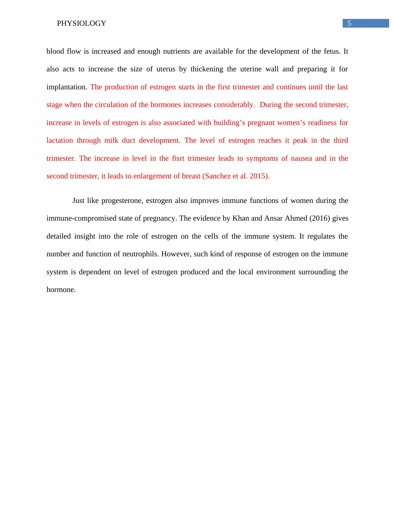
Estrogen:
Another steroid hormone that greatly regulates physiological mechanism during
pregnancy includes the estrogen hormone. Estrone, estradiod and estriol are three major forms of
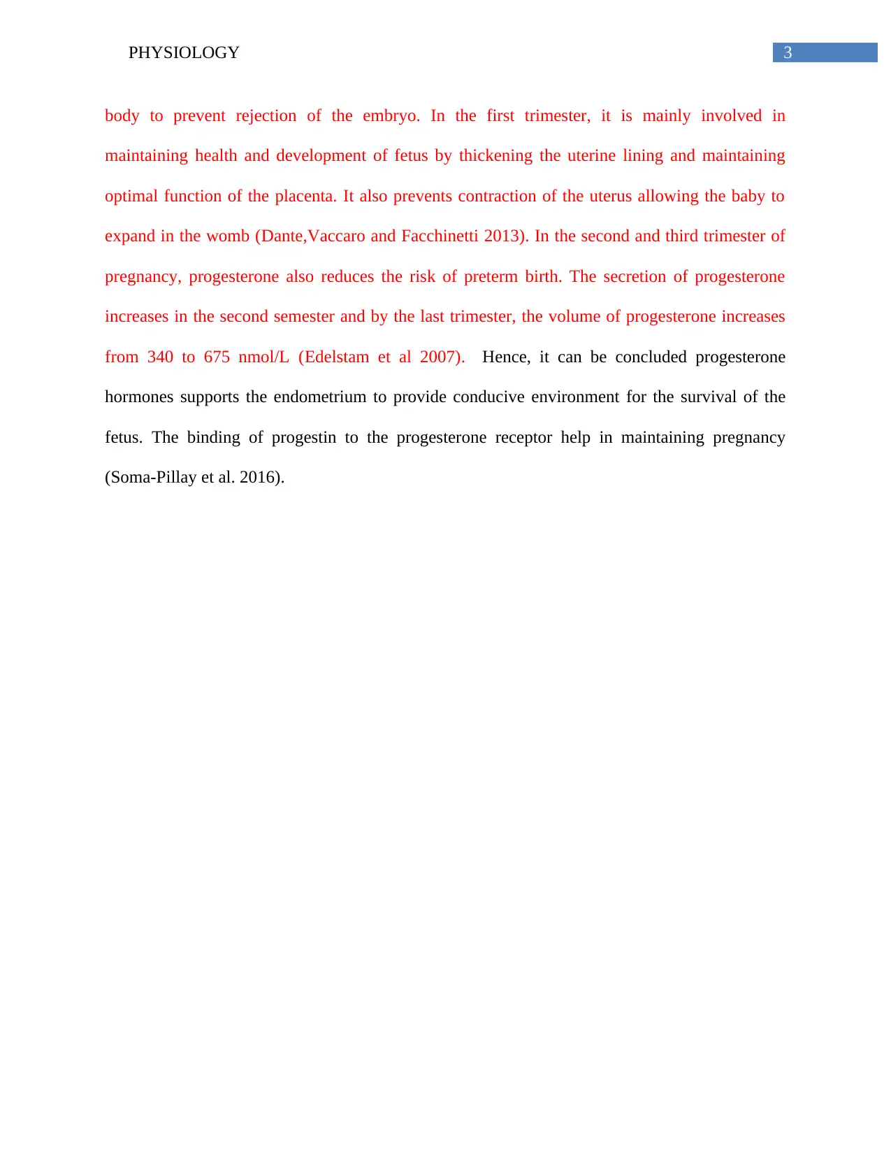
estrogen hormones and the synthesis of the hormone starts from the theca interna cells. These
cells convert cholesterol into androstenedione, which is then converted into estradiol in the
granulose cells (Kumar and Magon, 2012). Hence, granulose and theca cells are responsible for
the synthesis estrogen hormones. During the first stage of pregnancy, estrogen maintains a
healthy pregnancy by increasing the number of blood vessels going to the uterus. In this way,
Figure 2: Production of progesterone in the placenta Source: (Openi.nlm.nih.gov. 2018)
Estrogen:
Another steroid hormone that greatly regulates physiological mechanism during
pregnancy includes the estrogen hormone. Estrone, estradiod and estriol are three major forms of
estrogen hormones and the synthesis of the hormone starts from the theca interna cells. These
cells convert cholesterol into androstenedione, which is then converted into estradiol in the
granulose cells (Kumar and Magon, 2012). Hence, granulose and theca cells are responsible for
the synthesis estrogen hormones. During the first stage of pregnancy, estrogen maintains a
healthy pregnancy by increasing the number of blood vessels going to the uterus. In this way,
5PHYSIOLOGY
blood flow is increased and enough nutrients are available for the development of the fetus. It
also acts to increase the size of uterus by thickening the uterine wall and preparing it for
implantation. The production of estrogen starts in the first trimester and continues until the last
stage when the circulation of the hormones increases considerably. During the second trimester,
increase in levels of estrogen is also associated with building’s pregnant women’s readiness for
lactation through milk duct development. The level of estrogen reaches it peak in the third
trimester. The increase in level in the fisrt trimester leads to symptoms of nausea and in the
second trimester, it leads to enlargement of breast (Sanchez et al. 2015).
Just like progesterone, estrogen also improves immune functions of women during the
immune-compromised state of pregnancy. The evidence by Khan and Ansar Ahmed (2016) gives
detailed insight into the role of estrogen on the cells of the immune system. It regulates the
number and function of neutrophils. However, such kind of response of estrogen on the immune
system is dependent on level of estrogen produced and the local environment surrounding the
hormone.
⊘ This is a preview!⊘
Do you want full access?
Subscribe today to unlock all pages.
Trusted by 1+ million students worldwide
6PHYSIOLOGY
Figure 3: Role of theca and granulose cells in the production of estrogen hormones. Source:
(Doshi and Agarwal 2013)
hCG:
Apart from steroid hormones, there are also protein hormones like hCG that is produced
in large amount during early stage of pregnancy. The most vital role of hCG in pregnancy is that
it regulates the function of the corpus luteum which in turn facilitates the production of estrogen
and progesterone. The hCG hormone is produced by the trophoblast layer of the blastocyst
during the early phase of pregnancy and its level of secretion decreases during the second and
third stage of pregnancy. About 120 to 130 IU/ml of hCG is produced during pregnancy and its
amount decrease to about 20-30 IU/ ml by the 16th week of pregnancy (Korevaar et al. 2015).
During the first trimester of pregnancy, the main role of hCG is to keep the corpus luteum alive.
Paraphrase This Document
Need a fresh take? Get an instant paraphrase of this document with our AI Paraphraser
7PHYSIOLOGY
This occurs by the interaction of the hormone with the LHCG receptor of the ovary. This also
facilitates secreation of progesterone during the first trimester (Evans et al. 2015). In contrats to
first trimester, the level of estrogen decreases in the second and third trimester. Corpus luteum is
the organ which establishes and maintains pregnancy in women. Corpus luteum makes
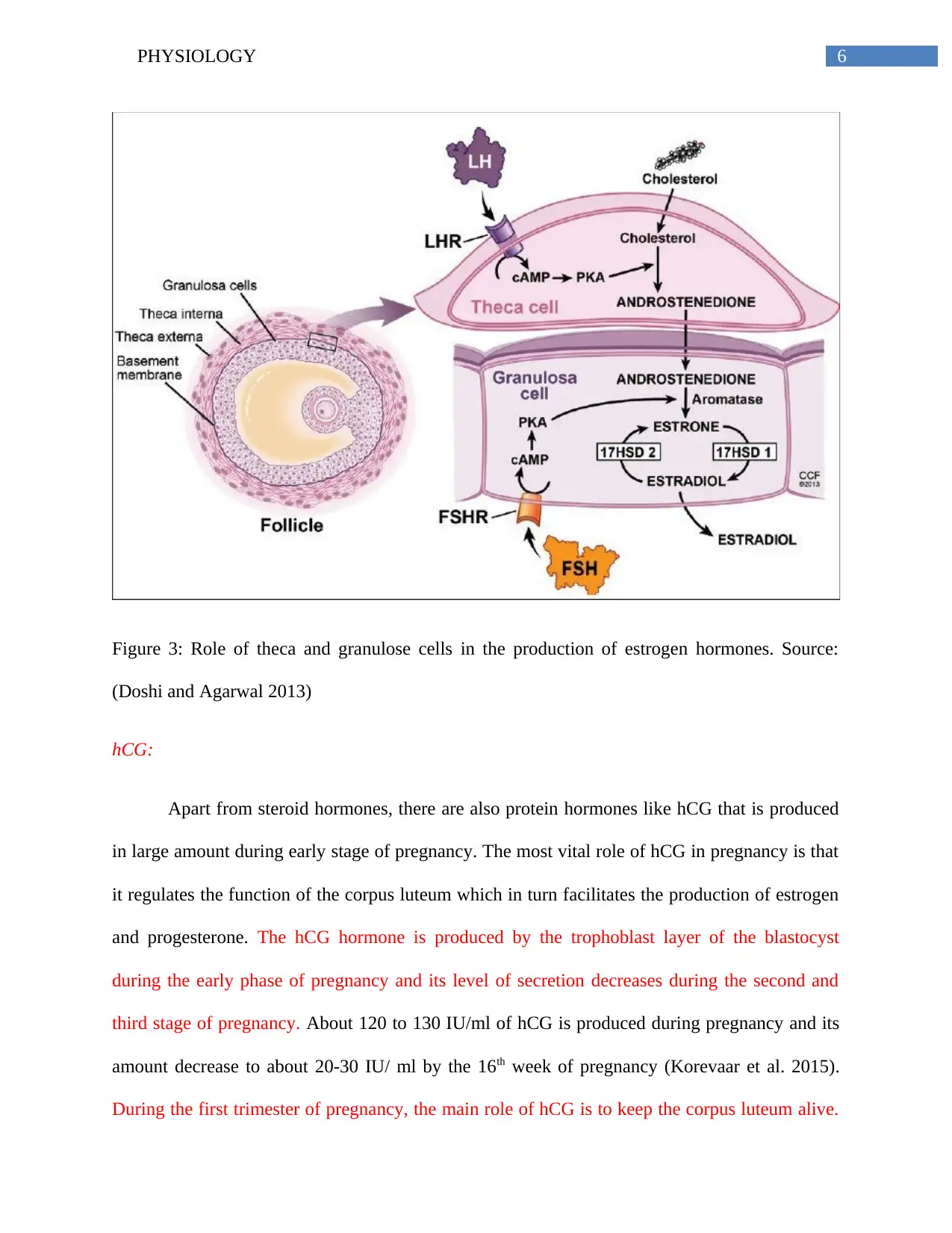
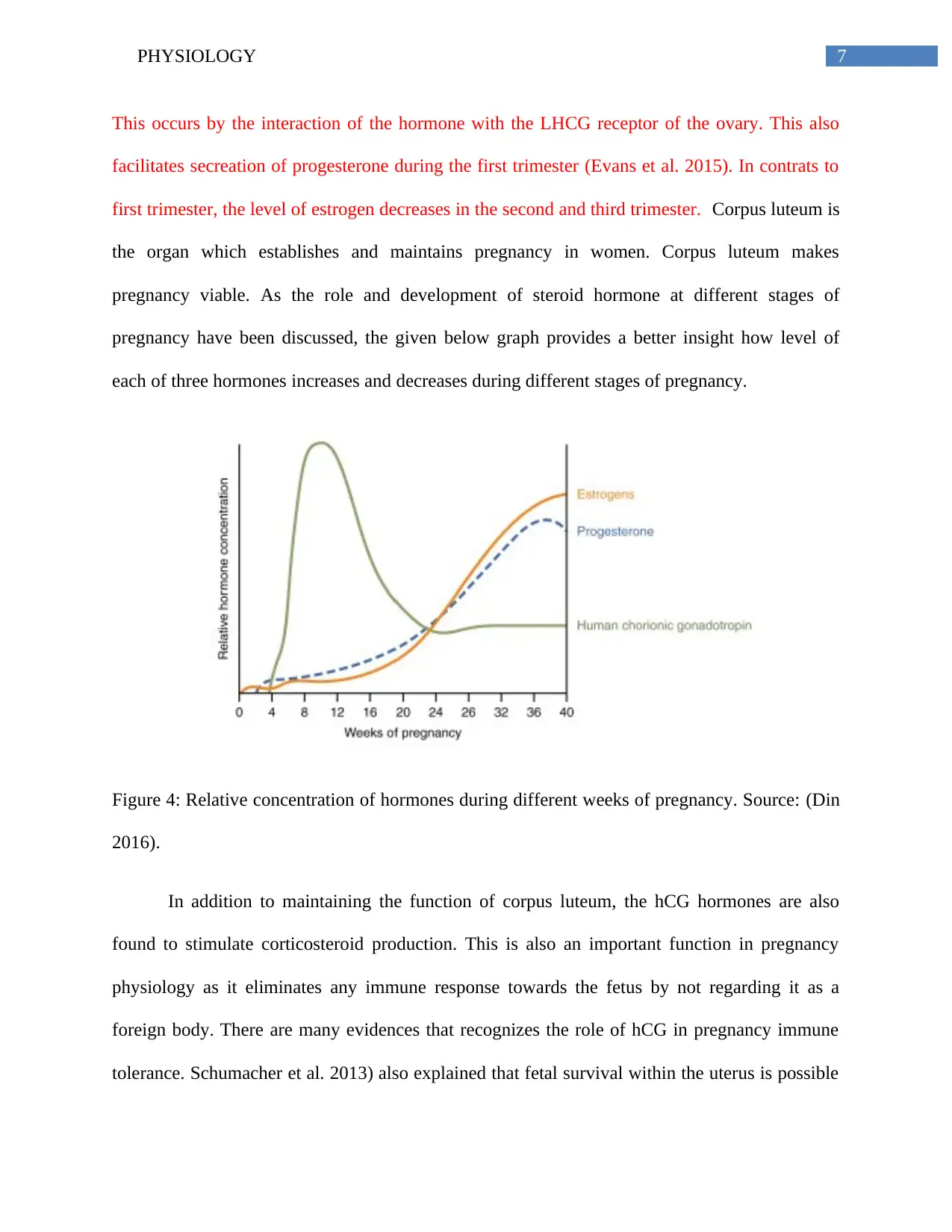
pregnancy viable. As the role and development of steroid hormone at different stages of
pregnancy have been discussed, the given below graph provides a better insight how level of
each of three hormones increases and decreases during different stages of pregnancy.
Figure 4: Relative concentration of hormones during different weeks of pregnancy. Source: (Din
2016).
In addition to maintaining the function of corpus luteum, the hCG hormones are also
found to stimulate corticosteroid production. This is also an important function in pregnancy
physiology as it eliminates any immune response towards the fetus by not regarding it as a
foreign body. There are many evidences that recognizes the role of hCG in pregnancy immune
tolerance. Schumacher et al. 2013) also explained that fetal survival within the uterus is possible
This occurs by the interaction of the hormone with the LHCG receptor of the ovary. This also
facilitates secreation of progesterone during the first trimester (Evans et al. 2015). In contrats to
first trimester, the level of estrogen decreases in the second and third trimester. Corpus luteum is
the organ which establishes and maintains pregnancy in women. Corpus luteum makes
pregnancy viable. As the role and development of steroid hormone at different stages of
pregnancy have been discussed, the given below graph provides a better insight how level of
each of three hormones increases and decreases during different stages of pregnancy.
Figure 4: Relative concentration of hormones during different weeks of pregnancy. Source: (Din
2016).
In addition to maintaining the function of corpus luteum, the hCG hormones are also
found to stimulate corticosteroid production. This is also an important function in pregnancy
physiology as it eliminates any immune response towards the fetus by not regarding it as a
foreign body. There are many evidences that recognizes the role of hCG in pregnancy immune
tolerance. Schumacher et al. 2013) also explained that fetal survival within the uterus is possible
8PHYSIOLOGY
because of many hormonal changes and their role in regulating maternal immune response
towards the foreign fetal antigens. Hence, it can be said that effective interaction between
hormonal and immunological factors results in fetal tolerance during pregnancy.
After the discussion on the function of hCG, estrogen and progesterone hormones, their
role in maintaining successful pregnancy outcome is even more clear. The hCG reaches its
maximum level during 9th to 12th week and declines thereafter until birth. After the 12th week
(second trimester), it mainly involved in supporting angiogenesis and ensuring nourishment of
the fetus. It manages to do so by attracting regulatory T cells (Tregs) to trophoblast. The more is
the expansion of the Treg cells, the higher is the establishment of pregnancy. The transfer of
Treg cells minimizes chances of fetal rejection. It creates a tolerant microenvironment at the FPU
unit (Muzzio, Zygmunt and Jensen 2014). Hence, hCG acts as the chemo-attractant of tregs that
triggers immune tolerance of the fetus during pregnancy and other steroid hormones act as the
source that confers immune suppressive capacity to B lymphocytes. By the third trimester, the
hormone is involved in promoting relaxation of the uterine contraction and initiating onset of
labour (Edelstam et al. 2007).
hPL:
Another protein hormone involved in regulating changes during pregnancy includes the
Human placental lactogen (hPL) hormone. This exclusive pregnancy hormone is also produced
in the placenta. In the first trimester, it maintains health of the fetus by maintaining energy
supply of the fetus. It is secreted by synctiotrophoblast at the time when the production of hCG
decreases. One of the important and vital attributes of this hormone is its anti-insulin properties.
Insulin resistance is the decrease in target tissues ability to respond to normal concentration of
⊘ This is a preview!⊘
Do you want full access?
Subscribe today to unlock all pages.
Trusted by 1+ million students worldwide
9PHYSIOLOGY
insulin. This is also an adaptive changes seen in pregnant women where mother utilizes more fats
and more carbohydrates is reserved for fetus. This condition ensures that the growing fetus gets
ample amount of carbohydrate as a form of energy. However, increased production of hPL in the
third trimester of pregnancy results in decline of insulin sensitivity by 50%. The increase in
estrogen and progesterone level also decreases insulin sensitivity (Sonagra et al. 2014). Hence,
increases insulin resistance should be controlled in patient as it results in premature labor as well
as fetal complications. Taking balanced diet and engaging in mild exercise can prevent insulin
related complications in patients.
The above discussion related to the role of hPL indicates the endocrinal mechanism
involves in insulin resistance during pregnancy. The discussion of different maternal adaptations
and hormonal changes during pregnancy also signifies how effective production and secretion of
hormones regulates the health of fetus. However, abnormal or undesired concentration of
hormones may also cause complications in pregnancy. For example, the concentration of thyroid
hormone also increases during 6-12 week of pregnancy. Abnormal concentration may disrupt the
function of thyroid hormone thus increasing the likelihood of thyroid disease during pregnancy
(Costantine 2014). Hence, it is necessary to monitor endocrinological parameters during early
gestation period. This may help to detect any complications in the developing fetus and chances
of abnormal pregnancies.
Conclusion:
The report gave an insight into the physiological changes seen in a pregnant woman due
to hormonal changes. Several adaptations take place during pregnancy. Some of the adaptations
are related to the production and secretion of hormones to support the developing fetus. The
Paraphrase This Document
Need a fresh take? Get an instant paraphrase of this document with our AI Paraphraser
10PHYSIOLOGY
discussion gave an insight into the role of steroid and protein hormones in the development and
nourishment of fetus. The increase and decline in production of each of these hormones at
different stages signify their specific role in maintaining the optimal health of fetus. Apart from
the role of different hormones in supporting the fetus, hormones like estrogen and hCL has also
been found to enhance immune response in pregnant women’s body. For instance, the role of
hCL in incorporating immune tolerance of pregnant women’s body to the developing fetus is
also one of the remarkable features that depicts its role in immune system. Hence, changes in
concentration of hormones serve different adaptive purpose for the healthy development of fetus
However, abnormal concentration increases chances of many diseases too. Therefore, it is
necessary to monitor such changes in pregnant women to ensure they have a normal pregnancy.
11PHYSIOLOGY
Reference
Bazer, F.W. ed., 2012. Endocrinology of pregnancy (Vol. 9). Springer Science & Business
Media.
Costantine, M., 2014. Physiologic and pharmacokinetic changes in pregnancy. Frontiers in
pharmacology, 5, p.65.
Dante, G., Vaccaro, V. and Facchinetti, F., 2013. Use of progestagens during early
pregnancy. Facts, views & vision in ObGyn, 5(1), p.66.
Din, J. 2016. Female Hormones: Types, Causes, Symptoms, Disorders And Cure. Health Units.
Retrieved 18 February 2018, from https://healthunits.com/hormones/female-hormones-types-
disorders-cure/
Doshi, S.B. and Agarwal, A., 2013. The role of oxidative stress in menopause. Journal of mid-
life health, 4(3), p.140.
Edelstam, G., Karlsson, C., Westgren, M., Löwbeer, C. and Swahn, M.L., 2007. Human
chorionic gonadatropin (hCG) during third trimester pregnancy. Scandinavian journal of clinical
and laboratory investigation, 67(5), pp.519-525.
Evans, J., Salamonsen, L.A., Menkhorst, E. and Dimitriadis, E., 2015. Dynamic changes in
hyperglycosylated human chorionic gonadotrophin throughout the first trimester of pregnancy
and its role in early placentation. Human Reproduction, 30(5), pp.1029-1038.
⊘ This is a preview!⊘
Do you want full access?
Subscribe today to unlock all pages.
Trusted by 1+ million students worldwide
1 out of 14
Related Documents


Your All-in-One AI-Powered Toolkit for Academic Success.
+13062052269
info@desklib.com
Available 24*7 on WhatsApp / Email
![[object Object]](/_next/static/media/star-bottom.7253800d.svg)
Unlock your academic potential
Copyright © 2020–2026 A2Z Services. All Rights Reserved. Developed and managed by ZUCOL.