MMED2931/8931 Human Physiology Assignment 2: Solutions Provided
VerifiedAdded on 2022/12/29
|5
|3039
|69
Homework Assignment
AI Summary
This document presents comprehensive solutions to Human Physiology Assignment 2. The answers cover various aspects of human physiology, including the mechanisms of peristalsis, the diagnosis and symptoms of diabetes, creatinine clearance and kidney function, the process of micturition, and the process of fertilization. The solutions detail the physiological processes, relevant formulas (like the Cockcroft-Gault equation), and the hormonal and nervous system controls involved. The assignment also includes discussion of hormone action, the female reproductive system, and the stages of meiosis. Furthermore, the solutions incorporate references to scientific literature, providing a strong foundation for the concepts discussed. The document is a valuable resource for students seeking to understand and excel in their human physiology coursework.

Assignment 2 Question # 1 Surname, First name
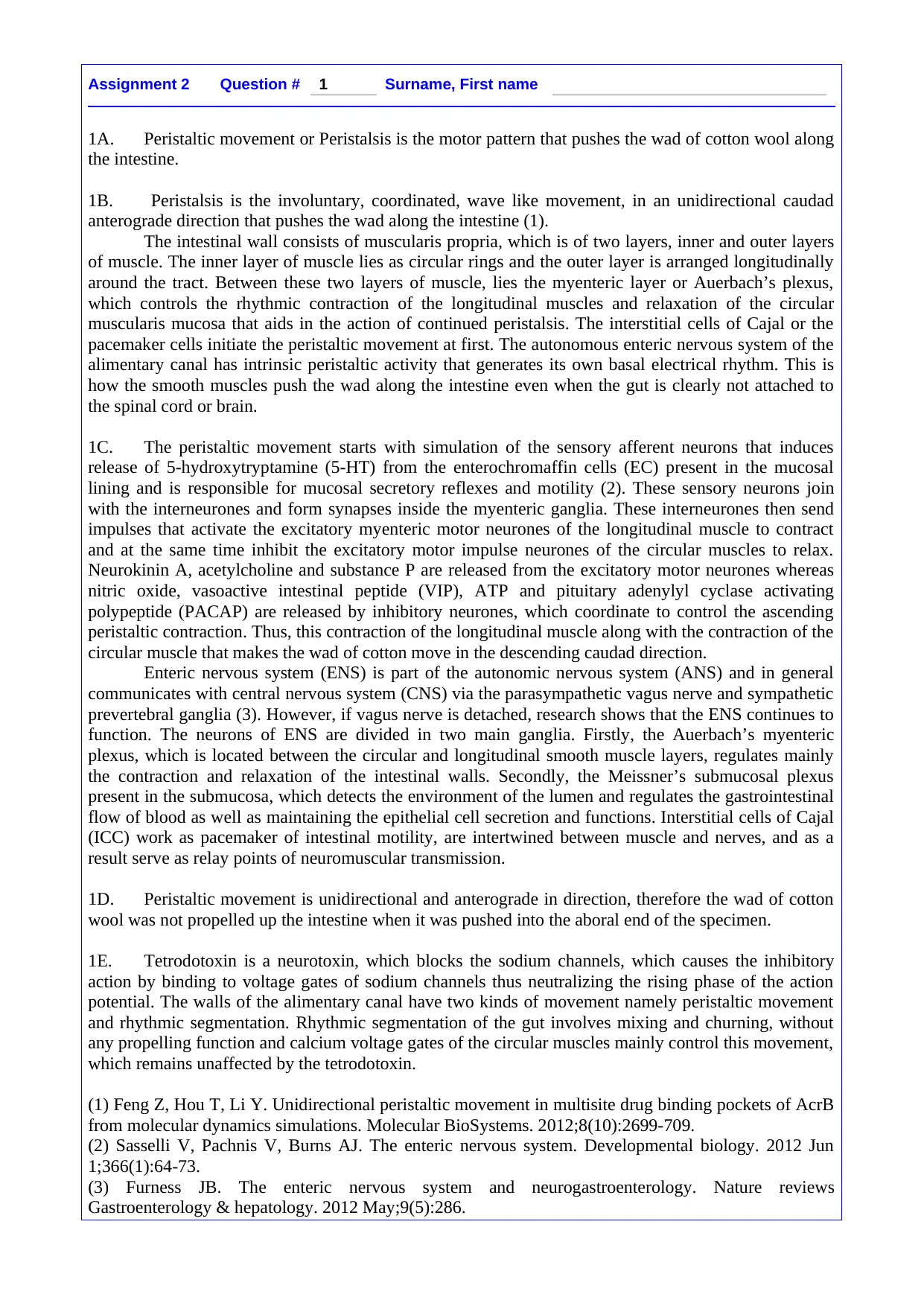
1A. Peristaltic movement or Peristalsis is the motor pattern that pushes the wad of cotton wool along
the intestine.
1B. Peristalsis is the involuntary, coordinated, wave like movement, in an unidirectional caudad
anterograde direction that pushes the wad along the intestine (1).
The intestinal wall consists of muscularis propria, which is of two layers, inner and outer layers
of muscle. The inner layer of muscle lies as circular rings and the outer layer is arranged longitudinally
around the tract. Between these two layers of muscle, lies the myenteric layer or Auerbach’s plexus,
which controls the rhythmic contraction of the longitudinal muscles and relaxation of the circular
muscularis mucosa that aids in the action of continued peristalsis. The interstitial cells of Cajal or the
pacemaker cells initiate the peristaltic movement at first. The autonomous enteric nervous system of the
alimentary canal has intrinsic peristaltic activity that generates its own basal electrical rhythm. This is
how the smooth muscles push the wad along the intestine even when the gut is clearly not attached to
the spinal cord or brain.
1C. The peristaltic movement starts with simulation of the sensory afferent neurons that induces
release of 5-hydroxytryptamine (5-HT) from the enterochromaffin cells (EC) present in the mucosal
lining and is responsible for mucosal secretory reflexes and motility (2). These sensory neurons join
with the interneurones and form synapses inside the myenteric ganglia. These interneurones then send
impulses that activate the excitatory myenteric motor neurones of the longitudinal muscle to contract
and at the same time inhibit the excitatory motor impulse neurones of the circular muscles to relax.
Neurokinin A, acetylcholine and substance P are released from the excitatory motor neurones whereas
nitric oxide, vasoactive intestinal peptide (VIP), ATP and pituitary adenylyl cyclase activating
polypeptide (PACAP) are released by inhibitory neurones, which coordinate to control the ascending
peristaltic contraction. Thus, this contraction of the longitudinal muscle along with the contraction of the
circular muscle that makes the wad of cotton move in the descending caudad direction.
Enteric nervous system (ENS) is part of the autonomic nervous system (ANS) and in general
communicates with central nervous system (CNS) via the parasympathetic vagus nerve and sympathetic
prevertebral ganglia (3). However, if vagus nerve is detached, research shows that the ENS continues to
function. The neurons of ENS are divided in two main ganglia. Firstly, the Auerbach’s myenteric
plexus, which is located between the circular and longitudinal smooth muscle layers, regulates mainly
the contraction and relaxation of the intestinal walls. Secondly, the Meissner’s submucosal plexus
present in the submucosa, which detects the environment of the lumen and regulates the gastrointestinal
flow of blood as well as maintaining the epithelial cell secretion and functions. Interstitial cells of Cajal
(ICC) work as pacemaker of intestinal motility, are intertwined between muscle and nerves, and as a
result serve as relay points of neuromuscular transmission.
1D. Peristaltic movement is unidirectional and anterograde in direction, therefore the wad of cotton
wool was not propelled up the intestine when it was pushed into the aboral end of the specimen.
1E. Tetrodotoxin is a neurotoxin, which blocks the sodium channels, which causes the inhibitory
action by binding to voltage gates of sodium channels thus neutralizing the rising phase of the action
potential. The walls of the alimentary canal have two kinds of movement namely peristaltic movement
and rhythmic segmentation. Rhythmic segmentation of the gut involves mixing and churning, without
any propelling function and calcium voltage gates of the circular muscles mainly control this movement,
which remains unaffected by the tetrodotoxin.
(1) Feng Z, Hou T, Li Y. Unidirectional peristaltic movement in multisite drug binding pockets of AcrB
from molecular dynamics simulations. Molecular BioSystems. 2012;8(10):2699-709.
(2) Sasselli V, Pachnis V, Burns AJ. The enteric nervous system. Developmental biology. 2012 Jun
1;366(1):64-73.
(3) Furness JB. The enteric nervous system and neurogastroenterology. Nature reviews
Gastroenterology & hepatology. 2012 May;9(5):286.
Paraphrase This Document
Need a fresh take? Get an instant paraphrase of this document with our AI Paraphraser

Assignment 2 Question # 2 Surname, First name
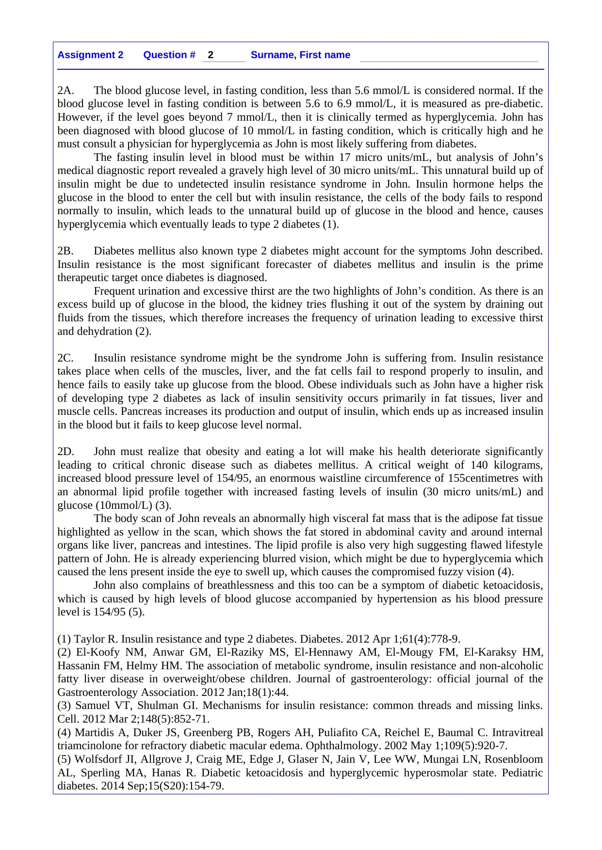
2A. The blood glucose level, in fasting condition, less than 5.6 mmol/L is considered normal. If the
blood glucose level in fasting condition is between 5.6 to 6.9 mmol/L, it is measured as pre-diabetic.
However, if the level goes beyond 7 mmol/L, then it is clinically termed as hyperglycemia. John has
been diagnosed with blood glucose of 10 mmol/L in fasting condition, which is critically high and he
must consult a physician for hyperglycemia as John is most likely suffering from diabetes.
The fasting insulin level in blood must be within 17 micro units/mL, but analysis of John’s
medical diagnostic report revealed a gravely high level of 30 micro units/mL. This unnatural build up of
insulin might be due to undetected insulin resistance syndrome in John. Insulin hormone helps the
glucose in the blood to enter the cell but with insulin resistance, the cells of the body fails to respond
normally to insulin, which leads to the unnatural build up of glucose in the blood and hence, causes
hyperglycemia which eventually leads to type 2 diabetes (1).
2B. Diabetes mellitus also known type 2 diabetes might account for the symptoms John described.
Insulin resistance is the most significant forecaster of diabetes mellitus and insulin is the prime
therapeutic target once diabetes is diagnosed.
Frequent urination and excessive thirst are the two highlights of John’s condition. As there is an
excess build up of glucose in the blood, the kidney tries flushing it out of the system by draining out
fluids from the tissues, which therefore increases the frequency of urination leading to excessive thirst
and dehydration (2).
2C. Insulin resistance syndrome might be the syndrome John is suffering from. Insulin resistance
takes place when cells of the muscles, liver, and the fat cells fail to respond properly to insulin, and
hence fails to easily take up glucose from the blood. Obese individuals such as John have a higher risk
of developing type 2 diabetes as lack of insulin sensitivity occurs primarily in fat tissues, liver and
muscle cells. Pancreas increases its production and output of insulin, which ends up as increased insulin
in the blood but it fails to keep glucose level normal.
2D. John must realize that obesity and eating a lot will make his health deteriorate significantly
leading to critical chronic disease such as diabetes mellitus. A critical weight of 140 kilograms,
increased blood pressure level of 154/95, an enormous waistline circumference of 155centimetres with
an abnormal lipid profile together with increased fasting levels of insulin (30 micro units/mL) and
glucose (10mmol/L) (3).
The body scan of John reveals an abnormally high visceral fat mass that is the adipose fat tissue
highlighted as yellow in the scan, which shows the fat stored in abdominal cavity and around internal
organs like liver, pancreas and intestines. The lipid profile is also very high suggesting flawed lifestyle
pattern of John. He is already experiencing blurred vision, which might be due to hyperglycemia which
caused the lens present inside the eye to swell up, which causes the compromised fuzzy vision (4).
John also complains of breathlessness and this too can be a symptom of diabetic ketoacidosis,
which is caused by high levels of blood glucose accompanied by hypertension as his blood pressure
level is 154/95 (5).
(1) Taylor R. Insulin resistance and type 2 diabetes. Diabetes. 2012 Apr 1;61(4):778-9.
(2) El-Koofy NM, Anwar GM, El-Raziky MS, El-Hennawy AM, El-Mougy FM, El-Karaksy HM,
Hassanin FM, Helmy HM. The association of metabolic syndrome, insulin resistance and non-alcoholic
fatty liver disease in overweight/obese children. Journal of gastroenterology: official journal of the
Gastroenterology Association. 2012 Jan;18(1):44.
(3) Samuel VT, Shulman GI. Mechanisms for insulin resistance: common threads and missing links.
Cell. 2012 Mar 2;148(5):852-71.
(4) Martidis A, Duker JS, Greenberg PB, Rogers AH, Puliafito CA, Reichel E, Baumal C. Intravitreal
triamcinolone for refractory diabetic macular edema. Ophthalmology. 2002 May 1;109(5):920-7.
(5) Wolfsdorf JI, Allgrove J, Craig ME, Edge J, Glaser N, Jain V, Lee WW, Mungai LN, Rosenbloom
AL, Sperling MA, Hanas R. Diabetic ketoacidosis and hyperglycemic hyperosmolar state. Pediatric
diabetes. 2014 Sep;15(S20):154-79.

Assignment 2 Question # 3 Surname, First name
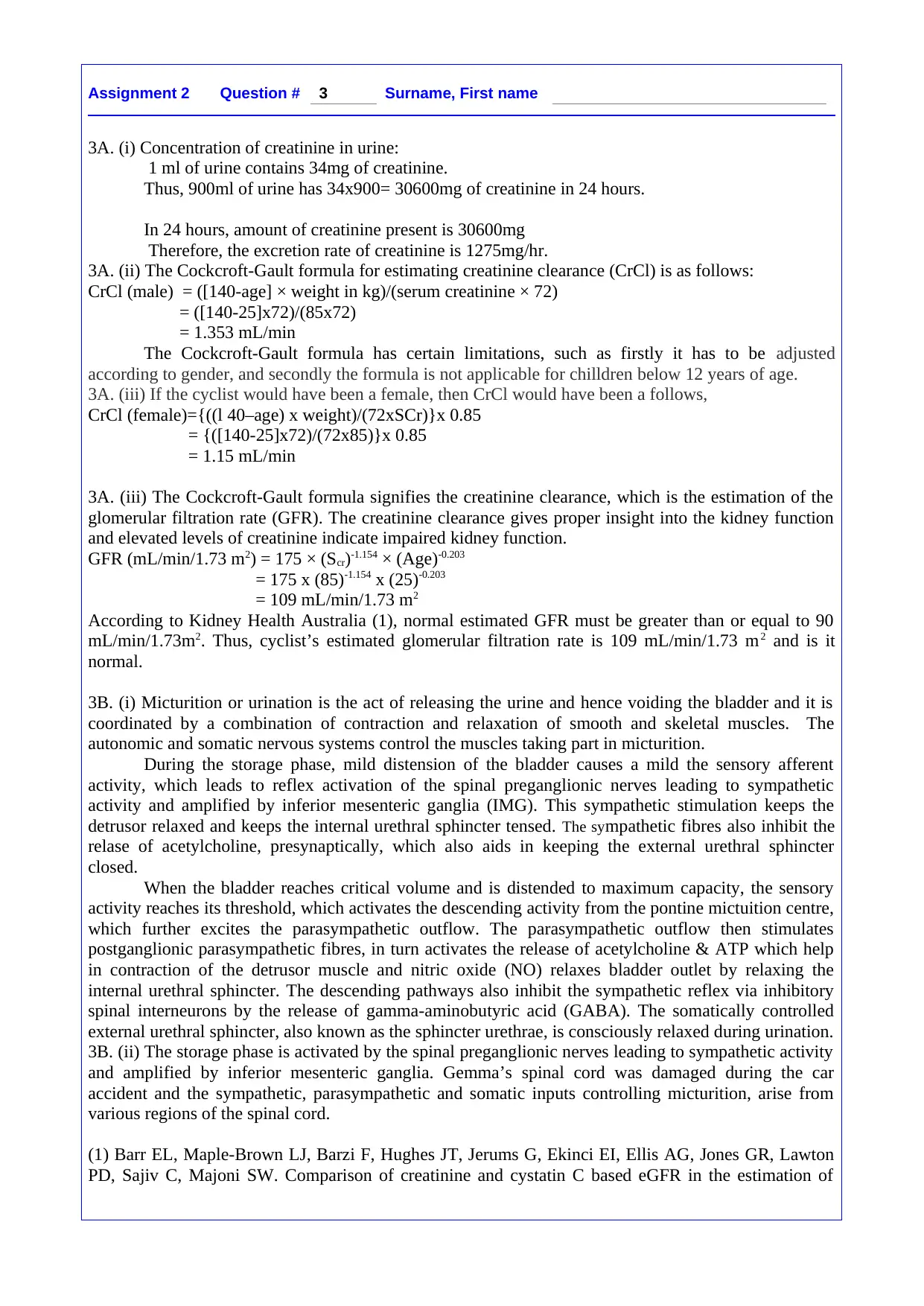
3A. (i) Concentration of creatinine in urine:
1 ml of urine contains 34mg of creatinine.
Thus, 900ml of urine has 34x900= 30600mg of creatinine in 24 hours.
In 24 hours, amount of creatinine present is 30600mg
Therefore, the excretion rate of creatinine is 1275mg/hr.
3A. (ii) The Cockcroft-Gault formula for estimating creatinine clearance (CrCl) is as follows:
CrCl (male) = ([140-age] × weight in kg)/(serum creatinine × 72)
= ([140-25]x72)/(85x72)
= 1.353 mL/min
The Cockcroft-Gault formula has certain limitations, such as firstly it has to be adjusted
according to gender, and secondly the formula is not applicable for chilldren below 12 years of age.
3A. (iii) If the cyclist would have been a female, then CrCl would have been a follows,
CrCl (female)={((l 40–age) x weight)/(72xSCr)}x 0.85
= {([140-25]x72)/(72x85)}x 0.85
= 1.15 mL/min
3A. (iii) The Cockcroft-Gault formula signifies the creatinine clearance, which is the estimation of the
glomerular filtration rate (GFR). The creatinine clearance gives proper insight into the kidney function
and elevated levels of creatinine indicate impaired kidney function.
GFR (mL/min/1.73 m2) = 175 × (Scr)-1.154 × (Age)-0.203
= 175 x (85)-1.154 x (25)-0.203
= 109 mL/min/1.73 m2
According to Kidney Health Australia (1), normal estimated GFR must be greater than or equal to 90
mL/min/1.73m2. Thus, cyclist’s estimated glomerular filtration rate is 109 mL/min/1.73 m2 and is it
normal.
3B. (i) Micturition or urination is the act of releasing the urine and hence voiding the bladder and it is
coordinated by a combination of contraction and relaxation of smooth and skeletal muscles. The
autonomic and somatic nervous systems control the muscles taking part in micturition.
During the storage phase, mild distension of the bladder causes a mild the sensory afferent
activity, which leads to reflex activation of the spinal preganglionic nerves leading to sympathetic
activity and amplified by inferior mesenteric ganglia (IMG). This sympathetic stimulation keeps the
detrusor relaxed and keeps the internal urethral sphincter tensed. The sympathetic fibres also inhibit the
relase of acetylcholine, presynaptically, which also aids in keeping the external urethral sphincter
closed.
When the bladder reaches critical volume and is distended to maximum capacity, the sensory
activity reaches its threshold, which activates the descending activity from the pontine mictuition centre,
which further excites the parasympathetic outflow. The parasympathetic outflow then stimulates
postganglionic parasympathetic fibres, in turn activates the release of acetylcholine & ATP which help
in contraction of the detrusor muscle and nitric oxide (NO) relaxes bladder outlet by relaxing the
internal urethral sphincter. The descending pathways also inhibit the sympathetic reflex via inhibitory
spinal interneurons by the release of gamma-aminobutyric acid (GABA). The somatically controlled
external urethral sphincter, also known as the sphincter urethrae, is consciously relaxed during urination.
3B. (ii) The storage phase is activated by the spinal preganglionic nerves leading to sympathetic activity
and amplified by inferior mesenteric ganglia. Gemma’s spinal cord was damaged during the car
accident and the sympathetic, parasympathetic and somatic inputs controlling micturition, arise from
various regions of the spinal cord.
(1) Barr EL, Maple-Brown LJ, Barzi F, Hughes JT, Jerums G, Ekinci EI, Ellis AG, Jones GR, Lawton
PD, Sajiv C, Majoni SW. Comparison of creatinine and cystatin C based eGFR in the estimation of
⊘ This is a preview!⊘
Do you want full access?
Subscribe today to unlock all pages.

Trusted by 1+ million students worldwide

glomerular filtration rate in Indigenous Australians: The eGFR Study. Clinical biochemistry. 2017 Apr
1;50(6):301-8.
1;50(6):301-8.
Paraphrase This Document
Need a fresh take? Get an instant paraphrase of this document with our AI Paraphraser

Assignment 2 Question # 4 Surname, First name
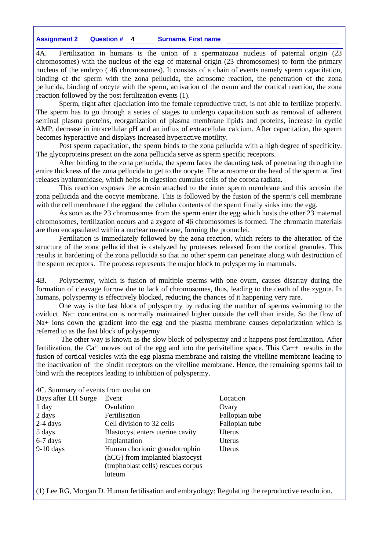
4A. Fertilization in humans is the union of a spermatozoa nucleus of paternal origin (23
chromosomes) with the nucleus of the egg of maternal origin (23 chromosomes) to form the primary
nucleus of the embryo ( 46 chromosomes). It consists of a chain of events namely sperm capacitation,
binding of the sperm with the zona pellucida, the acrosome reaction, the penetration of the zona
pellucida, binding of oocyte with the sperm, activation of the ovum and the cortical reaction, the zona
reaction followed by the post fertilization events (1).
Sperm, right after ejaculation into the female reproductive tract, is not able to fertilize properly.
The sperm has to go through a series of stages to undergo capacitation such as removal of adherent
seminal plasma proteins, reorganization of plasma membrane lipids and proteins, increase in cyclic
AMP, decrease in intracellular pH and an influx of extracellular calcium. After capacitation, the sperm
becomes hyperactive and displays increased hyperactive motility.
Post sperm capacitation, the sperm binds to the zona pellucida with a high degree of specificity.
The glycoproteins present on the zona pellucida serve as sperm specific receptors.
After binding to the zona pellucida, the sperm faces the daunting task of penetrating through the
entire thickness of the zona pellucida to get to the oocyte. The acrosome or the head of the sperm at first
releases hyaluronidase, which helps in digestion cumulus cells of the corona radiata.
This reaction exposes the acrosin attached to the inner sperm membrane and this acrosin the
zona pellucida and the oocyte membrane. This is followed by the fusion of the sperm’s cell membrane
with the cell membrane f the eggand the cellular contents of the sperm finally sinks into the egg.
As soon as the 23 chromosomes from the sperm enter the egg which hosts the other 23 maternal
chromosomes, fertilization occurs and a zygote of 46 chromosomes is formed. The chromatin materials
are then encapsulated within a nuclear membrane, forming the pronuclei.
Fertiliation is immediately followed by the zona reaction, which refers to the alteration of the
structure of the zona pellucid that is catalyzed by proteases released from the cortical granules. This
results in hardening of the zona pellucida so that no other sperm can penetrate along with destruction of
the sperm receptors. The process represents the major block to polyspermy in mammals.
4B. Polyspermy, which is fusion of multiple sperms with one ovum, causes disarray during the
formation of cleavage furrow due to lack of chromosomes, thus, leading to the death of the zygote. In
humans, polyspermy is effectively blocked, reducing the chances of it happening very rare.
One way is the fast block of polyspermy by reducing the number of sperms swimming to the
oviduct. Na+ concentration is normally maintained higher outside the cell than inside. So the flow of
Na+ ions down the gradient into the egg and the plasma membrane causes depolarization which is
referred to as the fast block of polyspermy.
The other way is known as the slow block of polyspermy and it happens post fertilization. After
fertilization, the Ca2+ moves out of the egg and into the perivitelline space. This Ca++ results in the
fusion of cortical vesicles with the egg plasma membrane and raising the vitelline membrane leading to
the inactivation of the bindin receptors on the vitelline membrane. Hence, the remaining sperms fail to
bind with the receptors leading to inhibition of polyspermy.
4C. Summary of events from ovulation
Days after LH Surge Event Location
1 day Ovulation Ovary
2 days Fertilisation Fallopian tube
2-4 days Cell division to 32 cells Fallopian tube
5 days Blastocyst enters uterine cavity Uterus
6-7 days Implantation Uterus
9-10 days Human chorionic gonadotrophin Uterus
(hCG) from implanted blastocyst
(trophoblast cells) rescues corpus
luteum
(1) Lee RG, Morgan D. Human fertilisation and embryology: Regulating the reproductive revolution.
1 out of 5
Your All-in-One AI-Powered Toolkit for Academic Success.
+13062052269
info@desklib.com
Available 24*7 on WhatsApp / Email
![[object Object]](/_next/static/media/star-bottom.7253800d.svg)
Unlock your academic potential
Copyright © 2020–2026 A2Z Services. All Rights Reserved. Developed and managed by ZUCOL.