Analysis of Human Respiratory and Cardiac Systems: Biology Report
VerifiedAdded on 2020/07/23
|15
|3951
|45
Report
AI Summary
This report provides a comprehensive overview of the human respiratory and cardiac systems. It begins with an exploration of the respiratory system, detailing its components, such as the nose, pharynx, larynx, trachea, bronchi, bronchioles, alveoli, lungs, and diaphragm, and their roles in ventilation and gas exchange. The report then delves into the components of blood, including plasma and red blood cells (RBCs), explaining their functions and the process of gas exchange in the blood. The structure and function of arteries, veins, and capillaries are described, followed by an in-depth analysis of heart structure, the cardiac cycle, and the electrical activity of the heart. The report also covers mechanisms for regulating ventilation and pulse rates, blood redistribution during exercise, the relationship between artery structure and circulatory diseases, and the impact of diet, blood pressure, and blood cholesterol on circulatory health. Finally, the report examines the effects of smoking on the respiratory and cardiac systems. The report is supported by illustrations and references to provide a thorough understanding of these vital human systems.
Human Respiratory
and
Cardiac Systems
and
Cardiac Systems
Paraphrase This Document
Need a fresh take? Get an instant paraphrase of this document with our AI Paraphraser
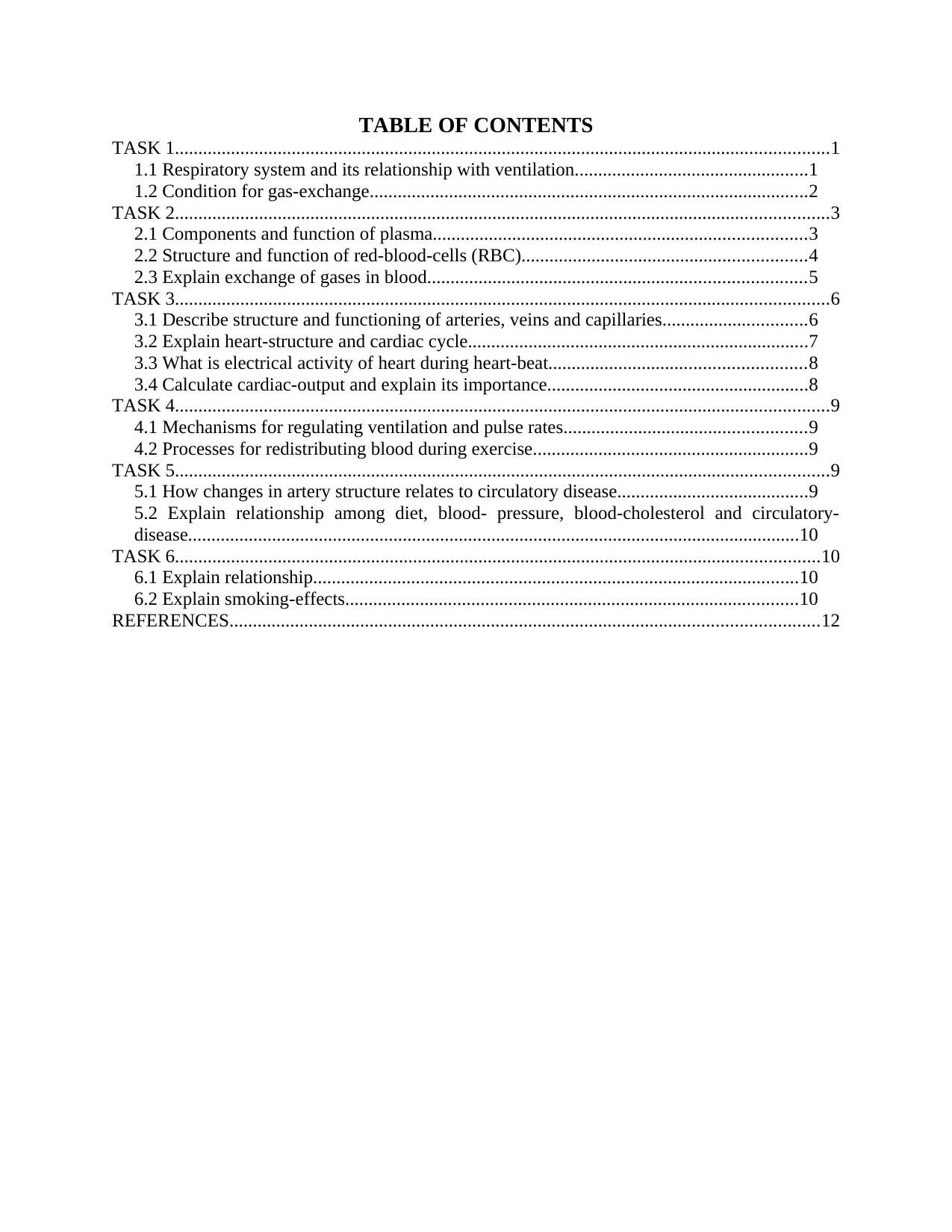
TABLE OF CONTENTS
TASK 1............................................................................................................................................1
1.1 Respiratory system and its relationship with ventilation..................................................1
1.2 Condition for gas-exchange..............................................................................................2
TASK 2............................................................................................................................................3
2.1 Components and function of plasma................................................................................3
2.2 Structure and function of red-blood-cells (RBC).............................................................4
2.3 Explain exchange of gases in blood.................................................................................5
TASK 3............................................................................................................................................6
3.1 Describe structure and functioning of arteries, veins and capillaries...............................6
3.2 Explain heart-structure and cardiac cycle.........................................................................7
3.3 What is electrical activity of heart during heart-beat.......................................................8
3.4 Calculate cardiac-output and explain its importance........................................................8
TASK 4............................................................................................................................................9
4.1 Mechanisms for regulating ventilation and pulse rates....................................................9
4.2 Processes for redistributing blood during exercise...........................................................9
TASK 5............................................................................................................................................9
5.1 How changes in artery structure relates to circulatory disease.........................................9
5.2 Explain relationship among diet, blood- pressure, blood-cholesterol and circulatory-
disease...................................................................................................................................10
TASK 6..........................................................................................................................................10
6.1 Explain relationship........................................................................................................10
6.2 Explain smoking-effects.................................................................................................10
REFERENCES..............................................................................................................................12
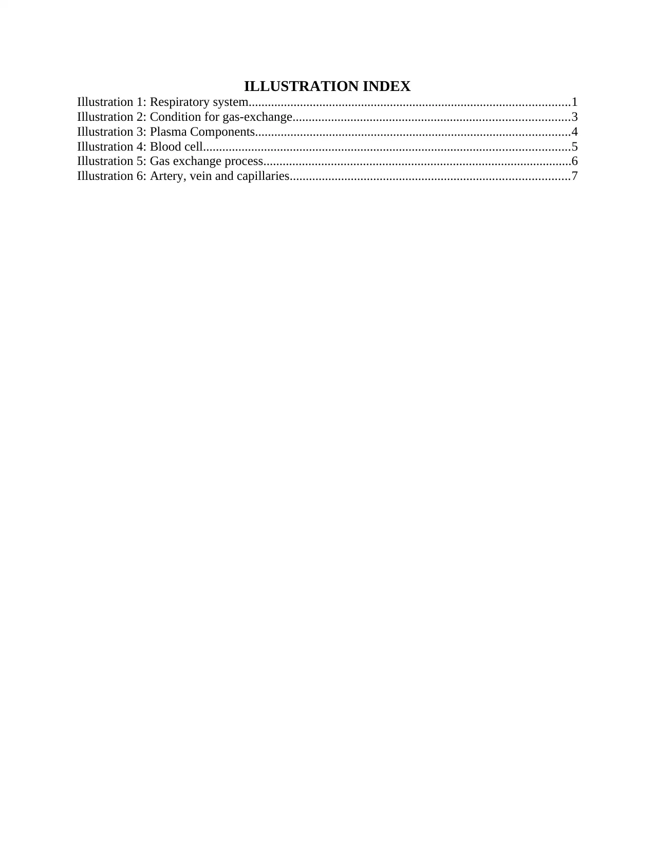
ILLUSTRATION INDEX
Illustration 1: Respiratory system....................................................................................................1
Illustration 2: Condition for gas-exchange......................................................................................3
Illustration 3: Plasma Components..................................................................................................4
Illustration 4: Blood cell..................................................................................................................5
Illustration 5: Gas exchange process................................................................................................6
Illustration 6: Artery, vein and capillaries.......................................................................................7
⊘ This is a preview!⊘
Do you want full access?
Subscribe today to unlock all pages.
Trusted by 1+ million students worldwide
TASK 1
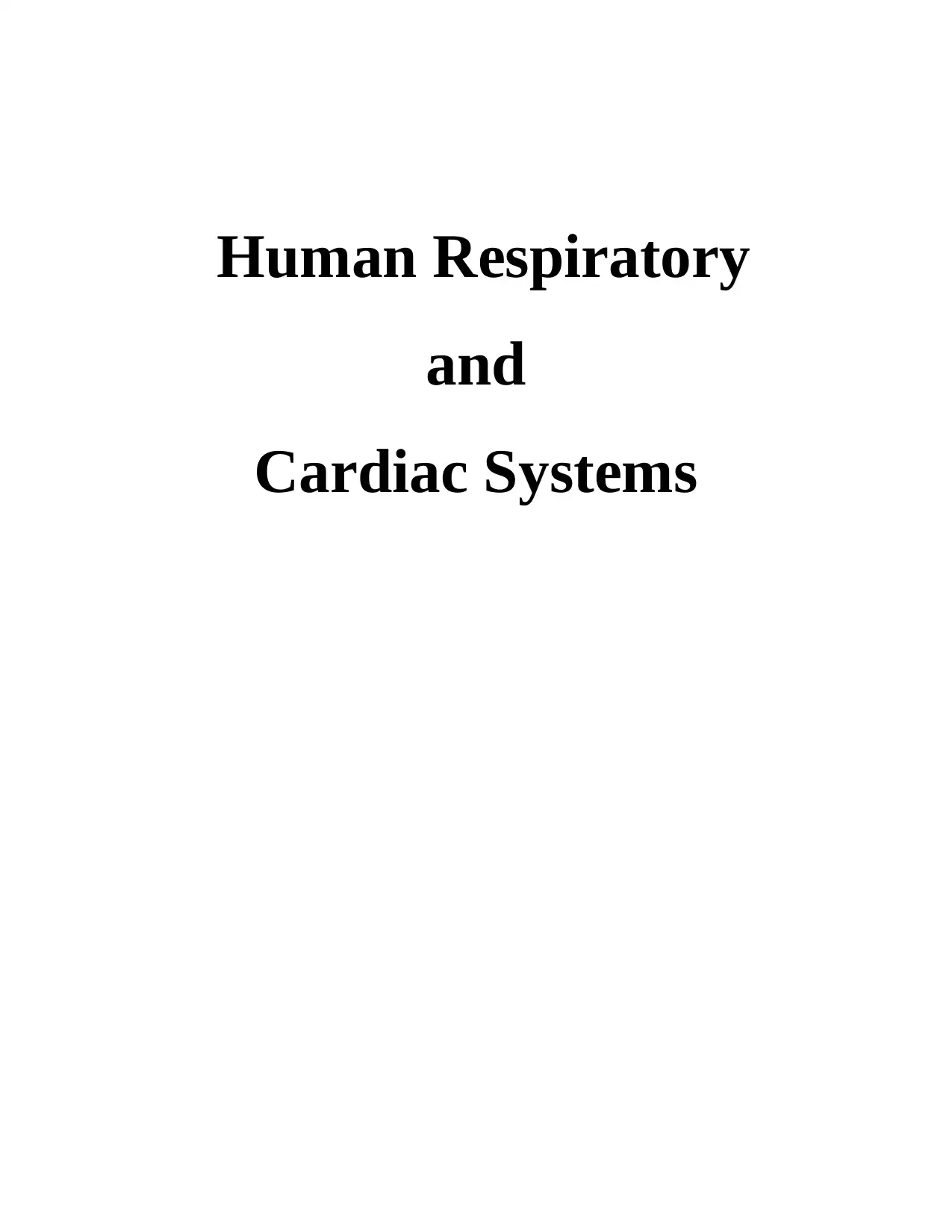
1.1 Respiratory system and its relationship with ventilation.
Respiratory system consists of 9-main parts starting from nose. It has Pharynx, Larynx,
Trachea, Bronchi, Bronchioles, Alveoli, Lungs and Diaphragm (Xing and et. al., 2016).
Illustration 1: Human respiratory system
(Sources: The Human Respiratory System,
2014)
Nose has two separated air passages known as nostrils. It is made-up of cartilage, fatty-
tissues and bone. Cilia, the tiny hair that are present in nose lining which assist keeping mucus
towards nose exit (Meili and et. al., 2016). Mucus is secreted by Goblet cells in the lining of the
nose. It is main part of respiratory system.
Pharynx is part of throat, made of fibromuscular tube extended from skull-base to
cricoid cartilage.
Larynx is connecting part of pharynx and trachea also known as voice box as it produces
the vocal sound. Its function is to allow the air to be directed so that gas-exchange can be made
possible into the respiratory organs.
Trachea is long elastic tissues that are supported by C shaped rings cartilage. Its length is
12-13 cm and 2.5 cm wide and helps in air flow.
1
Paraphrase This Document
Need a fresh take? Get an instant paraphrase of this document with our AI Paraphraser
Bronchi are smooth muscles that makes Y shape in trachea. It is known as cartilage-
ringed tubes that makes it venerable to foreign antigens. The left primary bronchus is smaller
than right, it protects it from being venerable. They expand when air enters the lungs and at the
time of expiration they contract.
Bronchioles are smaller tree shaped network tubes having diameter of 0.5-mm and
contains less cartilage than bronchi. Till than all the foreign antigens get removed and it gets sub-
divided into the alveolar ducts.
Alveoli appears like grapes bunch and aids in gaseous exchange. It is 1-cell thick and
there are millions of alveoli for gas-exchange. Through this, oxygen is transferred in
haemoglobin which with the help of blood-streams carries to whole body. carbon-di-oxide is
diffused back to blood-cells and through expiration it is breathed out of body.
Diaphragm and intercostals muscles helps in ventilation of alveoli. Diaphragm is large
skeletal muscle having tendon surrounding of other muscles. During contraction, they become
flatter. The rib part of body is covered externally and internally with the intercostals muscles.
They allow the ribcage to pivot in all ways.
The process of Ventilation:
The entry and exit of air is through nose which is primary part of respiratory system.
Inside the nose air gets moisture and warm after which it is send to lungs. After that through
pharynx and larynx air flows and rushes down the pipe. Bronchi and Bronchioles takes oxygen
down where Alveoli gets enlarged and exchange of gases takes place. Then function of lungs
starts. Due to presence of heart, left lungs have 2-lobes and right has 3. outer walls are exerted
outwards and upwards while lower are pulled downwards.
Inhalation process occurs when diaphragm and external-intercostal muscles undertake to
fill gases in chest cavity. After this inhalation and exhalation process starts and gaseous
exchange takes place.
1.2 Condition for gas-exchange.
The alveoli part of respiratory system is the position where gaseous-exchange takes place
inside the lungs. There is large surface area required so that efficient gas-exchange can be
completed, due to this reason, alveoli are tiny sacs that are bunched together so that more surface
area is available so that gases can diffuse easily. (Shida and et. al., 2017). If the surface area is
smaller than body would have to work harder for intake and supply. Due to this reason alveoli
2
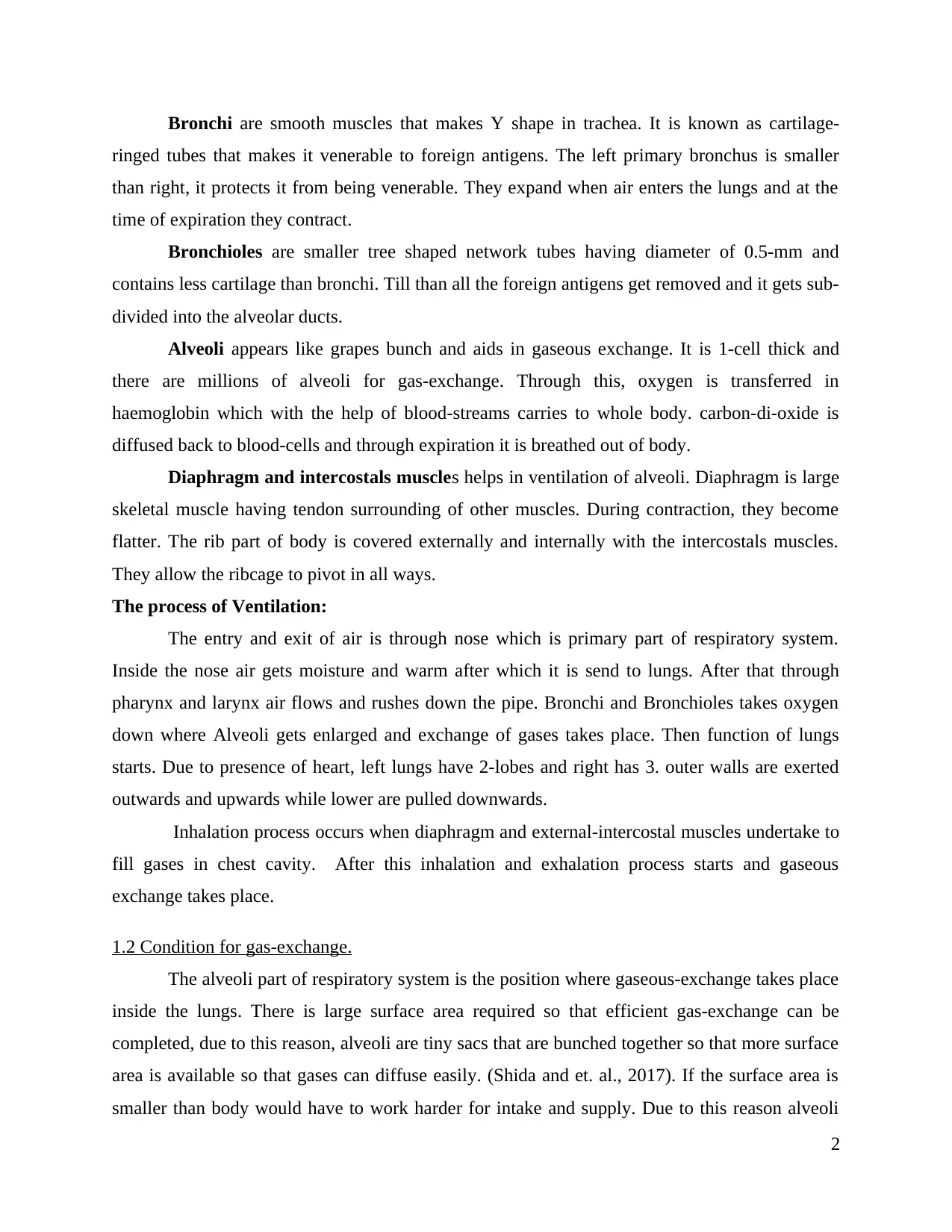
are thinner single flattened epithelial cells. Moreover, the capillaries too are 1-cell thick and the
distance between the alveoli and it is very small so that oxygen can be mixed with haemoglobin
to form oxy-haemoglobin. Meanwhile the carbon-di-oxide diffuses back to alveoli to get
exhaled. The oxygen is supplied to whole body. The region needs to be smaller, moist so that
diffusion can take place and can be dissolved in membrane. This makes water transportation
smaller easier or else water diffusion will be harder. In order to reduce the surface tension and to
prevent the alveoli-collapsing, water contains soapy surfactant.
To make the equilibrium condition, there is need to maintain steep concentration gradient
of the gases. The flow of high-concentration to low-concentration is maintained blood-flow that
carries oxygen away quickly. Using the concentration flow, oxygen flow from air to blood and
carbon-di-oxide from blood to air.
TASK 2
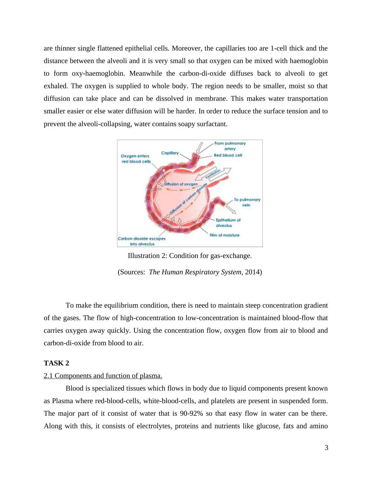
2.1 Components and function of plasma.
Blood is specialized tissues which flows in body due to liquid components present known
as Plasma where red-blood-cells, white-blood-cells, and platelets are present in suspended form.
The major part of it consist of water that is 90-92% so that easy flow in water can be there.
Along with this, it consists of electrolytes, proteins and nutrients like glucose, fats and amino
3
Illustration 2: Condition for gas-exchange.
(Sources: The Human Respiratory System, 2014)
distance between the alveoli and it is very small so that oxygen can be mixed with haemoglobin
to form oxy-haemoglobin. Meanwhile the carbon-di-oxide diffuses back to alveoli to get
exhaled. The oxygen is supplied to whole body. The region needs to be smaller, moist so that
diffusion can take place and can be dissolved in membrane. This makes water transportation
smaller easier or else water diffusion will be harder. In order to reduce the surface tension and to
prevent the alveoli-collapsing, water contains soapy surfactant.
To make the equilibrium condition, there is need to maintain steep concentration gradient
of the gases. The flow of high-concentration to low-concentration is maintained blood-flow that
carries oxygen away quickly. Using the concentration flow, oxygen flow from air to blood and
carbon-di-oxide from blood to air.
TASK 2
2.1 Components and function of plasma.
Blood is specialized tissues which flows in body due to liquid components present known
as Plasma where red-blood-cells, white-blood-cells, and platelets are present in suspended form.
The major part of it consist of water that is 90-92% so that easy flow in water can be there.
Along with this, it consists of electrolytes, proteins and nutrients like glucose, fats and amino
3
Illustration 2: Condition for gas-exchange.
(Sources: The Human Respiratory System, 2014)
⊘ This is a preview!⊘
Do you want full access?
Subscribe today to unlock all pages.
Trusted by 1+ million students worldwide
acids. There is also yellow coloured fluid that flows along with blood. Plasma consist of blood,
albumin, that prevents blood leaking into tissues (Serra and et. al., 2017). Moreover, it contains
hormones and drugs that that binds blood at the time of impinge. It contains fibrinogen which
acts as clotting factors and helps to control bleeding during an injury. Besides this, it carries
indigestible and unused cells to kidneys so they can be excreted in the form of urine. At the time
of exercise carbon-di-oxide is transported by plasma so that it can be exhaled-out.
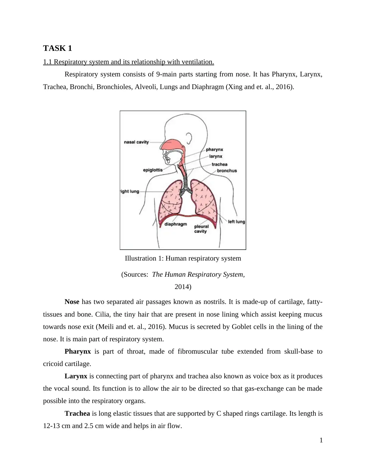
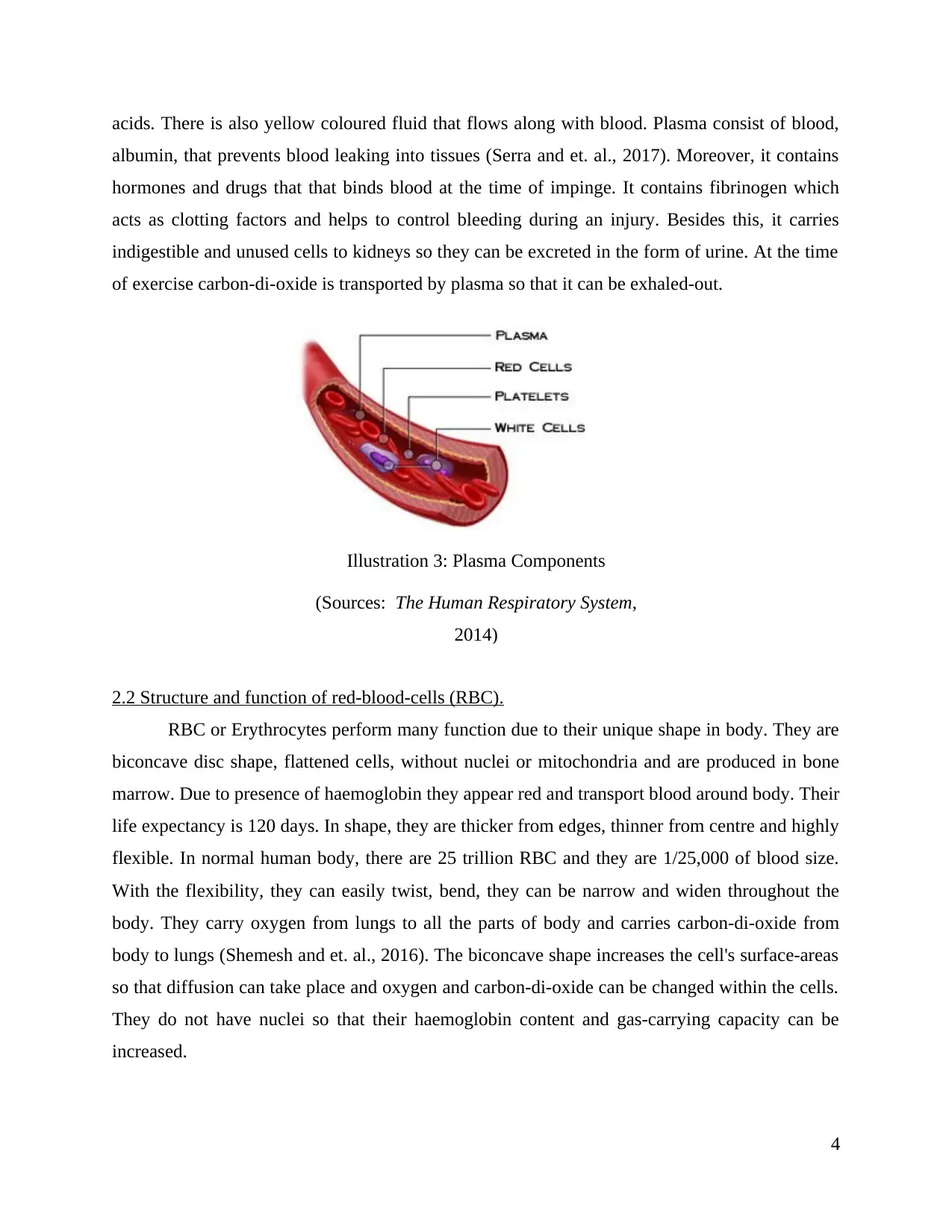
2.2 Structure and function of red-blood-cells (RBC).
RBC or Erythrocytes perform many function due to their unique shape in body. They are
biconcave disc shape, flattened cells, without nuclei or mitochondria and are produced in bone
marrow. Due to presence of haemoglobin they appear red and transport blood around body. Their
life expectancy is 120 days. In shape, they are thicker from edges, thinner from centre and highly
flexible. In normal human body, there are 25 trillion RBC and they are 1/25,000 of blood size.
With the flexibility, they can easily twist, bend, they can be narrow and widen throughout the
body. They carry oxygen from lungs to all the parts of body and carries carbon-di-oxide from
body to lungs (Shemesh and et. al., 2016). The biconcave shape increases the cell's surface-areas
so that diffusion can take place and oxygen and carbon-di-oxide can be changed within the cells.
They do not have nuclei so that their haemoglobin content and gas-carrying capacity can be
increased.
4
Illustration 3: Plasma Components
(Sources: The Human Respiratory System,
2014)
albumin, that prevents blood leaking into tissues (Serra and et. al., 2017). Moreover, it contains
hormones and drugs that that binds blood at the time of impinge. It contains fibrinogen which
acts as clotting factors and helps to control bleeding during an injury. Besides this, it carries
indigestible and unused cells to kidneys so they can be excreted in the form of urine. At the time
of exercise carbon-di-oxide is transported by plasma so that it can be exhaled-out.
2.2 Structure and function of red-blood-cells (RBC).
RBC or Erythrocytes perform many function due to their unique shape in body. They are
biconcave disc shape, flattened cells, without nuclei or mitochondria and are produced in bone
marrow. Due to presence of haemoglobin they appear red and transport blood around body. Their
life expectancy is 120 days. In shape, they are thicker from edges, thinner from centre and highly
flexible. In normal human body, there are 25 trillion RBC and they are 1/25,000 of blood size.
With the flexibility, they can easily twist, bend, they can be narrow and widen throughout the
body. They carry oxygen from lungs to all the parts of body and carries carbon-di-oxide from
body to lungs (Shemesh and et. al., 2016). The biconcave shape increases the cell's surface-areas
so that diffusion can take place and oxygen and carbon-di-oxide can be changed within the cells.
They do not have nuclei so that their haemoglobin content and gas-carrying capacity can be
increased.
4
Illustration 3: Plasma Components
(Sources: The Human Respiratory System,
2014)
Paraphrase This Document
Need a fresh take? Get an instant paraphrase of this document with our AI Paraphraser
2.3 Explain exchange of gases in blood.
With the help of RBC, oxygen is carried from lungs to other body-organs. Plasma carries
the dissolved gas that is oxygen (1.5%) in small amount. Almost all the oxygen is carried out
through the blood, between the protein haemoglobin and RBC. There are 4-oxygen molecules
attached to fully saturated oxy-haemoglobin. Transported oxygen helps in respiration however,
the solubility of carbon-di-oxide is more than oxygen. In blood 5% is transported as unchanged
there they combine with haemoglobin and form carb-amino-haemoglobin (Moschos, Usher, and
Lindsay, 2017). In Polypeptide-chains amino-groups binds 10% of carbon-di-oxide and forms
haemoglobin and plasma-proteins. It enters through tissue capillaries and forms carbonic-acids.
Though, it is slow process but enzyme like carbonic hydras accelerate RBC.
5
Illustration 4: Blood cell
(Sources: The Human Respiratory System, 2014)
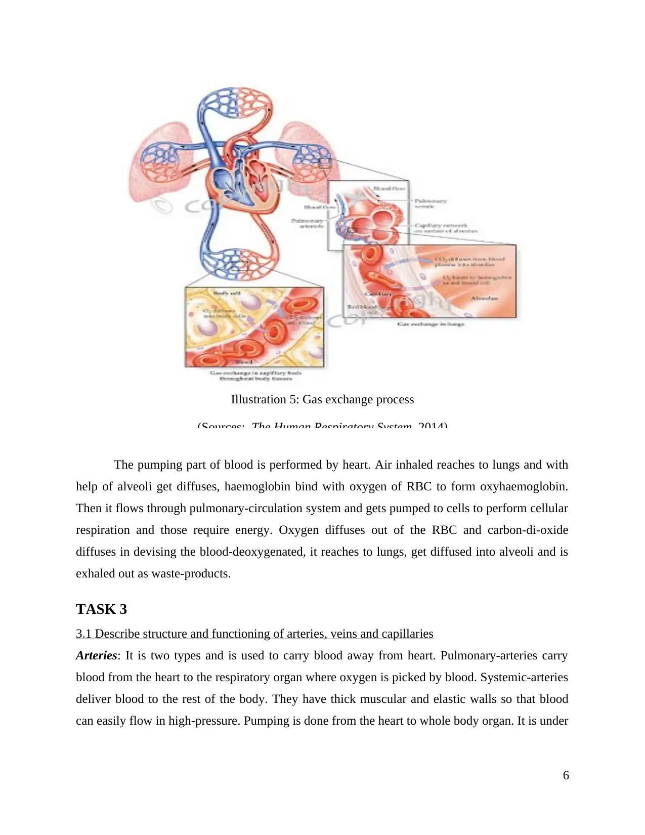
The pumping part of blood is performed by heart. Air inhaled reaches to lungs and with
help of alveoli get diffuses, haemoglobin bind with oxygen of RBC to form oxyhaemoglobin.
Then it flows through pulmonary-circulation system and gets pumped to cells to perform cellular
respiration and those require energy. Oxygen diffuses out of the RBC and carbon-di-oxide
diffuses in devising the blood-deoxygenated, it reaches to lungs, get diffused into alveoli and is
exhaled out as waste-products.
TASK 3
3.1 Describe structure and functioning of arteries, veins and capillaries
Arteries: It is two types and is used to carry blood away from heart. Pulmonary-arteries carry
blood from the heart to the respiratory organ where oxygen is picked by blood. Systemic-arteries
deliver blood to the rest of the body. They have thick muscular and elastic walls so that blood
can easily flow in high-pressure. Pumping is done from the heart to whole body organ. It is under
6
Illustration 5: Gas exchange process
(Sources: The Human Respiratory System, 2014)
⊘ This is a preview!⊘
Do you want full access?
Subscribe today to unlock all pages.
Trusted by 1+ million students worldwide
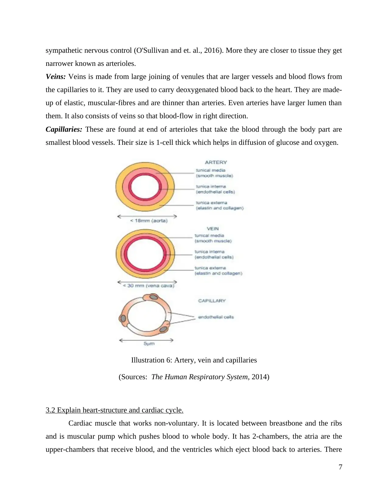
sympathetic nervous control (O'Sullivan and et. al., 2016). More they are closer to tissue they get
narrower known as arterioles.
Veins: Veins is made from large joining of venules that are larger vessels and blood flows from
the capillaries to it. They are used to carry deoxygenated blood back to the heart. They are made-
up of elastic, muscular-fibres and are thinner than arteries. Even arteries have larger lumen than
them. It also consists of veins so that blood-flow in right direction.
Capillaries: These are found at end of arterioles that take the blood through the body part are
smallest blood vessels. Their size is 1-cell thick which helps in diffusion of glucose and oxygen.
3.2 Explain heart-structure and cardiac cycle.
Cardiac muscle that works non-voluntary. It is located between breastbone and the ribs
and is muscular pump which pushes blood to whole body. It has 2-chambers, the atria are the
upper-chambers that receive blood, and the ventricles which eject blood back to arteries. There
7
Illustration 6: Artery, vein and capillaries
(Sources: The Human Respiratory System, 2014)
Paraphrase This Document
Need a fresh take? Get an instant paraphrase of this document with our AI Paraphraser
are 4-valves that separates the chambers, Tricuspid, Pulmonary-valve, Bicuspid-valve and
Aortic-valve. They perform different function such as the first separates the right-atrium from the
right-ventricle, second, separates the right-ventricle from the pulmonary-artery, separates the
left-atrium from the left-ventricle and last separates the right-ventricle from the aorta. It has 3-
layers, epicardium, myocardium and endocardium. They provide smooth texture, pumping
action, made up of muscle-fibres which connect to electrical-synapses and inner layer that
connect to large blood vessels. Deoxygenated-blood is received at right-side and oxygenated-
blood is reached at left-side.
The cardiac cycle is action performed by heart in contraction and relaxation. The right-
side-heart receives deoxygenated-blood from the vena cava, and fill up the right-atrium and then
flows through the tricuspid-valve and into the right-ventricle. From there it is forced shut by
ventricular-pressure, ventricular-systole-forces the blood up through the pulmonary-valve in to
the arterial-blood-vessel, the heart-ventricle at heartbeat-stage which makes the pulmonary-valve
close to prevent back flow. When blood reaches lungs it gets oxygenated and transported back to
the heart via the pulmonary-vein as-well-as from left-atrium. After that it travels through
bicuspid-valve into the left-ventricle. In Aortic-valve it gets pumped and through great-pressure
it exhaled from aorta. Further, it travels body and reaches to areas that require oxygen. It is
continuous cycle and repeats.
3.3 What is electrical activity of heart during heart-beat.
The heart is myogenic and its functioning is performed by 2-nervous system. The
sympathetic part accelerates the heart-beat, which completes bodies requirements while
exercising, and other body workouts. The rhythmic-sequence of contractions is completed by
Sino-atrial (SA), and atria-ventricular (AV) nodes. The SA node is present at right atrium at the
upper side of wall and provides electrical impulses for initiating atrial-contraction. As the
impulse reaches the AV node (lower right atrium) is situated, it remains static for a short period
of time, so that blood in the atria can fulfil the ventricles.
3.4 Calculate cardiac-output and explain its importance.
It is the amount of blood that is pumped by heart. At the time of ventricles-contract,
about 70-90 cm3 of blood is ejected into the pulmonary-artery and aorta. It shows the blood-
volume being pumped round the human-body. It is used to determine effectiveness of the heart
8
and to avoid heart-failure or poor-circulation (Lai and et. al., 2016). To measure cardiac output
there are two methods and heart rate (number of heart-loads being pumped round the body/time-
unit) and stroke-volume (blood-volume that fill heart/contraction).
Cardiac output = heart rate x stroke volume.
The stroke-volume is regulated by stimulant from the sympathetic nervous system. As
heart-rate increases there is hike in cardiac-output by accelerating blood-volume released in the
body. During diastole, there is increase in heart rate. However, if it is too low then it could lead
to heart-failure. It is measured using attached electrodes to a machine that can amplify and record
an ECG (electrocardiogram).
TASK 4
4.1 Mechanisms for regulating ventilation and pulse rates.
Heart-rate and respiratory-system is controlled by the sympathetic and parasympathetic
nervous system. Sympathetic nerves transmit signals when heavy exercise is done to the SA
node which releases adrenalin from the adrenal medulla present at upper side of kidneys and
released by the sympathetic neurons. It normally happens at high-pressure, the body part requires
oxygen so that energy needed is released. The heart-beat increases and intense breathing is an
attempt to get more oxygen through pumping. It also happens at the time of stressful situation or
frightening experience (Lee and et.al., 2017). Whenever, there is body-pressure the breathing and
the heart-rate increases leading to high BP. The messages are send to aorta and carotid-artery.
After that it is received by cardiac centre, which impulsive response to parasympathetic nerves
and then to the SA node and the heart-rate gets lower. When there is no fear, parasympathetic
nervous system kicks in striving for homoeostasis and then normal breathing as-well-as heart
rate starts.
4.2 Processes for redistributing blood during exercise.
Blood vessels dilate when exercise is done and making it flow better. While consuming
energy, the muscles produce CO2. It results the capillaries-muscles to expand and exaggerated
the blood-flow delivers for which large amount of oxygenated blood to the working-muscles. At
times of under-pressure from exercise, blood leaves organs and reaches to muscles that are in
action. It occurs through sympathetic nervous system stimulating the nerves-heart and blood-
9
⊘ This is a preview!⊘
Do you want full access?
Subscribe today to unlock all pages.
Trusted by 1+ million students worldwide
1 out of 15
Related Documents


Your All-in-One AI-Powered Toolkit for Academic Success.
+13062052269
info@desklib.com
Available 24*7 on WhatsApp / Email
![[object Object]](/_next/static/media/star-bottom.7253800d.svg)
Unlock your academic potential
Copyright © 2020–2026 A2Z Services. All Rights Reserved. Developed and managed by ZUCOL.