Functions of the Human Skeleton and Muscular System Report
VerifiedAdded on 2020/12/29
|9
|2257
|350
Report
AI Summary
This report provides a detailed overview of the human skeletal and muscular systems. It begins with an introduction to the musculoskeletal system, including the functions of the skeleton and the importance of maintaining the health of these systems. Task 1 explores the gross structure of the human skeleton, types of joints (fibrous, cartilaginous, and synovial), the structure and components of synovial joints, and the properties and functions of tendons, ligaments, and cartilage. Task 2 compares the properties of different muscle types (skeletal, cardiac, and smooth) and explains the sliding filament hypothesis of muscle contraction, along with the extension and flexion of the elbow joint by antagonistic muscles. Task 3 is covered in a PPT. The report concludes with a summary of the key findings and references relevant sources, providing a comprehensive understanding of the skeletal and muscular systems.
Skeleton & Muscles
Paraphrase This Document
Need a fresh take? Get an instant paraphrase of this document with our AI Paraphraser
Table of Contents
INTRODUCTION...........................................................................................................................3
TASK 1 ...........................................................................................................................................3
1.1 Gross structure of human skeleton and its functions........................................................3
1.2 Types of joints and importance of their properties...........................................................3
1.3 Structure of synovial joint and roles of component parts.................................................5
1.4 Properties and functions of tendons, ligaments and cartilage..........................................5
TASK 2 ...........................................................................................................................................6
2.1 Compare properties of different types of muscles and explain sliding filament hypothesis
of muscle contraction.............................................................................................................6
2.2 Extension and flexion of elbow joint by antagonistic muscles........................................7
TASK 3............................................................................................................................................8
Covered in PPT.......................................................................................................................8
CONCLUSION................................................................................................................................8
REFERENCES................................................................................................................................9
INTRODUCTION
Skeleton and muscles together form a musculoskeletal system which is an important part
of human body. It can be defined as collection of 206 bones in human body and rest of part helps
them to move. However, the skeleton along with muscles, ligaments and cartilage provide
framework and support to body (Ranganathan and et. al., 2014). The present report is based on
West London NHS Trust which provide health services and treatment to needy people to make
them well-being. It provide care and treatment for around 62570 people every year and serve
local population approximately 700000 residents in London boroughs of Ealing, Hammersmith
& Fulham and Hounslow. This assignment will focus on function of skeletal and muscular
system in human body. It will also include importance of maintaining health of muscular and
skeletal systems.
TASK 1
1.1 Gross structure of human skeleton and its functions
The of human skeleton system consist mainly bones and cartilage in order perform
several functions such as supports body, facilitate movement, protect internal organs, produce
blood cells and Stores & release the minerals and fat. However, gross structure of skeletal system
include various parts such as appendicular skeleton, axial skeleton, coccyx, ear ossicles, hyoid
bone, ribs, sacrum, skull, sternum, thoracic cage, vertebra and vertebral column. Moreover, these
parts of skeletal system has their own role according to their location in particular body part of
human body (Dixon, 2017). In addition to this, these different parts are helpful to connect with
each other to form overall skeleton of body and facilitate to conduct overall functions of skeletal
system. Meanwhile, the gross structure provide help to carrying out several functions like
support, protection, movement, storage and regulation of endocrine glands.
⊘ This is a preview!⊘
Do you want full access?
Subscribe today to unlock all pages.
Trusted by 1+ million students worldwide
1.2 Types of joints and importance of their properties
There are types of joints which has their own structure and function in human body
which facilitate to conduct respective functions properly. These joints has few of properties
which has their own importance in functioning of skeletal system.
Types of joints and Properties
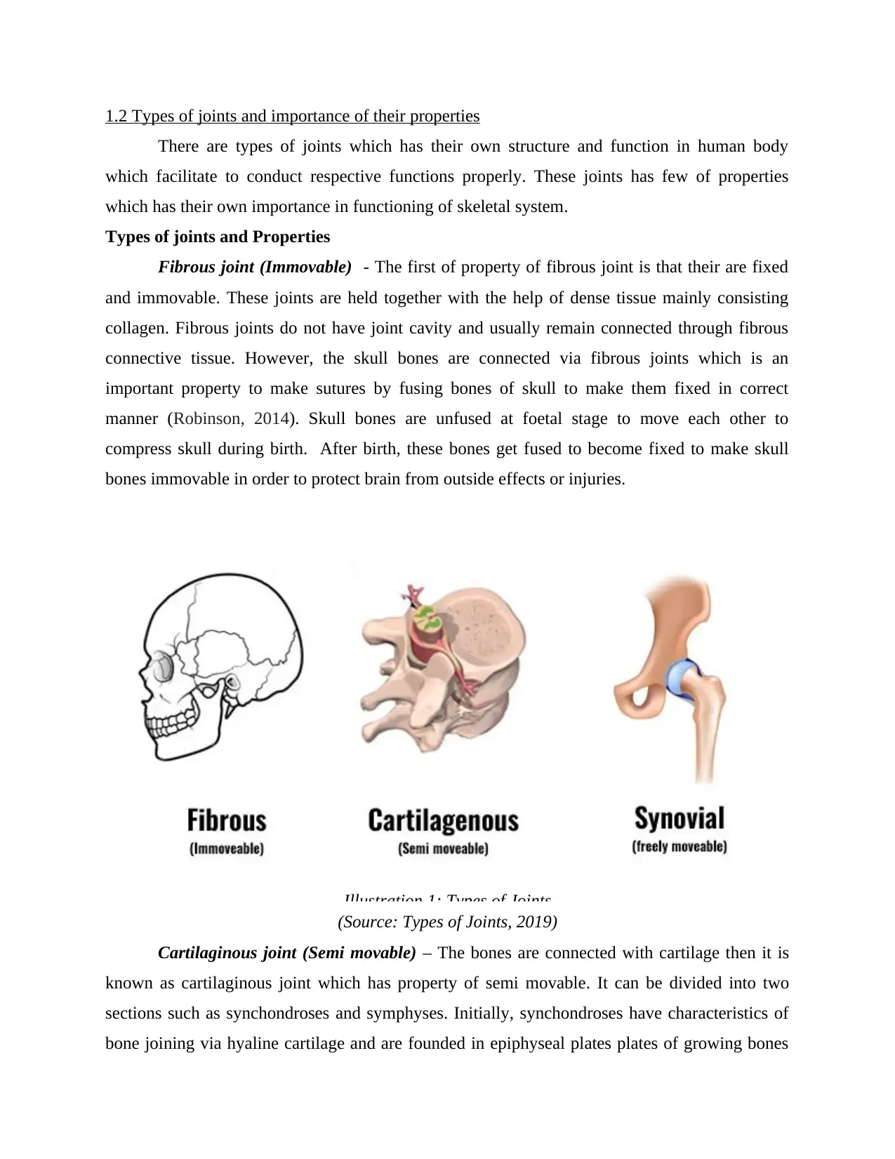
Fibrous joint (Immovable) - The first of property of fibrous joint is that their are fixed
and immovable. These joints are held together with the help of dense tissue mainly consisting
collagen. Fibrous joints do not have joint cavity and usually remain connected through fibrous
connective tissue. However, the skull bones are connected via fibrous joints which is an
important property to make sutures by fusing bones of skull to make them fixed in correct
manner (Robinson, 2014). Skull bones are unfused at foetal stage to move each other to
compress skull during birth. After birth, these bones get fused to become fixed to make skull
bones immovable in order to protect brain from outside effects or injuries.
(Source: Types of Joints, 2019)
Cartilaginous joint (Semi movable) – The bones are connected with cartilage then it is
known as cartilaginous joint which has property of semi movable. It can be divided into two
sections such as synchondroses and symphyses. Initially, synchondroses have characteristics of
bone joining via hyaline cartilage and are founded in epiphyseal plates plates of growing bones
Illustration 1: Types of Joints
There are types of joints which has their own structure and function in human body
which facilitate to conduct respective functions properly. These joints has few of properties
which has their own importance in functioning of skeletal system.
Types of joints and Properties
Fibrous joint (Immovable) - The first of property of fibrous joint is that their are fixed
and immovable. These joints are held together with the help of dense tissue mainly consisting
collagen. Fibrous joints do not have joint cavity and usually remain connected through fibrous
connective tissue. However, the skull bones are connected via fibrous joints which is an
important property to make sutures by fusing bones of skull to make them fixed in correct
manner (Robinson, 2014). Skull bones are unfused at foetal stage to move each other to
compress skull during birth. After birth, these bones get fused to become fixed to make skull
bones immovable in order to protect brain from outside effects or injuries.
(Source: Types of Joints, 2019)
Cartilaginous joint (Semi movable) – The bones are connected with cartilage then it is
known as cartilaginous joint which has property of semi movable. It can be divided into two
sections such as synchondroses and symphyses. Initially, synchondroses have characteristics of
bone joining via hyaline cartilage and are founded in epiphyseal plates plates of growing bones
Illustration 1: Types of Joints
Paraphrase This Document
Need a fresh take? Get an instant paraphrase of this document with our AI Paraphraser
in children. Moreover, hyaline cartilage covers end of bone and connection of bones occur via
fibrocartilage in symphyses. These properties of cartilaginous joints are important as they found
between vertebrae and pubic bones. Additionally, cartilaginous joints allow slight movement and
forms growth regions of immature bones and intervertebral discs in spinal column (Muscolino,
2016).
Synovial joint (freely movable) – The main property of synovial joints is their freely
movements having space between adjoining bones which is called as synovial cavity filled with
synovial fluid. This synovial fluid is important feature of synovial joints which is important to
lubricate the joint, reduce friction and allow the greater movement.
1.3 Structure of synovial joint and roles of component parts
The structure of synovial joint consist several important parts including articular capsule,
articular cartilage and synovial fluid. However, these components of synovial joints has their
own property and roles to facilitate overall function of the same.
Articular capsule – It has two layers including fibrous layer which is outer region and
synovial layer that is inner part. However, fibrous layer consist fibrous tissue considered as
capsular ligament which has role to hold articulating bones together and supports the underlying
synovium (Azaroual and et. al., 2014). Moreover, it is essential synovial layer can be defined as
highly vascularised of serious connective tissue in order to absorb and secretes. Additionally, this
layer plays an important role of mediation of nutrient exchange between blood and joint.
Articular cartilage – This include articulating surfaces of synovial joint are covered by a
thin layer of hyaline cartilage. It consist the surface which comes in contact of with each other as
joint moves. Moreover, articular cartilage plays an important role to minimise friction upon joint
movement and absorb shock.
Synovial Fluid – The synovial fluid is located in joint cavity of synovial joint which
conduct certain major roles lubrication, nutrient distribution and shock.
1.4 Properties and functions of tendons, ligaments and cartilage
Properties
Tendons - The complete tendons is built by building up and combining layer of multiple
layers of connective tissue. Tendon can be considered as a tissue which attach muscles to other
body parts generally bones. However, they are remarkable strong along with one of highest
tensile strengths found among soft tissues. Tendon has a hierarchical structure, parallel
orientation and tissue composition of fibres that is necessary for withstanding stressed created by
muscles (Balta, Lamb and Soames, 2015).
Ligaments – Ligaments are bundles of connective tissue that connect one bone to an
adjacent bone. These are make of collagen fibres which very strong, flexible and resistance to
damage from pulling or compressing stresses.
Cartilage – The cartilage have several properties such as homogeneous cellularity,
relatively low cell density, interstitial or oppositional growth, avascular, extensive extracellular
matrix and lack of innervation.
Functions
Tendons – The tendon are helpful to transmit tensile forces from muscle to bone,
maintain body posture and provide motor control. Moreover, they give an advantage mechanical
pulley enables muscle belly to be an optimal distance from joint without requiring an extended
length of muscle between origin and insertion.
Ligaments – They conduct function to resist external load and prevent excessive motion.
It also include to guide joint motion for facilitating relative movements of bones and passively
control maximum range of movement. However, it provides mechanical stability of joint and
provide motor control.
Cartilage – The cartilage plays an essential function of providing support but less rigid
than a bone. It allow some flexibility to movement but consist more stability as compared to
muscle.
TASK 2
2.1 Compare properties of different types of muscles and explain sliding filament hypothesis of
muscle contraction
Comparison between skeletal, cardiac and smooth muscles
Types of muscle Skeletal muscles Cardiac muscles Smooth muscles
Location These muscles remain
attached to bones to
facilitate movement of
body parts.
Cardiac muscles have
a location in heart to
facilitate several
functions this organ.
The smooth muscles
are located in walls of
internal organs and in
skin.
Function They conduct function They provide support They facilitate
⊘ This is a preview!⊘
Do you want full access?
Subscribe today to unlock all pages.
Trusted by 1+ million students worldwide
of moving bones
smoothly.
to conduct breathing
of heart.
movement of internal
organs.
Control mode They have voluntary
mode of control.
The mode of
controlling can be
considered as
involuntary for cardiac
muscles.
Smooth muscles
consist involuntary
mode of control.
Shape Skeletal muscles are
long and slender in
shape
They have a shape of
branching in which
these muscles exists in
body.
They have a spindle
shape.
characteristics They have several
features such as
striated- light and dark
bands. It has multiple
number of nuclei.
Cardiac muscle are
also striated but
having one or two
nuclei.
These muscles consist
only one nucleus
(visceral) and non
striated property
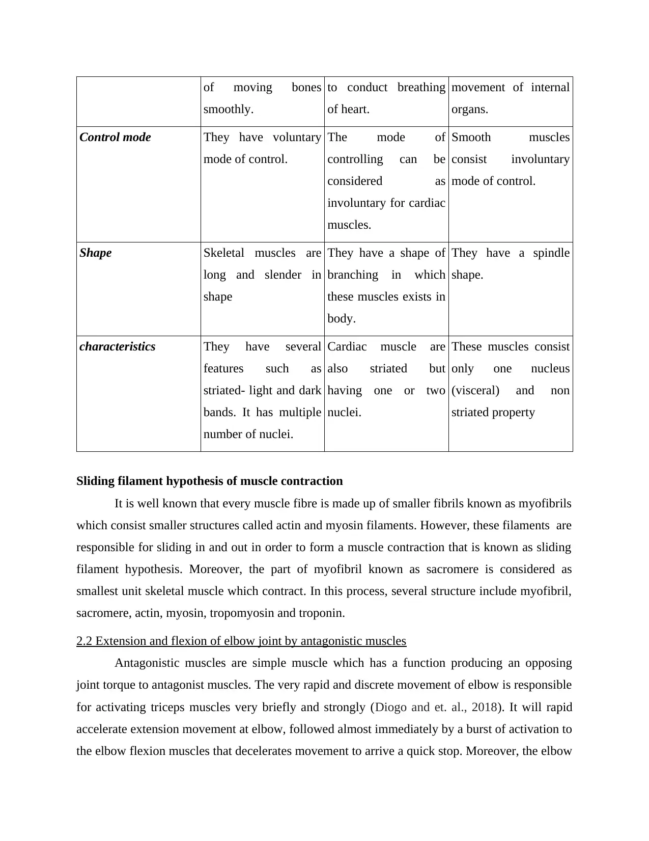
Sliding filament hypothesis of muscle contraction
It is well known that every muscle fibre is made up of smaller fibrils known as myofibrils
which consist smaller structures called actin and myosin filaments. However, these filaments are
responsible for sliding in and out in order to form a muscle contraction that is known as sliding
filament hypothesis. Moreover, the part of myofibril known as sacromere is considered as
smallest unit skeletal muscle which contract. In this process, several structure include myofibril,
sacromere, actin, myosin, tropomyosin and troponin.
2.2 Extension and flexion of elbow joint by antagonistic muscles
Antagonistic muscles are simple muscle which has a function producing an opposing
joint torque to antagonist muscles. The very rapid and discrete movement of elbow is responsible
for activating triceps muscles very briefly and strongly (Diogo and et. al., 2018). It will rapid
accelerate extension movement at elbow, followed almost immediately by a burst of activation to
the elbow flexion muscles that decelerates movement to arrive a quick stop. Moreover, the elbow
smoothly.
to conduct breathing
of heart.
movement of internal
organs.
Control mode They have voluntary
mode of control.
The mode of
controlling can be
considered as
involuntary for cardiac
muscles.
Smooth muscles
consist involuntary
mode of control.
Shape Skeletal muscles are
long and slender in
shape
They have a shape of
branching in which
these muscles exists in
body.
They have a spindle
shape.
characteristics They have several
features such as
striated- light and dark
bands. It has multiple
number of nuclei.
Cardiac muscle are
also striated but
having one or two
nuclei.
These muscles consist
only one nucleus
(visceral) and non
striated property
Sliding filament hypothesis of muscle contraction
It is well known that every muscle fibre is made up of smaller fibrils known as myofibrils
which consist smaller structures called actin and myosin filaments. However, these filaments are
responsible for sliding in and out in order to form a muscle contraction that is known as sliding
filament hypothesis. Moreover, the part of myofibril known as sacromere is considered as
smallest unit skeletal muscle which contract. In this process, several structure include myofibril,
sacromere, actin, myosin, tropomyosin and troponin.
2.2 Extension and flexion of elbow joint by antagonistic muscles
Antagonistic muscles are simple muscle which has a function producing an opposing
joint torque to antagonist muscles. The very rapid and discrete movement of elbow is responsible
for activating triceps muscles very briefly and strongly (Diogo and et. al., 2018). It will rapid
accelerate extension movement at elbow, followed almost immediately by a burst of activation to
the elbow flexion muscles that decelerates movement to arrive a quick stop. Moreover, the elbow
Paraphrase This Document
Need a fresh take? Get an instant paraphrase of this document with our AI Paraphraser
flexor muscles are antagonists at elbow during both up and down phase of movement while
conducting push ups. Additionally, during the dumbbell curl, elbow extensor are antagonists
while lifting and lowering phases.
TASK 3
Covered in PPT
CONCLUSION
From the above report, it has been concluded that skeleton and muscles are an important
pat of human body which facilitate to conduct various functions in proper manner. It include
fibrous, cartilaginous and synovial joints which has their own properties and facilitate the
functioning of skeletal system appropriately. However, it involve tendons, ligaments and
cartilage has their own features to conduct respective roles properly. In addition to this, poor
posture and improper lifting techniques may create skeletal and muscular injuries which can
become severe many times. Moreover, it consist that comparison of cardiac, skeletal and smooth
muscles on several basis like location, function, shape, mode of control etc.
REFERENCES
Books and journals
Ranganathan, K. and et. al., 2014. Temporalis muscle morphomics: the psoas of the craniofacial
skeleton. journal of surgical research. 186(1). pp.246-252.
Dixon, A. D., 2017. Prenatal development of the facial skeleton. In Fundamentals of
craniofacial growth (pp. 59-98). CRC Press.
Robinson, J., 2014. The muscles, body wall and valve-opening mechanism of extant craniid
(inarticulated) brachiopods. Journal of natural History. 48(21-22). pp.1231-1252.
Muscolino, J. E., 2016. The muscular system manual: The skeletal muscles of the human body.
Elsevier Health Sciences.
Azaroual, M. F. and et. al., 2014. Relationship between dimensions of muscles of mastication
(masseter and lateral pterygoid) and skeletal dimensions: study of 40 cases.
International orthodontics. 12(1). pp.111-124.
Balta, J. Y., Lamb, C. and Soames, R. W., 2015. A pilot study comparing the use of Thiel‐and
formalin‐embalmed cadavers in the teaching of human anatomy. Anatomical sciences
education. 8(1). pp.86-91.
Diogo, R. and et. al., 2018. Muscles of chordates: development, homologies, and evolution. CRC
Press.
Flack, N. A. M. S., Nicholson, H. D. and Woodley, S. J., 2014. The anatomy of the hip abductor
muscles. Clinical anatomy. 27(2). pp.241-253.
Online
Types of Joints. 2019. [Online]. Available through:
<https://www.teachpe.com/anatomy/joints.php>
⊘ This is a preview!⊘
Do you want full access?
Subscribe today to unlock all pages.
Trusted by 1+ million students worldwide
1 out of 9
Related Documents


Your All-in-One AI-Powered Toolkit for Academic Success.
+13062052269
info@desklib.com
Available 24*7 on WhatsApp / Email
![[object Object]](/_next/static/media/star-bottom.7253800d.svg)
Unlock your academic potential
Copyright © 2020–2026 A2Z Services. All Rights Reserved. Developed and managed by ZUCOL.