Literature Review on Hypovolaemia in Post-Anesthesia Care Unit (PACU)
VerifiedAdded on 2021/02/21
|15
|3461
|41
Report
AI Summary
This report presents a comprehensive literature review focusing on hypovolaemia, a critical emergency situation in the Post-Anesthesia Care Unit (PACU). The background and rationale highlight the importance of managing fluid loss during and after anesthesia, as hypovolaemia can lead to life-threatening complications like hypovolaemic shock. The literature search and critique describe the methodology used to gather and evaluate relevant sources, emphasizing the use of databases like Medline and Pubmed, and the application of the CRAPP test to ensure the quality of the secondary sources. The literature review delves into the definition, prevalence, consequences, causes, signs, symptoms, and risk factors associated with hypovolaemia. It examines the impact of anesthetic drugs on vascular tone, cardiac output, and the venous system, differentiating between absolute and relative hypovolaemia. The report explores the consequences for patients, including potential organ failure and cardiovascular issues. It outlines the various causes, such as venodilation, and symptoms like headache, dizziness, and low blood pressure. Risk factors, including patient positioning, use of IV fluids, and age, are analyzed to emphasize the importance of proactive interventions. Finally, the report discusses management interventions, including hemodynamic monitoring, fluid resuscitation, and the use of crystalloids, colloids, and blood products to restore circulating volume and maintain adequate tissue perfusion. The report emphasizes the need for nurses to customize interventions based on individual patient needs and to continuously monitor and adjust treatment strategies to ensure optimal patient outcomes.

Anaesthetics and Recovery
Paraphrase This Document
Need a fresh take? Get an instant paraphrase of this document with our AI Paraphraser
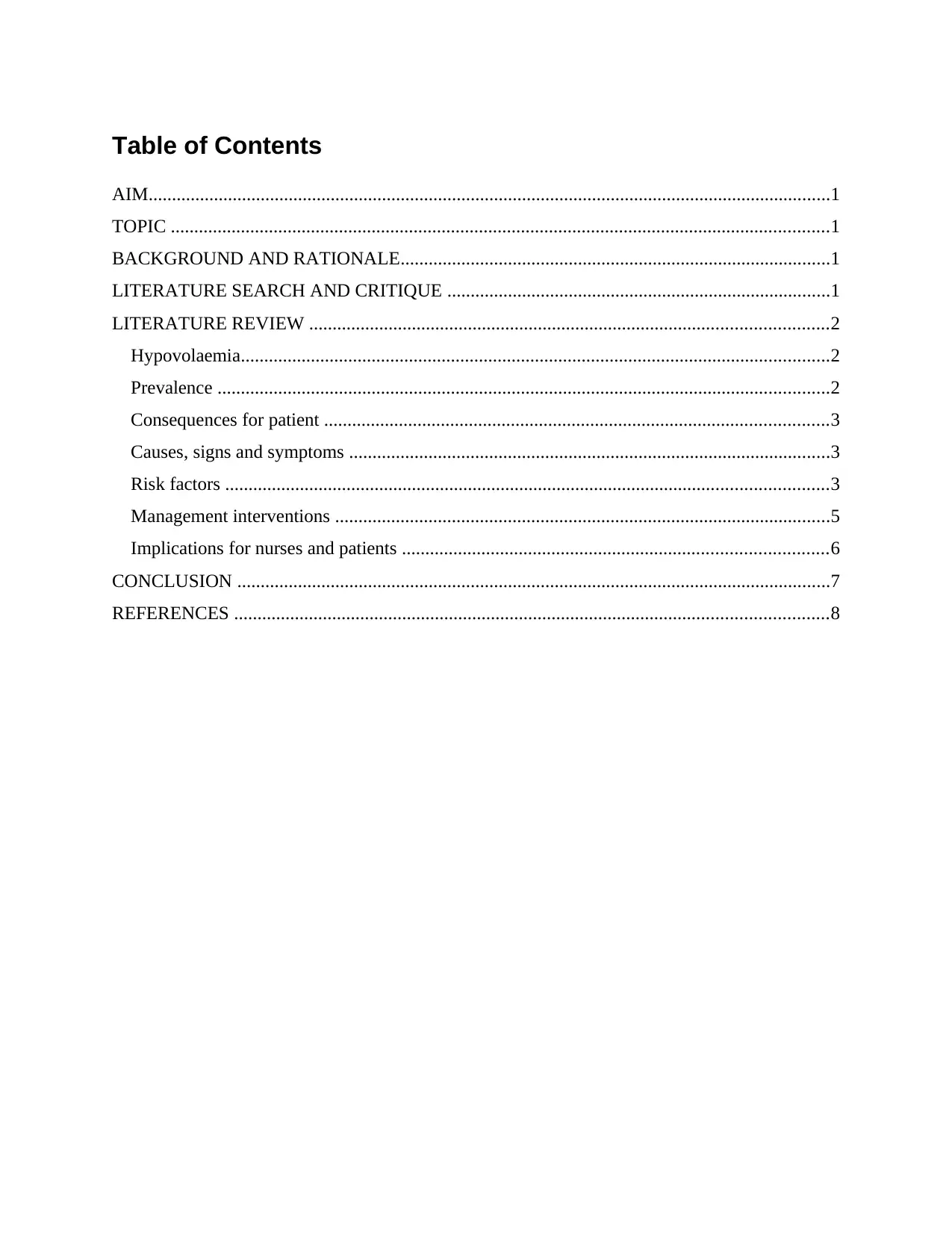
Table of Contents
AIM..................................................................................................................................................1
TOPIC .............................................................................................................................................1
BACKGROUND AND RATIONALE............................................................................................1
LITERATURE SEARCH AND CRITIQUE ..................................................................................1
LITERATURE REVIEW ...............................................................................................................2
Hypovolaemia..............................................................................................................................2
Prevalence ...................................................................................................................................2
Consequences for patient ............................................................................................................3
Causes, signs and symptoms .......................................................................................................3
Risk factors .................................................................................................................................3
Management interventions ..........................................................................................................5
Implications for nurses and patients ...........................................................................................6
CONCLUSION ...............................................................................................................................7
REFERENCES ...............................................................................................................................8
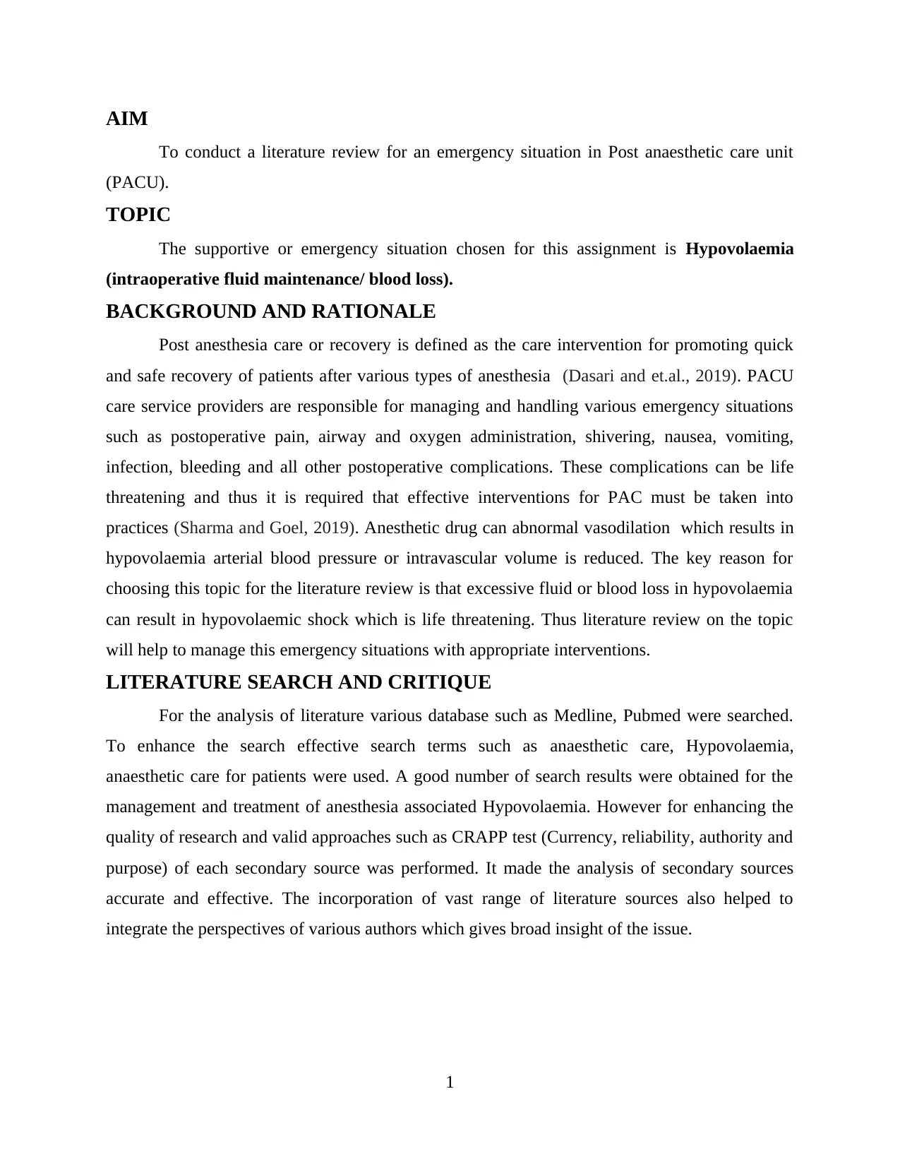
AIM
To conduct a literature review for an emergency situation in Post anaesthetic care unit
(PACU).
TOPIC
The supportive or emergency situation chosen for this assignment is Hypovolaemia
(intraoperative fluid maintenance/ blood loss).
BACKGROUND AND RATIONALE
Post anesthesia care or recovery is defined as the care intervention for promoting quick
and safe recovery of patients after various types of anesthesia (Dasari and et.al., 2019). PACU
care service providers are responsible for managing and handling various emergency situations
such as postoperative pain, airway and oxygen administration, shivering, nausea, vomiting,
infection, bleeding and all other postoperative complications. These complications can be life
threatening and thus it is required that effective interventions for PAC must be taken into
practices (Sharma and Goel, 2019). Anesthetic drug can abnormal vasodilation which results in
hypovolaemia arterial blood pressure or intravascular volume is reduced. The key reason for
choosing this topic for the literature review is that excessive fluid or blood loss in hypovolaemia
can result in hypovolaemic shock which is life threatening. Thus literature review on the topic
will help to manage this emergency situations with appropriate interventions.
LITERATURE SEARCH AND CRITIQUE
For the analysis of literature various database such as Medline, Pubmed were searched.
To enhance the search effective search terms such as anaesthetic care, Hypovolaemia,
anaesthetic care for patients were used. A good number of search results were obtained for the
management and treatment of anesthesia associated Hypovolaemia. However for enhancing the
quality of research and valid approaches such as CRAPP test (Currency, reliability, authority and
purpose) of each secondary source was performed. It made the analysis of secondary sources
accurate and effective. The incorporation of vast range of literature sources also helped to
integrate the perspectives of various authors which gives broad insight of the issue.
1
⊘ This is a preview!⊘
Do you want full access?
Subscribe today to unlock all pages.

Trusted by 1+ million students worldwide
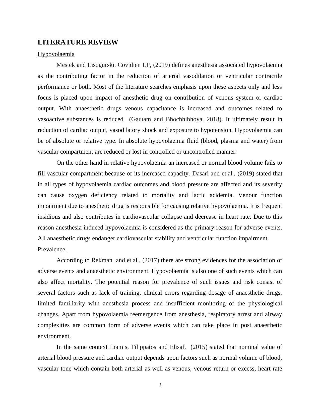
LITERATURE REVIEW
Hypovolaemia
Mestek and Lisogurski, Covidien LP, (2019) defines anesthesia associated hypovolaemia
as the contributing factor in the reduction of arterial vasodilation or ventricular contractile
performance or both. Most of the literature searches emphasis upon these aspects only and less
focus is placed upon impact of anesthetic drug on contribution of venous system or cardiac
output. With anaesthetic drugs venous capacitance is increased and outcomes related to
vasoactive substances is reduced (Gautam and Bhochhibhoya, 2018). It ultimately result in
reduction of cardiac output, vasodilatory shock and exposure to hypotension. Hypovolaemia can
be of absolute or relative type. In absolute hypovolaemia fluid (blood, plasma and water) from
vascular compartment are reduced or lost in controlled or uncontrolled manner.
On the other hand in relative hypovolaemia an increased or normal blood volume fails to
fill vascular compartment because of its increased capacity. Dasari and et.al., (2019) stated that
in all types of hypovolaemia cardiac outcomes and blood pressure are affected and its severity
can cause oxygen deficiency related to mortality and lactic acidemia. Venour function
impairment due to anesthetic drug is responsible for causing relative hypovolaemia. It is frequent
insidious and also contributes in cardiovascular collapse and decrease in heart rate. Due to this
reason anesthesia induced hypovolaemia is considered as the primary reason for adverse events.
All anaesthetic drugs endanger cardiovascular stability and ventricular function impairment.
Prevalence
According to Rekman and et.al., (2017) there are strong evidences for the association of
adverse events and anaesthetic environment. Hypovolaemia is also one of such events which can
also affect mortality. The potential reason for prevalence of such issues and risk consist of
several factors such as lack of training, clinical errors regarding dosage of anaesthetic drugs,
limited familiarity with anesthesia process and insufficient monitoring of the physiological
changes. Apart from hypovolaemia reemergence from anesthesia, respiratory arrest and airway
complexities are common form of adverse events which can take place in post anaesthetic
environment.
In the same context Liamis, Filippatos and Elisaf, (2015) stated that nominal value of
arterial blood pressure and cardiac output depends upon factors such as normal volume of blood,
vascular tone which contain both arterial as well as venous, venous return or excess, heart rate
2
Paraphrase This Document
Need a fresh take? Get an instant paraphrase of this document with our AI Paraphraser
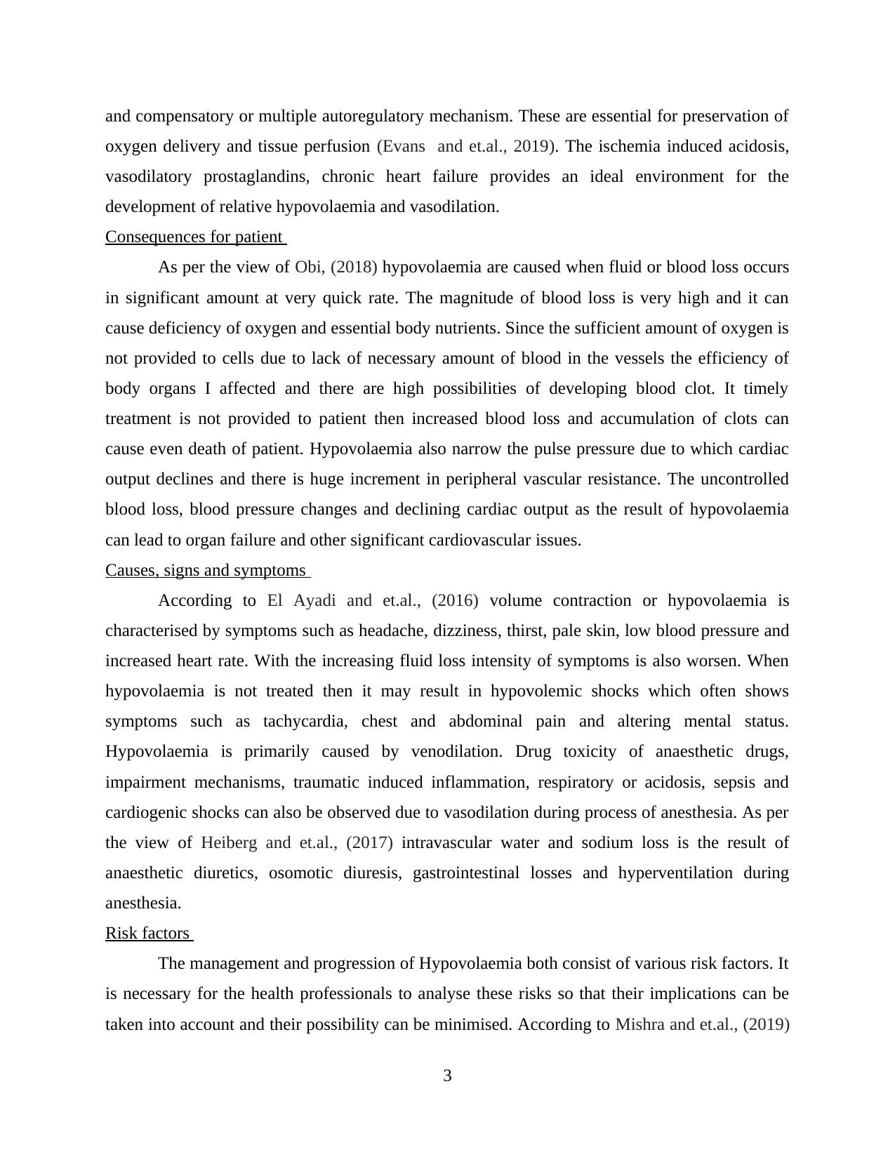
and compensatory or multiple autoregulatory mechanism. These are essential for preservation of
oxygen delivery and tissue perfusion (Evans and et.al., 2019). The ischemia induced acidosis,
vasodilatory prostaglandins, chronic heart failure provides an ideal environment for the
development of relative hypovolaemia and vasodilation.
Consequences for patient
As per the view of Obi, (2018) hypovolaemia are caused when fluid or blood loss occurs
in significant amount at very quick rate. The magnitude of blood loss is very high and it can
cause deficiency of oxygen and essential body nutrients. Since the sufficient amount of oxygen is
not provided to cells due to lack of necessary amount of blood in the vessels the efficiency of
body organs I affected and there are high possibilities of developing blood clot. It timely
treatment is not provided to patient then increased blood loss and accumulation of clots can
cause even death of patient. Hypovolaemia also narrow the pulse pressure due to which cardiac
output declines and there is huge increment in peripheral vascular resistance. The uncontrolled
blood loss, blood pressure changes and declining cardiac output as the result of hypovolaemia
can lead to organ failure and other significant cardiovascular issues.
Causes, signs and symptoms
According to El Ayadi and et.al., (2016) volume contraction or hypovolaemia is
characterised by symptoms such as headache, dizziness, thirst, pale skin, low blood pressure and
increased heart rate. With the increasing fluid loss intensity of symptoms is also worsen. When
hypovolaemia is not treated then it may result in hypovolemic shocks which often shows
symptoms such as tachycardia, chest and abdominal pain and altering mental status.
Hypovolaemia is primarily caused by venodilation. Drug toxicity of anaesthetic drugs,
impairment mechanisms, traumatic induced inflammation, respiratory or acidosis, sepsis and
cardiogenic shocks can also be observed due to vasodilation during process of anesthesia. As per
the view of Heiberg and et.al., (2017) intravascular water and sodium loss is the result of
anaesthetic diuretics, osomotic diuresis, gastrointestinal losses and hyperventilation during
anesthesia.
Risk factors
The management and progression of Hypovolaemia both consist of various risk factors. It
is necessary for the health professionals to analyse these risks so that their implications can be
taken into account and their possibility can be minimised. According to Mishra and et.al., (2019)
3
while making changes to position of patients for managing hypovolaemia care must be taken so
that it does not induce any complications (Tsetsou and et.al., 2017). Hypovolaemia lowers gut
perfusion and thus in positions such as trendelenburg reduced blood flow in abdominal area can
further increase aspiration, reflux and other possible risks related to gastric residuals. Hence in
cases when patient is able to receive enteral nutrition then tube feeding must be discontinue on
temporary basis.
Tánczos and et.al., (2015) stated that though I.V. Fluids are given for treatment of
hypovolaemia but these are also associated with the increased risk of heart failure. Nurses must
be careful that they perform a clear assessment of breathing sound, oxygenation, heart rate and
fluid volume so that this riks can be controlled. The events of low perfusion in the patients of
hypovolaemia is also one of the vulnerable factor for causing pressure ulcers. Though in
comparison to fluid resuscitation skin integrity is secondary priority but some of the
interventions can also be used to eliminate this risk (Rekman and et.al., 2017). For instance low
air loss mattress and restriction to use of hard surface for long period can minimise the pressure
on skin and thus risk of pressure ulcer can be diminished. There are several critical risk factors
related to hypovolaemia which can be life threatening for patients.
In the period when volume is not restored and bleeding is not controlled there are high
chances of developing coagulopathies. It can also worsen the resuscitation and acidosis. I.V.
Fluids are extensively used for the treatments. However if hypovolaemia is having extreme
negative impact upon acidosis then nurses must make interventions to increase the body
temperature of patient. As per the view of Lauronen and et.al., (2017) age factor is also one of
the risk factor which can affect the treatment outcomes. In certain cases even if patient is given
immediate treatment then due to higher age they may not respond quickly and thus it results in
their death.
While providing medications nurses must assure that some high and critical risk factors
does not affect add to progression of disease. When large bore I.V is used for the resuscitation
and management of body fluid then it can also cause systemic infection. Hence with
administration dosage and monitoring of vital symbols must be done on regular basis by nurses.
If blood transfusion is given to patients then it can also trigger the risk of transfusion reaction.
Stragier and et.al., (2019) stated that fluid therapy for treatment of relative hypovolaemia has
limited effectiveness thus it is recommended that for providing treatment for low blood flow
4
⊘ This is a preview!⊘
Do you want full access?
Subscribe today to unlock all pages.

Trusted by 1+ million students worldwide
anesthetic protocol must be adjusted and balanced crystalloid must be administered on the basis
of monitoring data and clinical symptoms. Vasoactive drugs such as norepinephrine for
vasodilation must also be regularly observed so that cardiac output can be retained within safe
ranges.
Management interventions
It may be challenging for anesthesiologists to identify and treat the tissue hypoperfusion
and hypovolaemia. The major concern for service providers is to sustain appropriate
management strategies and interventions so that hypovolaemia can be treated. According to
Pinsky, (2016) sophisticated assessment methods are effective in terms of analysing vascular
compliance but they cannot be applied to patients who receives vasoactive drugs. The nurses
must monitor hemodynamic and perfusion monitoring of cardiac parameters in regular intervals.
Due to decrease of circulating blood volume and fluid associated with anesthesia first line
therapies are proposed. The health outcomes in hypovolaemia can be improved significantly by
adopting early interventions. Suitable emphasis must also be given to the cause of hypovolaemia
shock and it must be ensured by nurses that airway, breathing and circulation is restored.
Gautam and Bhochhibhoya, (2018) stated that medications and interventions like IV line ,
fluid delivery by multiple lumen line, fluid administration by catheter along with IV medication
is very beneficial for managing the anesthesia induced hypovolaemia . Another popular
intervention used for the treatment is use of crystalloids and colloid infusion. Colloids are larger
molecules which gives higher extent of osmolarity as compare to crystalloids. Hence their
infusion play significant role in preventing fluid shift. Colloidal infusions stays within
intravascular compartment and retain a constant plasma colloid osmotic pressure. Nurses must
customise the intravenous therapies as per the requirement of each patient by taking in account
the consideration of associated risks and benefits for individual patient. Contrary to this Perner
and et.al., (2018) suggested that for electrolytic and fluid balance crystalloids are also suitable.
Crystalloids such as Ringer's Lactate and normal saline solution are smaller molecules than
colloids is used for patients which needs restoration of lost RBCs and for counteracting the
acidosis.
Mestek and Lisogurski, Covidien LP, (2019) elucidated that during surgery or situations
of trauma if blood losses are also occurred then PRBCs administration must be considered as
essential intervention. It will enhance the oxygen transportation capacity without increasing
5
Paraphrase This Document
Need a fresh take? Get an instant paraphrase of this document with our AI Paraphraser
volume. The use of frozen plasma is also validated in expansion of intravascular volume and in
restoring deficient clotting factors. For the treatment procedure platelet transfusion is also used if
the patient has thromocytopenia. In the same context it has been analysed by Kumar and et.al.,
(2016) that vasopressors usually results in the contraction of peripheral vasculature causing
blood shunting. However if they are used in small volumes they does not give satisfactory results
for improving the condition of patient. In cases when blood pressure of patients is not improved
by aggressive volume resuscitation then I.V. Fluids can be given with vasopressors. Intravenous
fluids (I.V.) are significantly helpful in compensation of blood volume lost in the bleeding.
However this intervention is used only up to a limited extent because these fluids are not able t o
transport oxygen in the same way as blood (Moulton and et.al., 2017). The colloid infusion
techniques is also preferred by health professionals for dilution of clotting factors present in the
blood which in turn increases bleeding risk.
Health care professionals also monitor and changes the position of patient. In
trendelenburg position blood is diverted from lower extremities to central circulation. Another
important treatment strategy for hypovolaemia shock is to control the bleeding. Since blood
supply reduces continuously health professionals must immediately provide emergency oxygen
to the patients so that oxygen deficiency can be avoided.
Implications for nurses and patients
For ensuring the safety of patient and to eliminate the risk factors of hypovolaemia nurses
must consider all necessary parameters such as cardiac fillings, adequate perfusion pressure,
micro-circulatory flow, and circulatory volume. Nurses and other health professionals use
macro-circulatory variable based perioperative monitoring which keeps relative hypovolaemia
undetected. The nurses must regularly monitor the factors which contributes the progression of
hypovolaemia. As per the view of Liamis, Filippatos and Elisaf, (2015) during monitoring
nurses must also consider factors such as available technology, status of patient and medical
history of the patient. The involvement of these factors will promote the quick recovery of
patient. Nurses must be aware of patient's medical history and requirements. There must be
effective communication and thorough examination of vital symptoms such as blood pressure,
pulse, urine output, and oxygenation.
6
According to Schiller and et.al., (2018) nurses are expected to provide ventilatory support
if there is need of tissue perfusion. For managing the blood pressure the position of patient can
also be adjusted. The flat or trendlenburg's position is effective in enhancing venous return from
legs. In trendelenburg position individual is supine and their head is tilted downward so that legs
and feet of patients are positioned above heart level. However nurses must adopt evidence based
approaches and associated risk factor with such positioning interventions. For instance with the
above discussed position of patient aspiration risk factors are also increased (Mestek and
Lisogurski, Covidien LP, 2019). The success and quality of health outcomes in the patients of
hypovolaemia can be increased by aggressive treatment strategies and prompt diagnosis. The
prognosis depends upon various factors such as degree and severity of hypovolaemia and how
early treatment is initiated.
CONCLUSION
It can be concluded that relative hypovolaemia caused by anaesthetic drugs can result in
tissue oxygen deficiency and thus has critical impact on mortality and morbidity. Thus during
anesthesia there is need of suitable diagnosis and clinical monitoring so that its risk can be
minimised. It has been also analysed that various approaches such as anesthetic plane,
therapeutic intervention like fluid administration and inotropic agents can be helpful in managing
deaths and other clinical complications related to hypovolaemia. It is also concluded that
effective care management strategies must be used by health professionals for providing safe
post anaesthetic environment.
7
⊘ This is a preview!⊘
Do you want full access?
Subscribe today to unlock all pages.

Trusted by 1+ million students worldwide
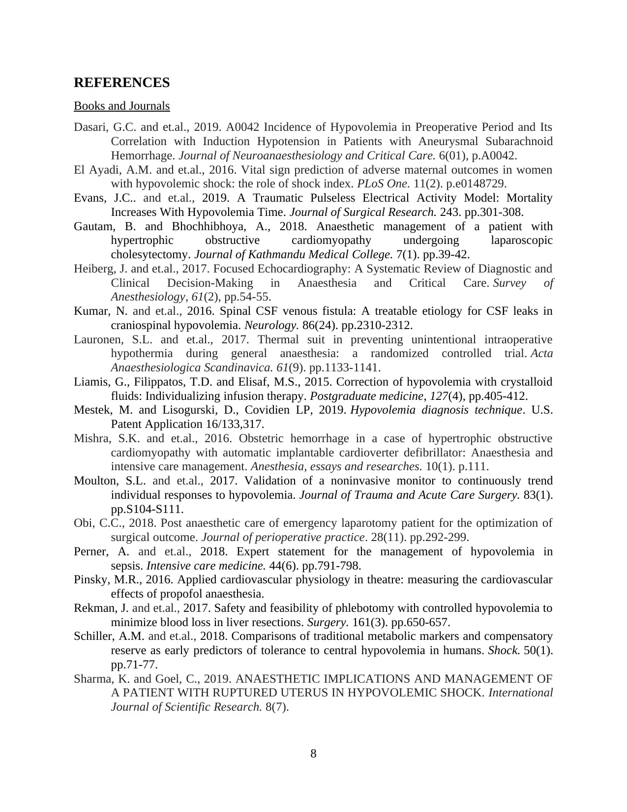
REFERENCES
Books and Journals
Dasari, G.C. and et.al., 2019. A0042 Incidence of Hypovolemia in Preoperative Period and Its
Correlation with Induction Hypotension in Patients with Aneurysmal Subarachnoid
Hemorrhage. Journal of Neuroanaesthesiology and Critical Care. 6(01), p.A0042.
El Ayadi, A.M. and et.al., 2016. Vital sign prediction of adverse maternal outcomes in women
with hypovolemic shock: the role of shock index. PLoS One. 11(2). p.e0148729.
Evans, J.C.. and et.al., 2019. A Traumatic Pulseless Electrical Activity Model: Mortality
Increases With Hypovolemia Time. Journal of Surgical Research. 243. pp.301-308.
Gautam, B. and Bhochhibhoya, A., 2018. Anaesthetic management of a patient with
hypertrophic obstructive cardiomyopathy undergoing laparoscopic
cholesytectomy. Journal of Kathmandu Medical College. 7(1). pp.39-42.
Heiberg, J. and et.al., 2017. Focused Echocardiography: A Systematic Review of Diagnostic and
Clinical Decision-Making in Anaesthesia and Critical Care. Survey of
Anesthesiology, 61(2), pp.54-55.
Kumar, N. and et.al., 2016. Spinal CSF venous fistula: A treatable etiology for CSF leaks in
craniospinal hypovolemia. Neurology. 86(24). pp.2310-2312.
Lauronen, S.L. and et.al., 2017. Thermal suit in preventing unintentional intraoperative
hypothermia during general anaesthesia: a randomized controlled trial. Acta
Anaesthesiologica Scandinavica. 61(9). pp.1133-1141.
Liamis, G., Filippatos, T.D. and Elisaf, M.S., 2015. Correction of hypovolemia with crystalloid
fluids: Individualizing infusion therapy. Postgraduate medicine, 127(4), pp.405-412.
Mestek, M. and Lisogurski, D., Covidien LP, 2019. Hypovolemia diagnosis technique. U.S.
Patent Application 16/133,317.
Mishra, S.K. and et.al., 2016. Obstetric hemorrhage in a case of hypertrophic obstructive
cardiomyopathy with automatic implantable cardioverter defibrillator: Anaesthesia and
intensive care management. Anesthesia, essays and researches. 10(1). p.111.
Moulton, S.L. and et.al., 2017. Validation of a noninvasive monitor to continuously trend
individual responses to hypovolemia. Journal of Trauma and Acute Care Surgery. 83(1).
pp.S104-S111.
Obi, C.C., 2018. Post anaesthetic care of emergency laparotomy patient for the optimization of
surgical outcome. Journal of perioperative practice. 28(11). pp.292-299.
Perner, A. and et.al., 2018. Expert statement for the management of hypovolemia in
sepsis. Intensive care medicine. 44(6). pp.791-798.
Pinsky, M.R., 2016. Applied cardiovascular physiology in theatre: measuring the cardiovascular
effects of propofol anaesthesia.
Rekman, J. and et.al., 2017. Safety and feasibility of phlebotomy with controlled hypovolemia to
minimize blood loss in liver resections. Surgery. 161(3). pp.650-657.
Schiller, A.M. and et.al., 2018. Comparisons of traditional metabolic markers and compensatory
reserve as early predictors of tolerance to central hypovolemia in humans. Shock. 50(1).
pp.71-77.
Sharma, K. and Goel, C., 2019. ANAESTHETIC IMPLICATIONS AND MANAGEMENT OF
A PATIENT WITH RUPTURED UTERUS IN HYPOVOLEMIC SHOCK. International
Journal of Scientific Research. 8(7).
8
Paraphrase This Document
Need a fresh take? Get an instant paraphrase of this document with our AI Paraphraser

Stragier, H. and et.al., 2019. General Anaesthesia versus Monitored Anaesthesia Care for
Transfemoral Transcatheter Aortic Valve Implantation. A retrospective study in a single
Belgian referral centre. Journal of Cardiothoracic and Vascular Anesthesia.
Tánczos, K. and et.al., 2015. Goal-directed resuscitation aiming cardiac index masks residual
hypovolemia: an animal experiment. BioMed research international, 2015.
Tsetsou, S. and et.al., 2017. Severe cerebral hypovolemia on perfusion CT and lower body
weight are associated with parenchymal haemorrhage after
thrombolysis. Neuroradiology. 59(1). pp.23-29.
9

10
⊘ This is a preview!⊘
Do you want full access?
Subscribe today to unlock all pages.

Trusted by 1+ million students worldwide
1 out of 15
Your All-in-One AI-Powered Toolkit for Academic Success.
+13062052269
info@desklib.com
Available 24*7 on WhatsApp / Email
![[object Object]](/_next/static/media/star-bottom.7253800d.svg)
Unlock your academic potential
Copyright © 2020–2025 A2Z Services. All Rights Reserved. Developed and managed by ZUCOL.