Identification of Unknown Bacterial Samples: Microbiology Practical
VerifiedAdded on 2023/04/19
|13
|2688
|454
Practical Assignment
AI Summary
This microbiology practical assignment details the identification of unknown bacterial samples using various techniques. The student performed gram staining to differentiate between gram-positive and gram-negative bacteria, followed by oxidase and catalase tests to further characterize the samples. The assignment also includes antibiotic susceptibility testing using the disk diffusion assay to determine the bacteria's resistance to different antibiotics. The results identified two colonies: one as Enterobacteriaceae and the other as Bacillus species. The practical highlights the methodologies, results, and discussion of the experiment, including the limitations and future implications of the study. The document provides detailed procedures, results tables, and figures to support the findings, making it a comprehensive resource for understanding bacterial identification techniques.
Running head: MICROBIOLOGY
Topic: MICROBIOLOGY
Name of the Student:
Name of the University:
Author’s Note:
Topic: MICROBIOLOGY
Name of the Student:
Name of the University:
Author’s Note:
Paraphrase This Document
Need a fresh take? Get an instant paraphrase of this document with our AI Paraphraser
1MICROBIOLOGY
Standard Operating Procedure (SOP):
Title: Identification of unknown bacterial samples through the procedure of gram staining
Standard operating procedure is defined as the set of stepwise instructions which are
compiled for proper carrying out the complex routine operations. The SOP is important as it
fruitfully explains the practices of the practices involved. They are needed for ultimately ensuring
the success of the practical.
The rationale of the experiment is the identification of the organisms from the unknown
samples containing the cultures 1 and 2. Identification is done on the basis of differentiation
between the gram positive and gram negative organism. The difference in the colors of the bacteria
in microscopy is on the basis of the stain retained through the various layers of the bacteria. There
is a distinct difference between the various composition of the cell walls of the gram negative as
well as gram positive bacteria. There is the presence of a thick layer of peptidoglycan which contain
numerous cross linking between the techoic acids. This results in the decolourization of the cell
wall. Among the aqueous solutions, there are various primary stains. In the given procedure of
gram staining, crystal violet (CV) is used. CV+ as well as Cl- ions are there which usually penetrate
through the walls and is present in the membrane of both gram positive as well as gram negative
bacteria. With addition of iodine, there is interaction of the CV+ with the various negatively
charged components present in the bacterial cells as a result of which the cells are stained with
purple dye (Gao et al. 2014).
There are four steps to be done regarding the procedures of gram staining. The first step
involves the application of a primary stain like the crystal violet which is done through heat fixation
of the bacterial culture forming a smear. It is followed by the addition of iodine, which usually
binds to the crystal violet and usually tarps in the cell. This is followed by decolourisation with
acetone as well as ethanol. Then the last procedure is counterstaining with saffranin. Sometime,
carbol fuschin is used in place of safranin as it stains more intensely than the anaerobic bacteria.
2MICROBIOLOGY
However, it is less commonly utilized a counter stain in the process of gram staining (Hall,
McGillicuddy and Kaplan 2014).
Thus, identification of the type of gram stain retained by the bacteria helps in determining
the gram character of the unknown samples. In the given experiment since the sample has taken up
the purple color it is confirmed that the unknown bacterial sample is gram positive in nature.
Flow chart of the methodologies used in the practical:
Day 1: Plate pouring
technique using BH11 and
CLED plates
Streak plating with the
quadrant method.
Day 2: Biochemical tests
are peroformed; Gram tests,
Oxidase and Catalase tests
Spread plating is done
followed by disk diffusion
assays
⊘ This is a preview!⊘
Do you want full access?
Subscribe today to unlock all pages.
Trusted by 1+ million students worldwide
3MICROBIOLOGY
Title: Identification of unknown bacterial samples through gram staining and catalase as well
as oxidase tests and identification of their resistance to antibiotics through antibiotic
susceptibility tests.
Abstract:
The following practical aims in the identification of bacteria on the basis of biochemical
tests. The processes done are biochemical tests like the catalase and oxidase test and lactose
fermentation tests, disk diffusion assays. It has been seen that the colony 1 is Enterobacteriaceae
and colony 2 is Bacillus species.
Introduction:
The aims of the given practical in the first day has been the successful preparation of the
BHI as well as CLED agar plates. The Day 2 practical would focus around the successful
performing of the oxidase, gram and the catalase tests. Thus the hypothesis of the experiment can
be formulated as the difference between the gram and the gram negative bacteria on the basis of
various biochemical tests like the oxidase and the catalase test. Gram staining is defined as the
procedure for the differentiation of the gram negative and positive organism on the basis of the
differences between their cell wall constituents (Burillo et al. 2014). The oxidase test is another
biochemical examination for the identification of organism on the basis of production of
cytochrome C oxidase (Ali et al. 2014). Moreover, the catalase tests would also be used for the
differentiation of the aerotoleratnt strains of Clostridium species mainly. Catalase is the enzyme
present in the bacteria which is used for converting hydrogen peroxide to water whereas Oxidase C
is usually a terminal enzyme which is usually used in the respiratory chain (Tulumoglu, Kaya and
Simsek 2014).
Paraphrase This Document
Need a fresh take? Get an instant paraphrase of this document with our AI Paraphraser

4MICROBIOLOGY
Materials and methods:
For the antibiotic assay, organisms need to be plated on the nutrient/BHI agar for the
antibiotic assay. 2 CLED plates would be done for the lactose determining ability for the bacteria.
Proper setting of the agar plates should be ensured by melting the gar and pouring them on the
plates at the correct temperature to prevent coagulation of the agar. Moreover, the pouring of the
agar and streaking should be done within the laminar air flow in an aseptic manner to prevent
contamination. This would be followed by a successful plating of the organisms for the production
of single colonies of the bacteria used using the streak plate method. Streak plating would involve
the proper labelling of the plates and streaking on the plate after heat sterilization of the inoculum
loop. After heat sterilization a loopful of inoculum would be taken and then streaked using the
quadrant streaking method. For the day 2 biochemical tests involved, gram staining methods along
with oxidase as well as catalase test would be done by inoculation of 1.5 mL of sterile sample. The
absorbance of the sample would be measured at 0.01 nm. Then the sterile swab would be dipped
into the centrifuge tube and spread over the indicated agar plate containing PBS. The experiments
would also involve performing an antimicrobial disk diffusion assay. 4-5 antibiotic disks would be
used which would be spread in the pate at equal distances and then embedded gently and the plates
should be turned down during incubation at room temperature. This would be followed by
identification of the mixture of samples.
5MICROBIOLOGY
Results:
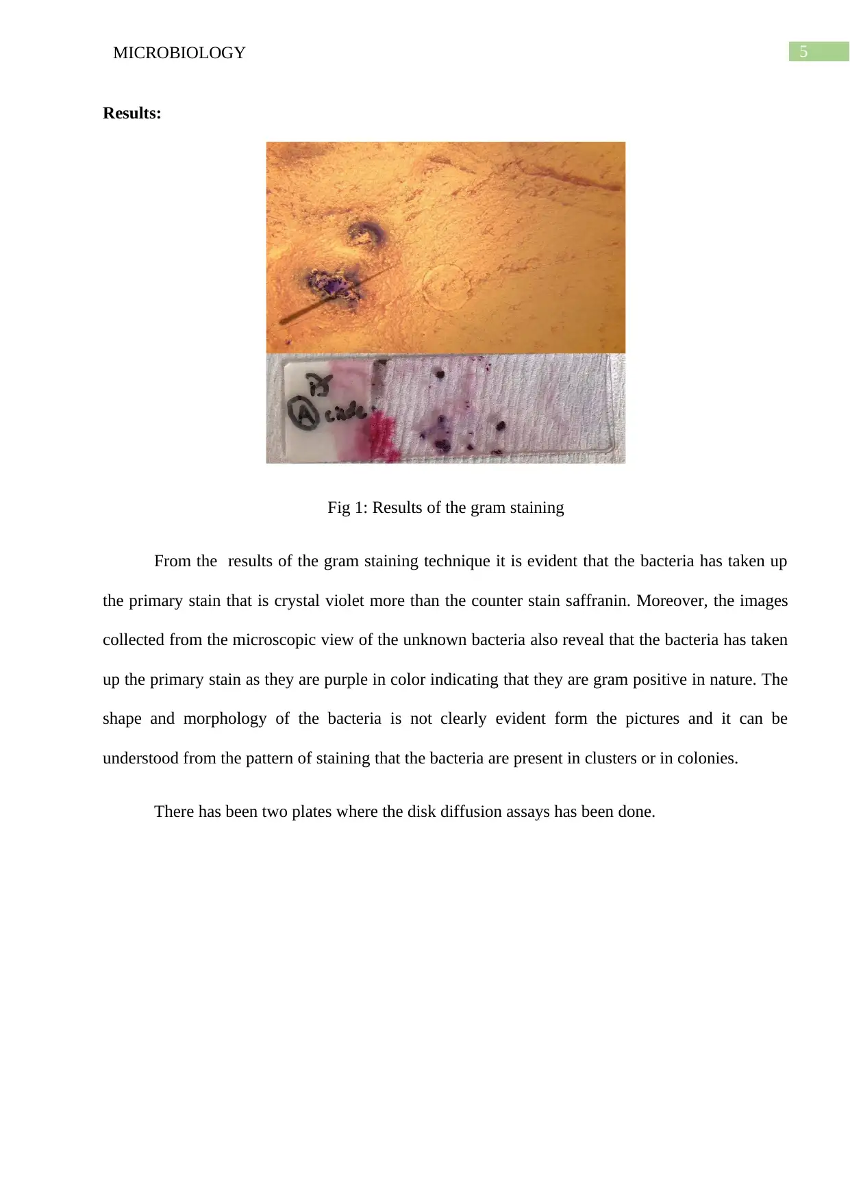
Fig 1: Results of the gram staining
From the results of the gram staining technique it is evident that the bacteria has taken up
the primary stain that is crystal violet more than the counter stain saffranin. Moreover, the images
collected from the microscopic view of the unknown bacteria also reveal that the bacteria has taken
up the primary stain as they are purple in color indicating that they are gram positive in nature. The
shape and morphology of the bacteria is not clearly evident form the pictures and it can be
understood from the pattern of staining that the bacteria are present in clusters or in colonies.
There has been two plates where the disk diffusion assays has been done.
Results:
Fig 1: Results of the gram staining
From the results of the gram staining technique it is evident that the bacteria has taken up
the primary stain that is crystal violet more than the counter stain saffranin. Moreover, the images
collected from the microscopic view of the unknown bacteria also reveal that the bacteria has taken
up the primary stain as they are purple in color indicating that they are gram positive in nature. The
shape and morphology of the bacteria is not clearly evident form the pictures and it can be
understood from the pattern of staining that the bacteria are present in clusters or in colonies.
There has been two plates where the disk diffusion assays has been done.
⊘ This is a preview!⊘
Do you want full access?
Subscribe today to unlock all pages.
Trusted by 1+ million students worldwide
6MICROBIOLOGY
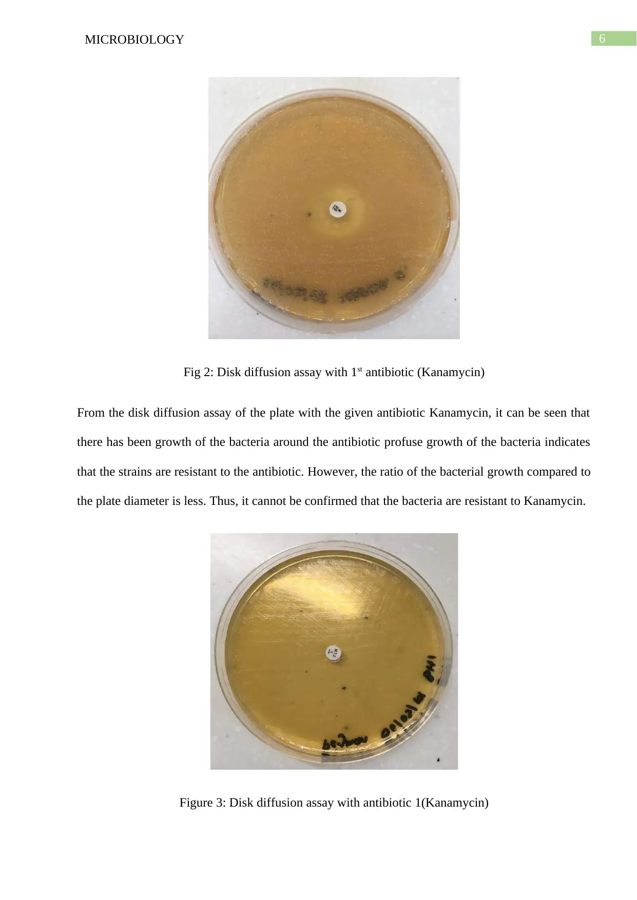
Fig 2: Disk diffusion assay with 1st antibiotic (Kanamycin)
From the disk diffusion assay of the plate with the given antibiotic Kanamycin, it can be seen that
there has been growth of the bacteria around the antibiotic profuse growth of the bacteria indicates
that the strains are resistant to the antibiotic. However, the ratio of the bacterial growth compared to
the plate diameter is less. Thus, it cannot be confirmed that the bacteria are resistant to Kanamycin.
Figure 3: Disk diffusion assay with antibiotic 1(Kanamycin)
Paraphrase This Document
Need a fresh take? Get an instant paraphrase of this document with our AI Paraphraser
7MICROBIOLOGY
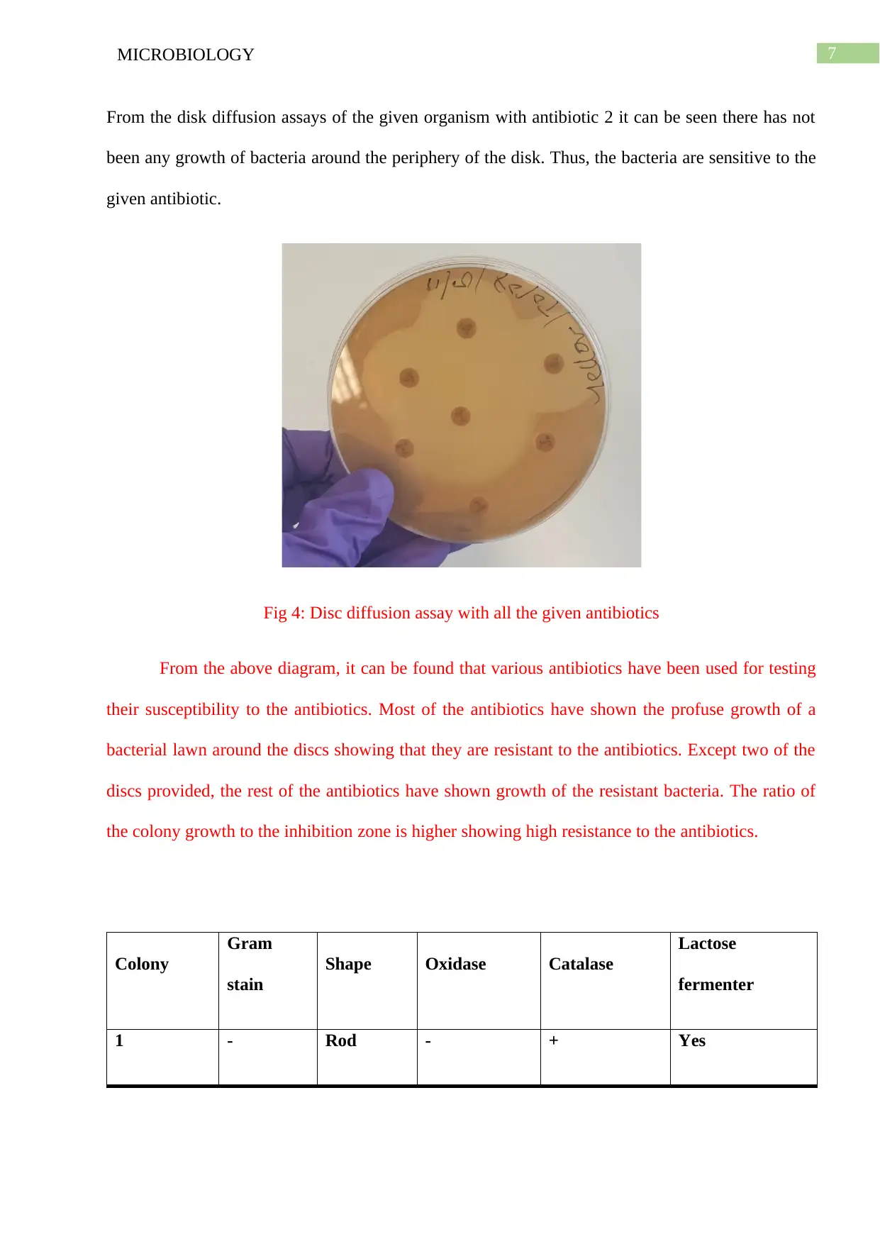
From the disk diffusion assays of the given organism with antibiotic 2 it can be seen there has not
been any growth of bacteria around the periphery of the disk. Thus, the bacteria are sensitive to the
given antibiotic.
Fig 4: Disc diffusion assay with all the given antibiotics
From the above diagram, it can be found that various antibiotics have been used for testing
their susceptibility to the antibiotics. Most of the antibiotics have shown the profuse growth of a
bacterial lawn around the discs showing that they are resistant to the antibiotics. Except two of the
discs provided, the rest of the antibiotics have shown growth of the resistant bacteria. The ratio of
the colony growth to the inhibition zone is higher showing high resistance to the antibiotics.
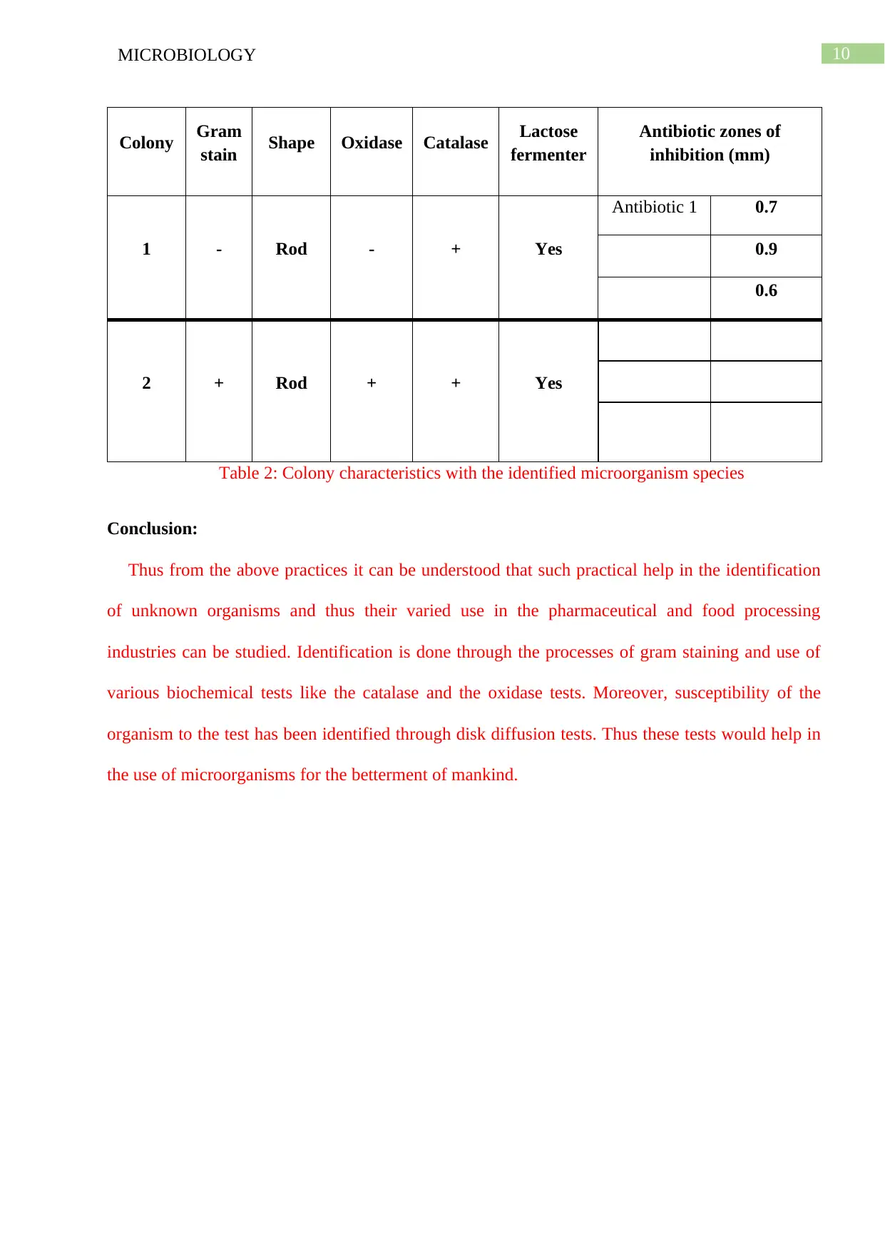
Colony
Gram
stain
Shape Oxidase Catalase
Lactose
fermenter
1 - Rod - + Yes
From the disk diffusion assays of the given organism with antibiotic 2 it can be seen there has not
been any growth of bacteria around the periphery of the disk. Thus, the bacteria are sensitive to the
given antibiotic.
Fig 4: Disc diffusion assay with all the given antibiotics
From the above diagram, it can be found that various antibiotics have been used for testing
their susceptibility to the antibiotics. Most of the antibiotics have shown the profuse growth of a
bacterial lawn around the discs showing that they are resistant to the antibiotics. Except two of the
discs provided, the rest of the antibiotics have shown growth of the resistant bacteria. The ratio of
the colony growth to the inhibition zone is higher showing high resistance to the antibiotics.
Colony
Gram
stain
Shape Oxidase Catalase
Lactose
fermenter
1 - Rod - + Yes
8MICROBIOLOGY
2 + Rod + + Yes
Table 1: Colony characteristics of the unknown sample
From the examination of the biochemical tests of the two colonies it can be seen that the 1st
colony has not taken up the gram staining. However, the morphology has been found to be rod
shaped. The catalase tests and the oxidase tests has been contradictory for the colony 1 bacteria as
they has not given any results for the oxidase test whereas it has been positive for the catalase test.
The species correlating with the above characteristics is Enterobacteria as it the only gram negative
rod capable of fermenting lactose and giving the required oxidase and catalase tests. From the
results of the 2nd colony it can be seen that the bacterial colonies has taken up the gram stain and are
rod shaped according to the morphology considered. The samples has given positive results for the
catalase and the oxidase tests which again aligns with the characteristics of Bacillus species in
terms of morphology and the kind of biochemical tests done. Moreover both the colonies has the
ability to ferment lactose.
Discussion:
The results of the above experiments has shown the presence of Gram positive and gram
negative species like Enterobacteriaceae and Bacillus species. The catalase tests has been positive
showing the presence of the enzyme catalase in bacteria. Similarly, negative catalase test indicate
⊘ This is a preview!⊘
Do you want full access?
Subscribe today to unlock all pages.
Trusted by 1+ million students worldwide
9MICROBIOLOGY
the absence of the bacteria to produce catalase, there is no bubble formation as the hydrogen
peroxide is not broken down (Chah et al. 2014). From the results of the staining procedure, it can be
seen that the bacteria has taken up the crystal violet. The color is retained in gram positive bacteria
due to the various layers of peptidoglycan which trap the stain between the layers and thus the
bacteria purple color in the microscope (Jiang et al. 2015). The disk diffusion assays are based on
the rates of growth of the bacteria around the antibiotic disk. The rates of susceptibility of the
bacteria are present on the diameter of the inhibition zone formed by the colonies (Gupta et al.
2015). Here it can be seen that there has not been any distinguished zone around the bacteria
claiming that they are resistant and their susceptibility is very low. Oxidase negative bacteria is an
indication of anaerobic bacteria as there is no oxidation by the test reagent. Regarding the clinical
implications, bacillus species are used in various agricultural, industrial processes for their special
ability of producing a vast range of antibiotics and metabolites. Similarly, Enterobacter species are
used for their mobility as well as the ability of fixation of various resistant genes. The limitations
involved in the study is the lack of appropriate lab apparatus for carrying out the experiments.
Moreover contamination has occurred while performing the experiment which should be performed
under a laminar air flow to reduce possible sources of contamination. The significance of this
experiment is to identify the microorganism which has a varied use in the medical industry. Thus,
exploitation of these microorganism would be beneficial for industrial uses especially in the
pharmaceutical industry. Future implications of this experiment include use of the techniques like
16srRNA for the exact identification of the unknown organisms and MALDI-TOF procedures for
characterization on the basis of their molecular mass.
Paraphrase This Document
Need a fresh take? Get an instant paraphrase of this document with our AI Paraphraser
10MICROBIOLOGY
Colony Gram
stain Shape Oxidase Catalase Lactose
fermenter
Antibiotic zones of
inhibition (mm)
1 - Rod - + Yes
Antibiotic 1 0.7
0.9
0.6
2 + Rod + + Yes
Table 2: Colony characteristics with the identified microorganism species
Conclusion:
Thus from the above practices it can be understood that such practical help in the identification
of unknown organisms and thus their varied use in the pharmaceutical and food processing
industries can be studied. Identification is done through the processes of gram staining and use of
various biochemical tests like the catalase and the oxidase tests. Moreover, susceptibility of the
organism to the test has been identified through disk diffusion tests. Thus these tests would help in
the use of microorganisms for the betterment of mankind.
11MICROBIOLOGY
References
Ali, S., Ali, Q., Melzer, F., Khan, I., Akhter, S., Neubauer, H. and Jamal, S.M., 2014. Isolation and
identification of bovine Brucella isolates from Pakistan by biochemical tests and PCR. Tropical
animal health and production, 46(1), pp.73-78.
Burillo, A., Rodríguez-Sánchez, B., Ramiro, A., Cercenado, E., Rodríguez-Créixems, M. and
Bouza, E., 2014. Gram-stain plus MALDI-TOF MS (matrix-assisted laser desorption ionization-
time of flight mass spectrometry) for a rapid diagnosis of urinary tract infection. PloS one, 9(1),
p.e86915.
Chah, K.F., Gómez-Sanz, E., Nwanta, J.A., Asadu, B., Agbo, I.C., Lozano, C., Zarazaga, M. and
Torres, C., 2014. Methicillin-resistant coagulase-negative staphylococci from healthy dogs in
Nsukka, Nigeria. Brazilian Journal of Microbiology, 45(1), pp.215-220.
Gao, S., Lewis, G. D., Ashokkumar, M., & Hemar, Y. (2014). Inactivation of microorganisms by
low-frequency high-power ultrasound: 2. A simple model for the inactivation
mechanism. Ultrasonics sonochemistry, 21(1), 454-460.
Gupta, P., Khare, V., Kumar, D., Ahmad, A., Banerjee, G. and Singh, M., 2015. Comparative
evaluation of disc diffusion and E-test with broth micro-dilution in susceptibility testing of
amphotericin B, voriconazole and caspofungin against clinical Aspergillus isolates. Journal of
clinical and diagnostic research: JCDR, 9(1), p.DC04.
Hall, M. R., McGillicuddy, E., & Kaplan, L. J. (2014). Biofilm: basic principles, pathophysiology,
and implications for clinicians. Surgical Infections, 15(1), 1-7.
Jiang, C.H., Wu, F., Yu, Z.Y., Xie, P., Ke, H.J., Li, H.W., Yu, Y.Y. and Guo, J.H., 2015. Study on
screening and antagonistic mechanisms of Bacillus amyloliquefaciens 54 against bacterial fruit
⊘ This is a preview!⊘
Do you want full access?
Subscribe today to unlock all pages.
Trusted by 1+ million students worldwide
1 out of 13
Related Documents


Your All-in-One AI-Powered Toolkit for Academic Success.
+13062052269
info@desklib.com
Available 24*7 on WhatsApp / Email
![[object Object]](/_next/static/media/star-bottom.7253800d.svg)
Unlock your academic potential
Copyright © 2020–2026 A2Z Services. All Rights Reserved. Developed and managed by ZUCOL.