Diagnostic Accuracy of Imaging Skeletal Metastases in Neuroblastoma
VerifiedAdded on 2022/11/15
|23
|9507
|453
Report
AI Summary
This report provides a detailed overview of neuroblastoma, a cancer affecting children, focusing on skeletal metastases. It discusses the disease's causes, symptoms, and risk factors, as well as various treatment options like surgery, chemotherapy, and immunotherapy. The report emphasizes the importance of early detection of bone metastases for effective treatment and accurate staging. It explores different imaging modalities, including bone scans, MRI, PET scans, and CT scans, highlighting their roles in diagnosing and monitoring the condition. The primary objective of this systematic review is to identify all published definitions of skeletal metastases on PET imaging; and also determine the diagnostic accuracy of detecting bone metastases, which is published on Nuclear Medicine scans. The report also addresses the limitations of conventional imaging techniques and the benefits of functional imaging modalities like MIBG scintiscans and PET-CT scans. The report aims to evaluate the most recent papers that have used PET scan with NaF and FDG tracers and Tc-99m MDP bone scan, which will evaluate the skeletal metastases of neuroblastoma using a systematic review of the literature and data meta-analysis.
Chapter 1: Introduction
Bone metastases are also known as “Bone Mets” that occurs when the cancerous cells
relocate to the bone from the area of primary tumour formation. Neuroblastoma is
considered one of the most common form of cancer that develops from the undeveloped
nerve cells located in different parts of the human body (Macedo et al. 2017).
Neuroblastoma generally arises nearby the adrenal gland that has the same nerve cell
origin. The diagnosis of the disease is done by the neurological examinations that includes
scanning of the spinal cord, nervous tissue and brain. The examination can be performed by
using different methods like CT scan, MRI, X-rays, MIBG and PET scan
(Stanfordchildrens.org 2019).
Neuroblastoma is also known as the “sympathetic nervous system” that is a rare cancer
condition affecting children. This cancer is considered very dangerous because it affects the
sympathetic nervous system, which is responsible for transporting the message from the
brain of an individual to the other parts of their body (Louis and Shohet 2015). The condition
of neuroblastoma arises due to a mutation that affects the undeveloped nerve cells of the
foetus within the womb. The cells are further known as neuroblast, and as the child develops
with age, the neuroblast cells also develop and affect the normal functioning of their body. It
is very important to identify if the child has neuroblastoma or not because as the child can
develop neuroblastoma before their birth. Therefore, it will affect the development and
growth of bone among the infants and further result in cancer (Van et al. 2017)
Bone metastases are considered very common for the patient who is suffering from
advanced solid tumours. Early detection or identification of the bone metastases is crucial for
optimal treatment and accurate staging. Effective monitoring of bone metastases will be
crucial for detection, treatment planning, diagnosis and follow-up of the health condition
(Langsteger et al. 2016). The different modalities used for detecting bone metastases
include bone scan, magnetic resonance imaging (MRI), positron emission tomography (PET)
scan and computed tomography (CT) scan. The systematic review method will help in
identifying which modality is best for detecting the presence of bone metastases by
summarising the result available from the trails associated with an extremely high level of
evidence (Santoni et al. 2015). There is a need to do the systemic review and meta-analysis
of the different scanning techniques that are available for the neurological diagnosis of
neuroblastoma in young children.
Paraphrase This Document
Need a fresh take? Get an instant paraphrase of this document with our AI Paraphraser
Hence, the primary objective of this systematic review was to identify all published definitions
of skeletal metastases on PET imaging; and also determine the diagnostic accuracy of
detecting bone metastases, which is published on Nuclear Medicine scans.
The review attempts to evaluate the most recent papers that have used PET scan with NaF
and FDG tracers and Tc-99m MDP bone scan, which will evaluate the skeletal metastases
of neuroblastoma using a systematic review of the literature and data meta‐analysis.
1.1 Thesis outline
In this study, a detailed description is provided on the disease condition of
neuroblastoma. Neuroblastoma is considered one of the most common form of cancer
that develops from the undeveloped nerve cells located in different parts of the human
body. Neuroblastoma generally arises nearby the adrenal gland that has the same nerve
cell origin. This report is also includes strategies that will assist in detecting the presence
of tumour, like PET scan and bone scintigraphy (Luksch et al. 2016). Both the detection
process are included under nuclear methods as the element which are used to trace the
location of tumour are radioactive in nature. In the report the statistics about the
detection of cancer is also briefly explained. Hence, complete report is prepared by
including the analysis obtained from the journal articles which were prepared by the
experts, and the data and information present in the journal articles regarding this
disease and methods to diagnose is also mentioned.
Chapter 2: Background
2.1 Target condition being diagnosed
Neuroblastoma is a cancer which usually occurs in the children of age group 5 years or less,
affecting mostly the adrenal gland. This cancer usually occurs during the initial stage of
nerve cells formation, it also founds in embryo or foetus. In neuroblastoma the term neuro
refers to nerve and blastoma refers to cancer which affects the developing cells (Chen et al
2019). The disorder mainly occurs in sympathetic nerve ganglia which are located close to
the spine or neck. In very rare occasion, the neuroblastoma spread in wide range, which is
not recognized effectively by the experts and they fail to appropriately examine the origin
location of this cancer.
2.1.1 Symptoms of neuroblastoma
Symptoms of neuroblastoma totally depends on the areas being affected due to cancer.
Some of the symptoms of neuroblastoma are included below:
1
1. Neuroblastoma in abdomen: Abdominal neuroblastoma is the most common form of
this disorder, and the symptoms observed in this type of Neuroblastoma are included
below:
• Abdominal pain
• Diarrhoea or constipation
• The mass present under skin is not soft when it is touched.
2. Neuroblastoma in chest: The symptoms of this form of neuroblastoma are as follows:
• Wheezing
• Pain in chest
• Unequal size of Pupil, or dropping of eyelids
3. Other associated symptoms which can be observed in neuroblastoma are as follows:
• Clustering of tissue under skin
• Dark circles
• Back pain
• Fever
• Loss of weight
• Pain in bone (Neviani et al. 2019).
2.1.2 Cause of neuroblastoma
Generally, cancer is primarily caused due to mutation affecting the cells that grow and
develop normally. The cancerous cells get multiplied and grow continuously, thereby
resulting in tumour formation within the human body. The condition of neuroblastoma arises
due to a mutation that affects the undeveloped nerve cells of the foetus within the womb.
The cells are further known as neuroblast, and as the child develops with age, the
neuroblast cells also develop and affect the normal functioning of their body. This disorder
arises in immature cells and affect the development of the foetus (Hwang, and Perry, 2019).
2.1.3 Risk Factors
After the detection of cancer, the risk level is classified as Low, medium or high risks. These
risk levels are determined after analysing following factors as mentioned below:
2
⊘ This is a preview!⊘
Do you want full access?
Subscribe today to unlock all pages.
Trusted by 1+ million students worldwide

• Stage of Cancer
• Histology of the Tumour
• Biology of Tumour ‘
• Age of the patient (child) (Nguyen, and Dyer, 2019)
2.1.4 Treatment of Neuroblastoma
Treatment of Neuroblastoma primarily depend upon the age of child and stage of the cancer.
The different types of treatment used for preventing the condition of neuroblastoma are
included as follows:
i. Surgery
ii. Chemotherapy
iii. Radiation Therapy
iv. Immunotherapy
v. Transplant of Stem cell
Chemotherapy: In this type of therapy, anti-cancer drugs are provided to the patient, which
starts killing or destroying the cancerous cells. These drugs are administrated either orally or
through injection as well. There are few side-effects of using this therapy that go away once
the treatments get finished. The side-effects included are as follows: (Ocak, et al. 2019).
• Loss of Hair
• Mouth Sores
• Nausea
• Vomiting
• Immune System will become weak
• Fatigue
Radiation Therapy: In this type of treatment the infected areas are targeted using X-rays,
which are aimed to kill the infected or cancerous cells. This treatment causes many side-
effect which are related to skin i.e. skin irritation diarrhoea, fatigue etc.
Immunotherapy: It is a biological therapy which is used to improve the immune system of
the body in order to fight with disease.
3
Paraphrase This Document
Need a fresh take? Get an instant paraphrase of this document with our AI Paraphraser
Stem Cell transplantation: It is also known as transplantation of bone marrow, after the
treatment of radiation and chemotherapy a replacement of stem cell is injected in the body
directly into the bloodstream of the children (Rodrigo et al. 2019).
2.2Skeletal Metastases for neuroblastoma
Neuroblastoma is defined as the health condition, which develops in the immature nerve
cells and then spreads all over the body. Research studies suggest that the tumour grows
rapidly and metastasizes within the human body that can be fatal for the patient. In addition
to this, it should also be noted that an average span of the patient after the diagnosis is less
than six months. The tumour is said to be neurogenic in origin, because the originating cells
can be traced to the primitive neural crest of the ectoderm. The tumour generally originates
from the medulla of the adrenal gland which then proceeds to the other parts of the body.
The original tumour is derived from the neural crest. Research studies suggest that
neuroblastoma cancer cells possess the ability to metastasize and spread to different parts
of the body which includes the areas of lymph nodes, lungs, bones, liver, central nervous
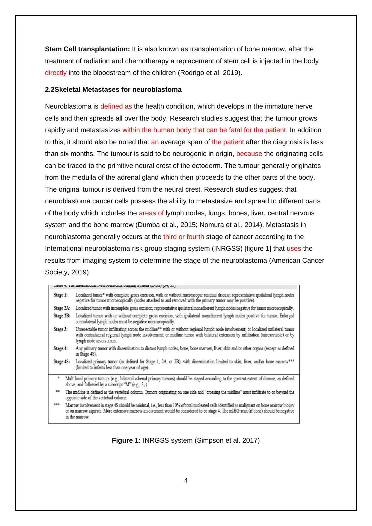
system and the bone marrow (Dumba et al., 2015; Nomura et al., 2014). Metastasis in
neuroblastoma generally occurs at the third or fourth stage of cancer according to the
International neuroblastoma risk group staging system (INRGSS) [figure 1] that uses the
results from imaging system to determine the stage of the neuroblastoma (American Cancer
Society, 2019).
Figure 1: INRGSS system (Simpson et al. 2017)
4
treatment of radiation and chemotherapy a replacement of stem cell is injected in the body
directly into the bloodstream of the children (Rodrigo et al. 2019).
2.2Skeletal Metastases for neuroblastoma
Neuroblastoma is defined as the health condition, which develops in the immature nerve
cells and then spreads all over the body. Research studies suggest that the tumour grows
rapidly and metastasizes within the human body that can be fatal for the patient. In addition
to this, it should also be noted that an average span of the patient after the diagnosis is less
than six months. The tumour is said to be neurogenic in origin, because the originating cells
can be traced to the primitive neural crest of the ectoderm. The tumour generally originates
from the medulla of the adrenal gland which then proceeds to the other parts of the body.
The original tumour is derived from the neural crest. Research studies suggest that
neuroblastoma cancer cells possess the ability to metastasize and spread to different parts
of the body which includes the areas of lymph nodes, lungs, bones, liver, central nervous
system and the bone marrow (Dumba et al., 2015; Nomura et al., 2014). Metastasis in
neuroblastoma generally occurs at the third or fourth stage of cancer according to the
International neuroblastoma risk group staging system (INRGSS) [figure 1] that uses the
results from imaging system to determine the stage of the neuroblastoma (American Cancer
Society, 2019).
Figure 1: INRGSS system (Simpson et al. 2017)
4
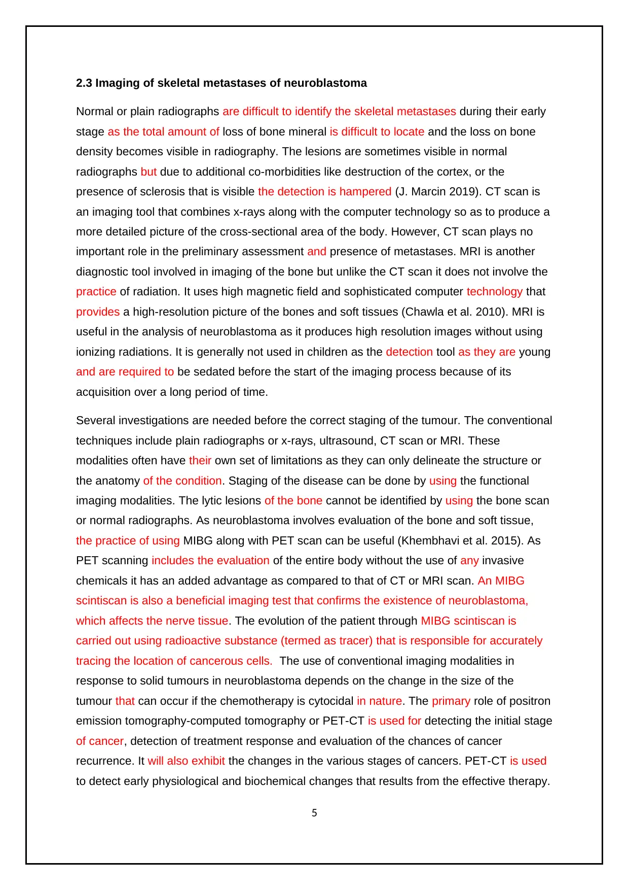
2.3 Imaging of skeletal metastases of neuroblastoma
Normal or plain radiographs are difficult to identify the skeletal metastases during their early
stage as the total amount of loss of bone mineral is difficult to locate and the loss on bone
density becomes visible in radiography. The lesions are sometimes visible in normal
radiographs but due to additional co-morbidities like destruction of the cortex, or the
presence of sclerosis that is visible the detection is hampered (J. Marcin 2019). CT scan is
an imaging tool that combines x-rays along with the computer technology so as to produce a
more detailed picture of the cross-sectional area of the body. However, CT scan plays no
important role in the preliminary assessment and presence of metastases. MRI is another
diagnostic tool involved in imaging of the bone but unlike the CT scan it does not involve the
practice of radiation. It uses high magnetic field and sophisticated computer technology that
provides a high-resolution picture of the bones and soft tissues (Chawla et al. 2010). MRI is
useful in the analysis of neuroblastoma as it produces high resolution images without using
ionizing radiations. It is generally not used in children as the detection tool as they are young
and are required to be sedated before the start of the imaging process because of its
acquisition over a long period of time.
Several investigations are needed before the correct staging of the tumour. The conventional
techniques include plain radiographs or x-rays, ultrasound, CT scan or MRI. These
modalities often have their own set of limitations as they can only delineate the structure or
the anatomy of the condition. Staging of the disease can be done by using the functional
imaging modalities. The lytic lesions of the bone cannot be identified by using the bone scan
or normal radiographs. As neuroblastoma involves evaluation of the bone and soft tissue,
the practice of using MIBG along with PET scan can be useful (Khembhavi et al. 2015). As
PET scanning includes the evaluation of the entire body without the use of any invasive
chemicals it has an added advantage as compared to that of CT or MRI scan. An MIBG
scintiscan is also a beneficial imaging test that confirms the existence of neuroblastoma,
which affects the nerve tissue. The evolution of the patient through MIBG scintiscan is
carried out using radioactive substance (termed as tracer) that is responsible for accurately
tracing the location of cancerous cells. The use of conventional imaging modalities in
response to solid tumours in neuroblastoma depends on the change in the size of the
tumour that can occur if the chemotherapy is cytocidal in nature. The primary role of positron
emission tomography-computed tomography or PET-CT is used for detecting the initial stage
of cancer, detection of treatment response and evaluation of the chances of cancer
recurrence. It will also exhibit the changes in the various stages of cancers. PET-CT is used
to detect early physiological and biochemical changes that results from the effective therapy.
5
⊘ This is a preview!⊘
Do you want full access?
Subscribe today to unlock all pages.
Trusted by 1+ million students worldwide
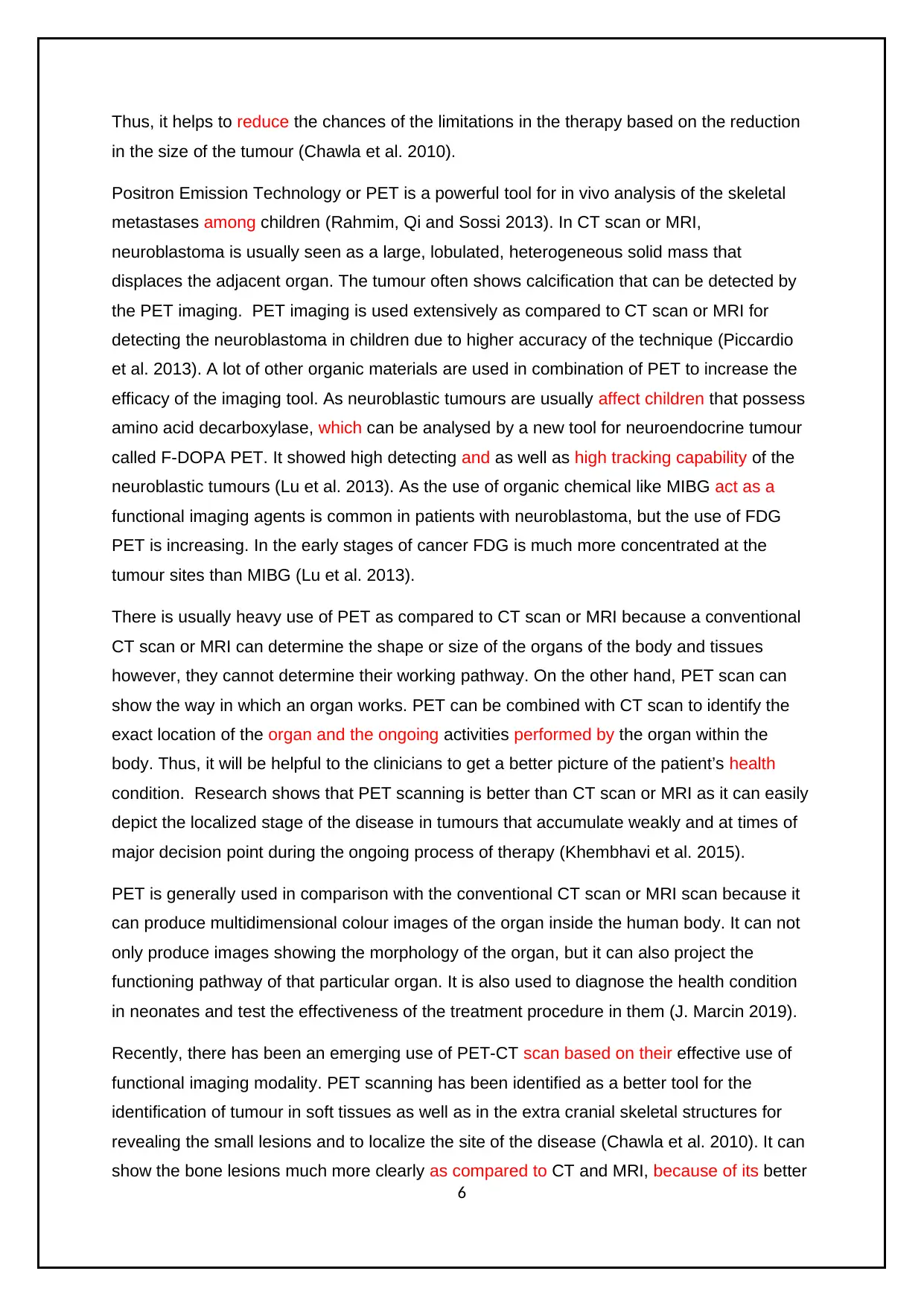
Thus, it helps to reduce the chances of the limitations in the therapy based on the reduction
in the size of the tumour (Chawla et al. 2010).
Positron Emission Technology or PET is a powerful tool for in vivo analysis of the skeletal
metastases among children (Rahmim, Qi and Sossi 2013). In CT scan or MRI,
neuroblastoma is usually seen as a large, lobulated, heterogeneous solid mass that
displaces the adjacent organ. The tumour often shows calcification that can be detected by
the PET imaging. PET imaging is used extensively as compared to CT scan or MRI for
detecting the neuroblastoma in children due to higher accuracy of the technique (Piccardio
et al. 2013). A lot of other organic materials are used in combination of PET to increase the
efficacy of the imaging tool. As neuroblastic tumours are usually affect children that possess
amino acid decarboxylase, which can be analysed by a new tool for neuroendocrine tumour
called F-DOPA PET. It showed high detecting and as well as high tracking capability of the
neuroblastic tumours (Lu et al. 2013). As the use of organic chemical like MIBG act as a
functional imaging agents is common in patients with neuroblastoma, but the use of FDG
PET is increasing. In the early stages of cancer FDG is much more concentrated at the
tumour sites than MIBG (Lu et al. 2013).
There is usually heavy use of PET as compared to CT scan or MRI because a conventional
CT scan or MRI can determine the shape or size of the organs of the body and tissues
however, they cannot determine their working pathway. On the other hand, PET scan can
show the way in which an organ works. PET can be combined with CT scan to identify the
exact location of the organ and the ongoing activities performed by the organ within the
body. Thus, it will be helpful to the clinicians to get a better picture of the patient’s health
condition. Research shows that PET scanning is better than CT scan or MRI as it can easily
depict the localized stage of the disease in tumours that accumulate weakly and at times of
major decision point during the ongoing process of therapy (Khembhavi et al. 2015).
PET is generally used in comparison with the conventional CT scan or MRI scan because it
can produce multidimensional colour images of the organ inside the human body. It can not
only produce images showing the morphology of the organ, but it can also project the
functioning pathway of that particular organ. It is also used to diagnose the health condition
in neonates and test the effectiveness of the treatment procedure in them (J. Marcin 2019).
Recently, there has been an emerging use of PET-CT scan based on their effective use of
functional imaging modality. PET scanning has been identified as a better tool for the
identification of tumour in soft tissues as well as in the extra cranial skeletal structures for
revealing the small lesions and to localize the site of the disease (Chawla et al. 2010). It can
show the bone lesions much more clearly as compared to CT and MRI, because of its better
6
Paraphrase This Document
Need a fresh take? Get an instant paraphrase of this document with our AI Paraphraser
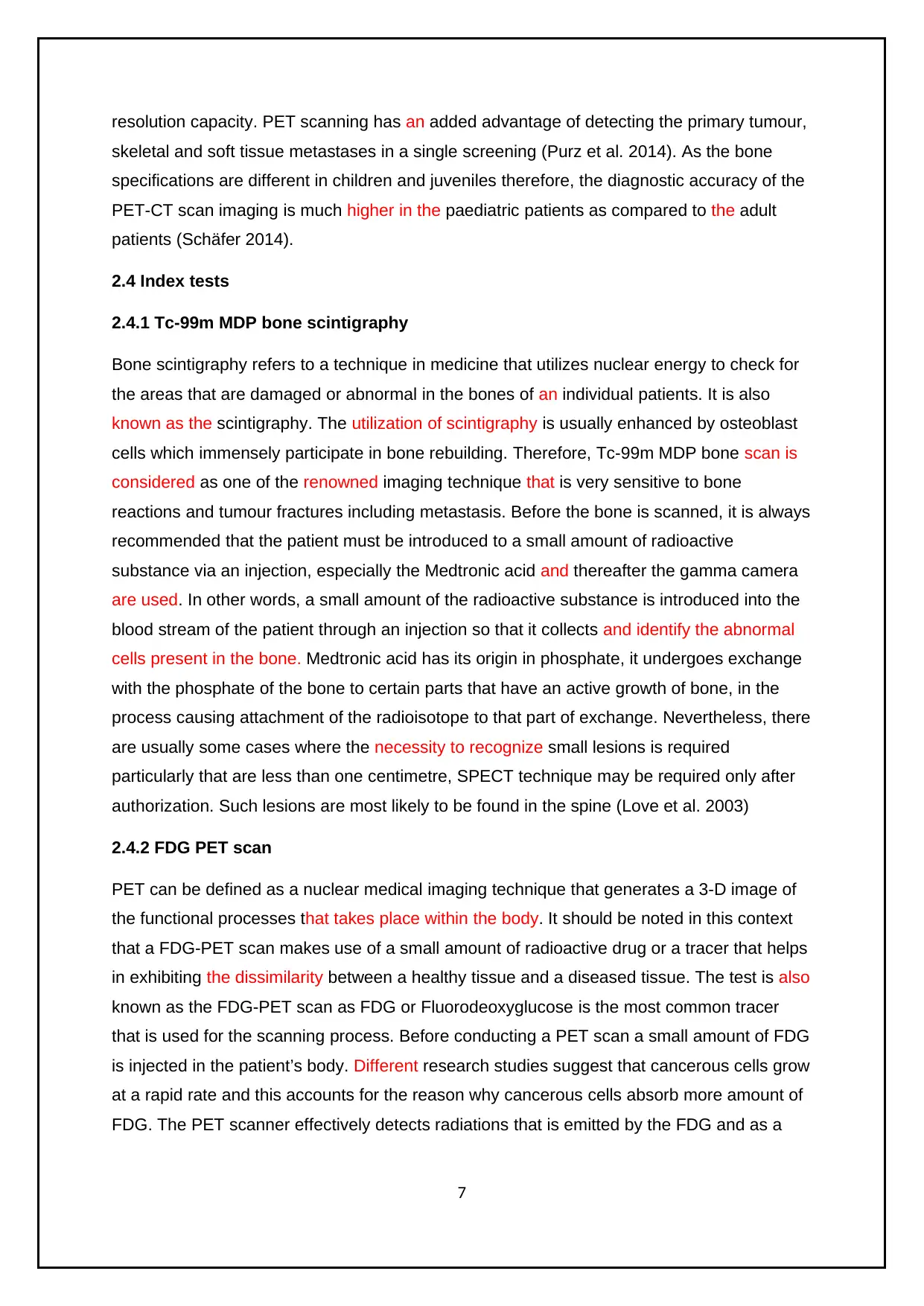
resolution capacity. PET scanning has an added advantage of detecting the primary tumour,
skeletal and soft tissue metastases in a single screening (Purz et al. 2014). As the bone
specifications are different in children and juveniles therefore, the diagnostic accuracy of the
PET-CT scan imaging is much higher in the paediatric patients as compared to the adult
patients (Schäfer 2014).
2.4 Index tests
2.4.1 Tc-99m MDP bone scintigraphy
Bone scintigraphy refers to a technique in medicine that utilizes nuclear energy to check for
the areas that are damaged or abnormal in the bones of an individual patients. It is also
known as the scintigraphy. The utilization of scintigraphy is usually enhanced by osteoblast
cells which immensely participate in bone rebuilding. Therefore, Tc-99m MDP bone scan is
considered as one of the renowned imaging technique that is very sensitive to bone
reactions and tumour fractures including metastasis. Before the bone is scanned, it is always
recommended that the patient must be introduced to a small amount of radioactive
substance via an injection, especially the Medtronic acid and thereafter the gamma camera
are used. In other words, a small amount of the radioactive substance is introduced into the
blood stream of the patient through an injection so that it collects and identify the abnormal
cells present in the bone. Medtronic acid has its origin in phosphate, it undergoes exchange
with the phosphate of the bone to certain parts that have an active growth of bone, in the
process causing attachment of the radioisotope to that part of exchange. Nevertheless, there
are usually some cases where the necessity to recognize small lesions is required
particularly that are less than one centimetre, SPECT technique may be required only after
authorization. Such lesions are most likely to be found in the spine (Love et al. 2003)
2.4.2 FDG PET scan
PET can be defined as a nuclear medical imaging technique that generates a 3-D image of
the functional processes that takes place within the body. It should be noted in this context
that a FDG-PET scan makes use of a small amount of radioactive drug or a tracer that helps
in exhibiting the dissimilarity between a healthy tissue and a diseased tissue. The test is also
known as the FDG-PET scan as FDG or Fluorodeoxyglucose is the most common tracer
that is used for the scanning process. Before conducting a PET scan a small amount of FDG
is injected in the patient’s body. Different research studies suggest that cancerous cells grow
at a rapid rate and this accounts for the reason why cancerous cells absorb more amount of
FDG. The PET scanner effectively detects radiations that is emitted by the FDG and as a
7
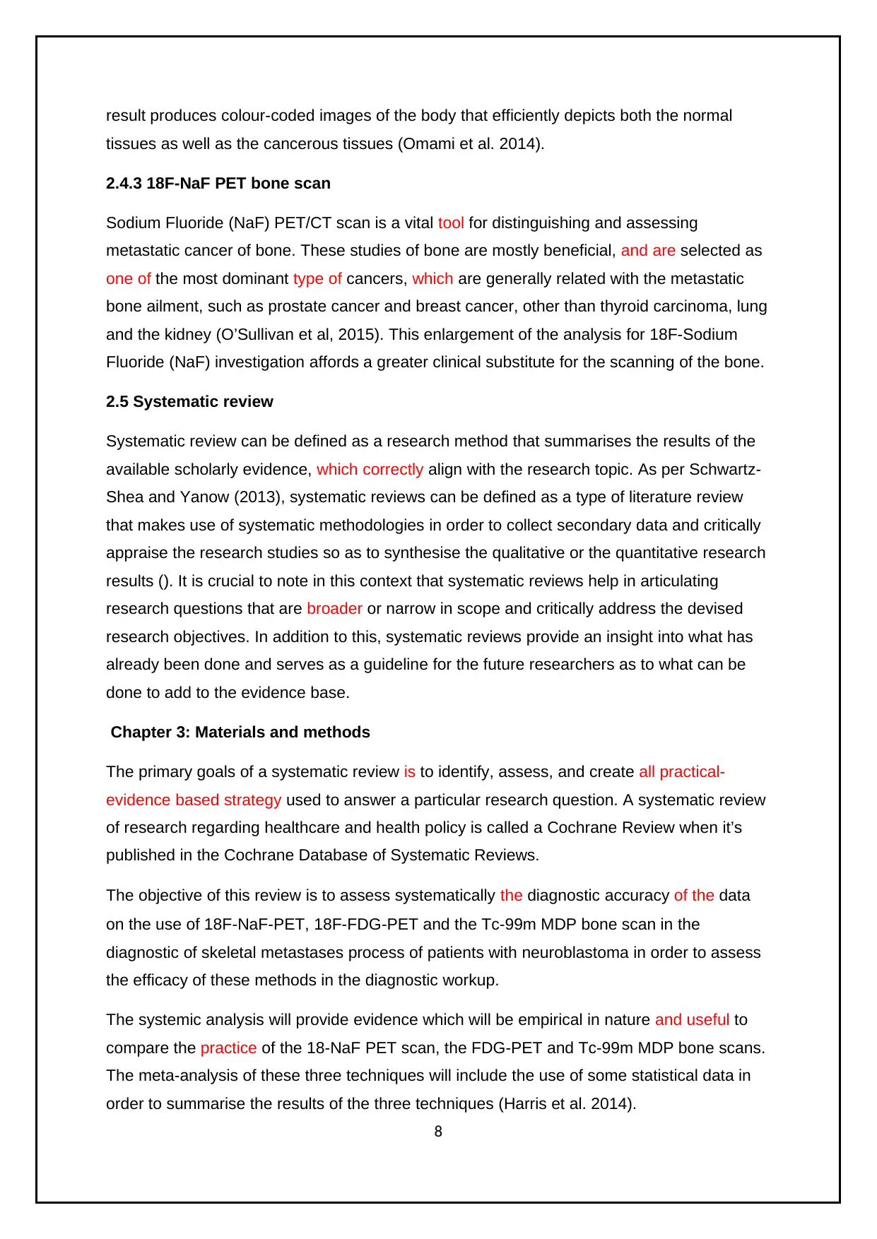
result produces colour-coded images of the body that efficiently depicts both the normal
tissues as well as the cancerous tissues (Omami et al. 2014).
2.4.3 18F-NaF PET bone scan
Sodium Fluoride (NaF) PET/CT scan is a vital tool for distinguishing and assessing
metastatic cancer of bone. These studies of bone are mostly beneficial, and are selected as
one of the most dominant type of cancers, which are generally related with the metastatic
bone ailment, such as prostate cancer and breast cancer, other than thyroid carcinoma, lung
and the kidney (O’Sullivan et al, 2015). This enlargement of the analysis for 18F-Sodium
Fluoride (NaF) investigation affords a greater clinical substitute for the scanning of the bone.
2.5 Systematic review
Systematic review can be defined as a research method that summarises the results of the
available scholarly evidence, which correctly align with the research topic. As per Schwartz-
Shea and Yanow (2013), systematic reviews can be defined as a type of literature review
that makes use of systematic methodologies in order to collect secondary data and critically
appraise the research studies so as to synthesise the qualitative or the quantitative research
results (). It is crucial to note in this context that systematic reviews help in articulating
research questions that are broader or narrow in scope and critically address the devised
research objectives. In addition to this, systematic reviews provide an insight into what has
already been done and serves as a guideline for the future researchers as to what can be
done to add to the evidence base.
Chapter 3: Materials and methods
The primary goals of a systematic review is to identify, assess, and create all practical-
evidence based strategy used to answer a particular research question. A systematic review
of research regarding healthcare and health policy is called a Cochrane Review when it’s
published in the Cochrane Database of Systematic Reviews.
The objective of this review is to assess systematically the diagnostic accuracy of the data
on the use of 18F‐NaF-PET, 18F-FDG‐PET and the Tc-99m MDP bone scan in the
diagnostic of skeletal metastases process of patients with neuroblastoma in order to assess
the efficacy of these methods in the diagnostic workup.
The systemic analysis will provide evidence which will be empirical in nature and useful to
compare the practice of the 18-NaF PET scan, the FDG-PET and Tc-99m MDP bone scans.
The meta-analysis of these three techniques will include the use of some statistical data in
order to summarise the results of the three techniques (Harris et al. 2014).
8
⊘ This is a preview!⊘
Do you want full access?
Subscribe today to unlock all pages.
Trusted by 1+ million students worldwide
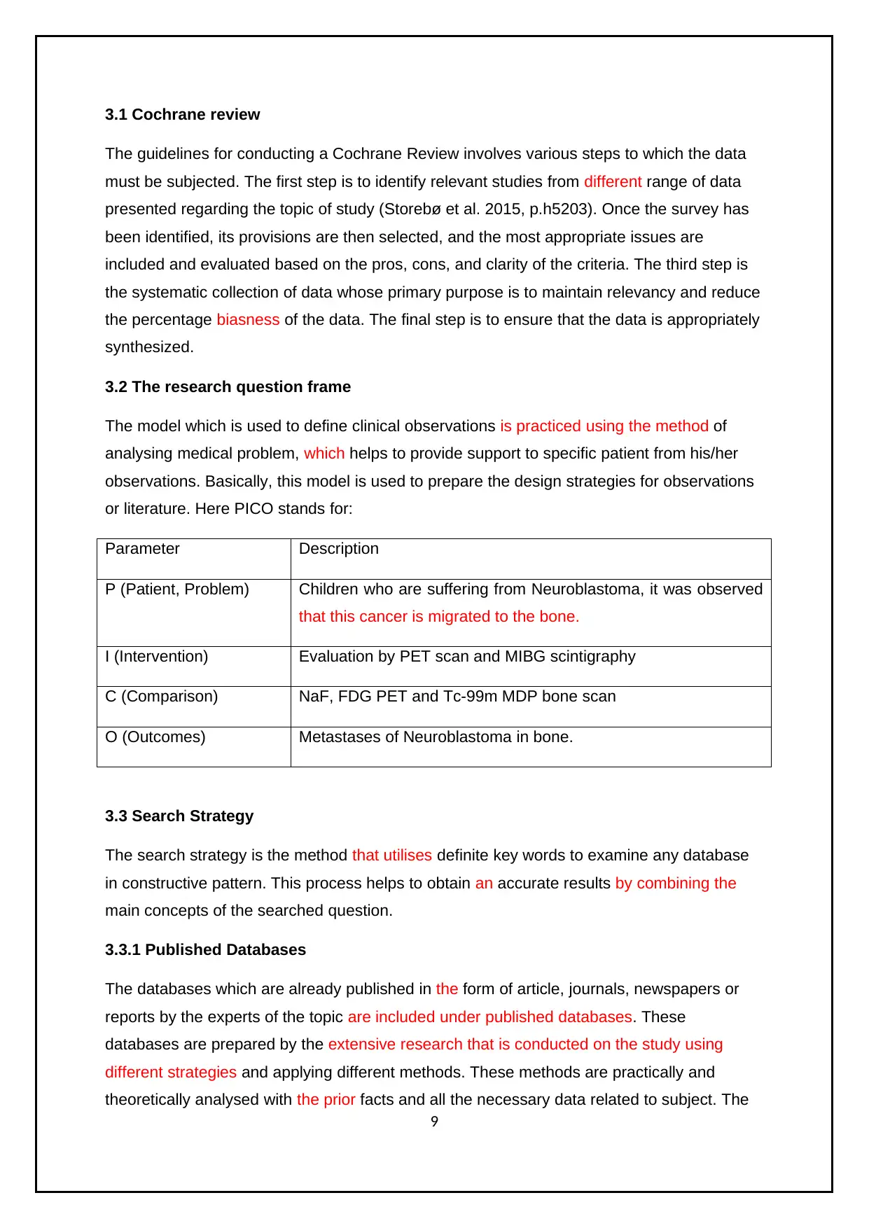
3.1 Cochrane review
The guidelines for conducting a Cochrane Review involves various steps to which the data
must be subjected. The first step is to identify relevant studies from different range of data
presented regarding the topic of study (Storebø et al. 2015, p.h5203). Once the survey has
been identified, its provisions are then selected, and the most appropriate issues are
included and evaluated based on the pros, cons, and clarity of the criteria. The third step is
the systematic collection of data whose primary purpose is to maintain relevancy and reduce
the percentage biasness of the data. The final step is to ensure that the data is appropriately
synthesized.
3.2 The research question frame
The model which is used to define clinical observations is practiced using the method of
analysing medical problem, which helps to provide support to specific patient from his/her
observations. Basically, this model is used to prepare the design strategies for observations
or literature. Here PICO stands for:
Parameter Description
P (Patient, Problem) Children who are suffering from Neuroblastoma, it was observed
that this cancer is migrated to the bone.
I (Intervention) Evaluation by PET scan and MIBG scintigraphy
C (Comparison) NaF, FDG PET and Tc-99m MDP bone scan
O (Outcomes) Metastases of Neuroblastoma in bone.
3.3 Search Strategy
The search strategy is the method that utilises definite key words to examine any database
in constructive pattern. This process helps to obtain an accurate results by combining the
main concepts of the searched question.
3.3.1 Published Databases
The databases which are already published in the form of article, journals, newspapers or
reports by the experts of the topic are included under published databases. These
databases are prepared by the extensive research that is conducted on the study using
different strategies and applying different methods. These methods are practically and
theoretically analysed with the prior facts and all the necessary data related to subject. The
9
Paraphrase This Document
Need a fresh take? Get an instant paraphrase of this document with our AI Paraphraser
information or data which are available in these databases are valid because they are
prepared under guidance and complete supervision of the researcher.
The used databases are as follows:
o PubMed
o Elsevier
o Cochrane library
o Embase
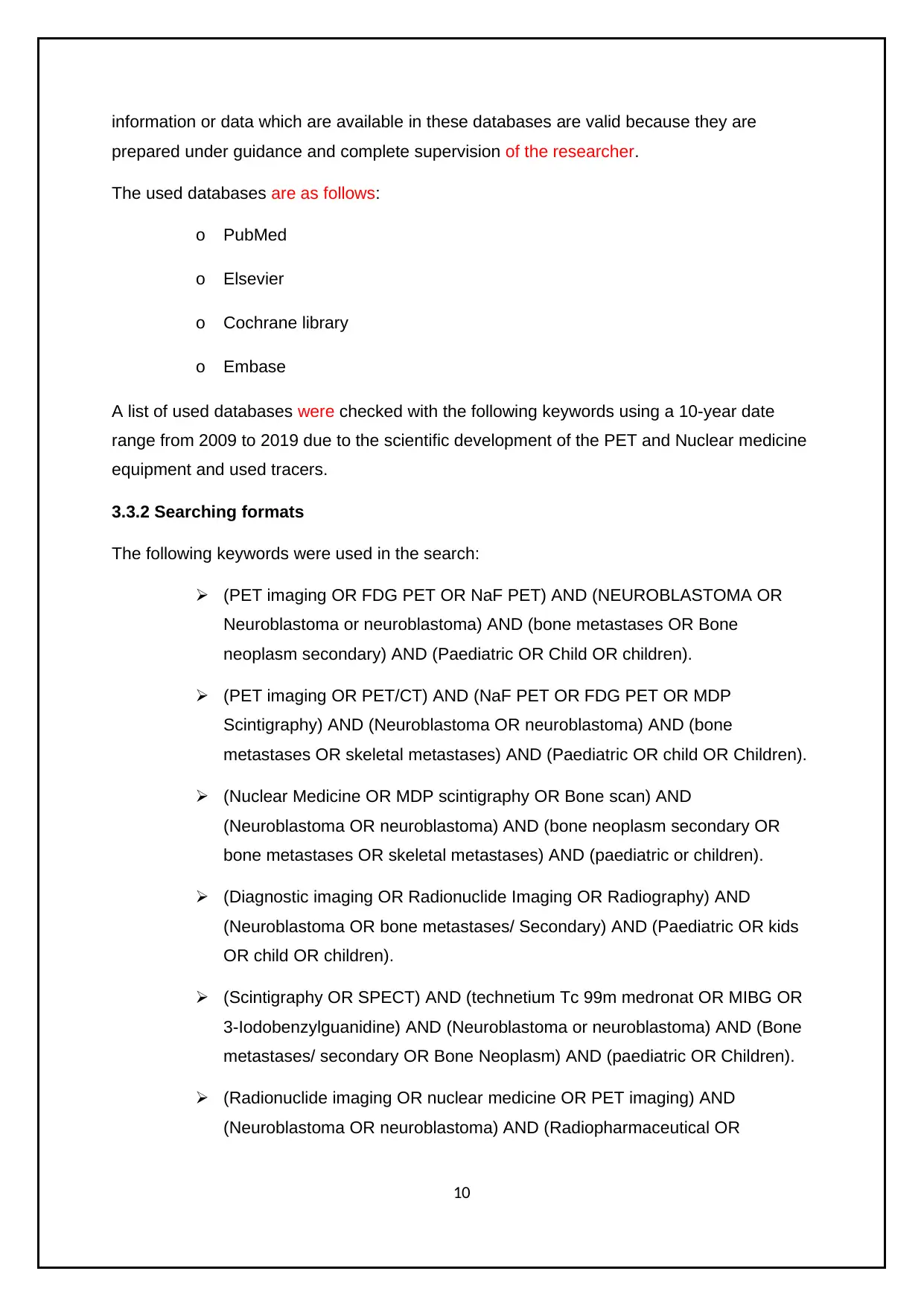
A list of used databases were checked with the following keywords using a 10-year date
range from 2009 to 2019 due to the scientific development of the PET and Nuclear medicine
equipment and used tracers.
3.3.2 Searching formats
The following keywords were used in the search:
(PET imaging OR FDG PET OR NaF PET) AND (NEUROBLASTOMA OR
Neuroblastoma or neuroblastoma) AND (bone metastases OR Bone
neoplasm secondary) AND (Paediatric OR Child OR children).
(PET imaging OR PET/CT) AND (NaF PET OR FDG PET OR MDP
Scintigraphy) AND (Neuroblastoma OR neuroblastoma) AND (bone
metastases OR skeletal metastases) AND (Paediatric OR child OR Children).
(Nuclear Medicine OR MDP scintigraphy OR Bone scan) AND
(Neuroblastoma OR neuroblastoma) AND (bone neoplasm secondary OR
bone metastases OR skeletal metastases) AND (paediatric or children).
(Diagnostic imaging OR Radionuclide Imaging OR Radiography) AND
(Neuroblastoma OR bone metastases/ Secondary) AND (Paediatric OR kids
OR child OR children).
(Scintigraphy OR SPECT) AND (technetium Tc 99m medronat OR MIBG OR
3-Iodobenzylguanidine) AND (Neuroblastoma or neuroblastoma) AND (Bone
metastases/ secondary OR Bone Neoplasm) AND (paediatric OR Children).
(Radionuclide imaging OR nuclear medicine OR PET imaging) AND
(Neuroblastoma OR neuroblastoma) AND (Radiopharmaceutical OR
10
prepared under guidance and complete supervision of the researcher.
The used databases are as follows:
o PubMed
o Elsevier
o Cochrane library
o Embase
A list of used databases were checked with the following keywords using a 10-year date
range from 2009 to 2019 due to the scientific development of the PET and Nuclear medicine
equipment and used tracers.
3.3.2 Searching formats
The following keywords were used in the search:
(PET imaging OR FDG PET OR NaF PET) AND (NEUROBLASTOMA OR
Neuroblastoma or neuroblastoma) AND (bone metastases OR Bone
neoplasm secondary) AND (Paediatric OR Child OR children).
(PET imaging OR PET/CT) AND (NaF PET OR FDG PET OR MDP
Scintigraphy) AND (Neuroblastoma OR neuroblastoma) AND (bone
metastases OR skeletal metastases) AND (Paediatric OR child OR Children).
(Nuclear Medicine OR MDP scintigraphy OR Bone scan) AND
(Neuroblastoma OR neuroblastoma) AND (bone neoplasm secondary OR
bone metastases OR skeletal metastases) AND (paediatric or children).
(Diagnostic imaging OR Radionuclide Imaging OR Radiography) AND
(Neuroblastoma OR bone metastases/ Secondary) AND (Paediatric OR kids
OR child OR children).
(Scintigraphy OR SPECT) AND (technetium Tc 99m medronat OR MIBG OR
3-Iodobenzylguanidine) AND (Neuroblastoma or neuroblastoma) AND (Bone
metastases/ secondary OR Bone Neoplasm) AND (paediatric OR Children).
(Radionuclide imaging OR nuclear medicine OR PET imaging) AND
(Neuroblastoma OR neuroblastoma) AND (Radiopharmaceutical OR
10
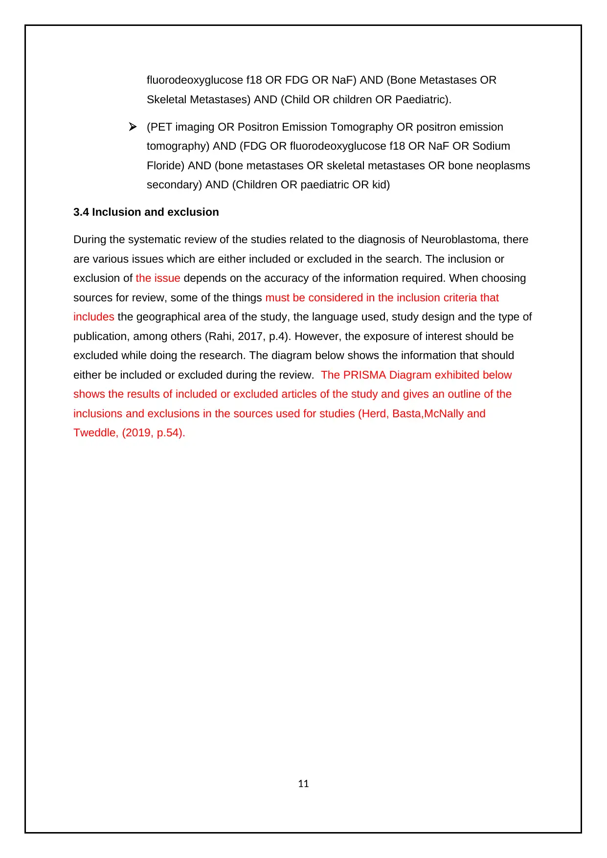
fluorodeoxyglucose f18 OR FDG OR NaF) AND (Bone Metastases OR
Skeletal Metastases) AND (Child OR children OR Paediatric).
(PET imaging OR Positron Emission Tomography OR positron emission
tomography) AND (FDG OR fluorodeoxyglucose f18 OR NaF OR Sodium
Floride) AND (bone metastases OR skeletal metastases OR bone neoplasms
secondary) AND (Children OR paediatric OR kid)
3.4 Inclusion and exclusion
During the systematic review of the studies related to the diagnosis of Neuroblastoma, there
are various issues which are either included or excluded in the search. The inclusion or
exclusion of the issue depends on the accuracy of the information required. When choosing
sources for review, some of the things must be considered in the inclusion criteria that
includes the geographical area of the study, the language used, study design and the type of
publication, among others (Rahi, 2017, p.4). However, the exposure of interest should be
excluded while doing the research. The diagram below shows the information that should
either be included or excluded during the review. The PRISMA Diagram exhibited below
shows the results of included or excluded articles of the study and gives an outline of the
inclusions and exclusions in the sources used for studies (Herd, Basta,McNally and
Tweddle, (2019, p.54).
11
⊘ This is a preview!⊘
Do you want full access?
Subscribe today to unlock all pages.
Trusted by 1+ million students worldwide
1 out of 23
Related Documents


Your All-in-One AI-Powered Toolkit for Academic Success.
+13062052269
info@desklib.com
Available 24*7 on WhatsApp / Email
![[object Object]](/_next/static/media/star-bottom.7253800d.svg)
Unlock your academic potential
Copyright © 2020–2026 A2Z Services. All Rights Reserved. Developed and managed by ZUCOL.