Comprehensive Immunology Report: Immune Responses and Hypersensitivity
VerifiedAdded on 2023/06/03
|25
|5983
|266
Report
AI Summary
This report delves into the intricacies of immunology, exploring both innate and adaptive immune responses. It begins by examining Type II hypersensitivity reactions, using blood transfusions as a prime example to illustrate antibody-mediated cell destruction and the importance of ABO and Rh compatibility. The report then dissects the components of the innate immune system, including physical barriers, phagocytic cells, and various chemical defenses, and explains how these elements respond to viral pathogens. Furthermore, it details how innate cells alert the adaptive immune system about viral invasions. The report also discusses T cell development, differentiation, and their responses to bacterial and viral pathogens. Finally, it explores the cellular phenomena that can be studied using flow cytometry with DNA-intercalating dyes or CFSE, describing the principles of both techniques. The report incorporates insights from the provided assignment brief, covering topics such as B cell interactions, isotype switching, and the role of T cells in immune responses. It also addresses hypersensitivity types and the mechanisms involved.

Running head: IMMUNOLOGY 1
IMMUNOLOGY
Student’s Name
Institutional Affiliation
Course
Instructor
Date
IMMUNOLOGY
Student’s Name
Institutional Affiliation
Course
Instructor
Date
Paraphrase This Document
Need a fresh take? Get an instant paraphrase of this document with our AI Paraphraser
IMMUNOLOGY 2
Question 1: Type 2 immediate hypersensitivity reactions are mediated by antibodies.
Please explain, using blood transfusion as an example, the mechanism of action.
Immunology refers to the study of the mechanism used by the human body to defend
itself against pathogens or invading microorganisms. The invaders can be external or internal
for example tumors. Immunology forms an integral aspect of life since and human beings
and medical studies. Hypersensitivity reactions are conditions whereby the impacts of the
immune system are harmful.
A wide range of autoimmune disorders and allergies fall under the same umbrella of
the hypersensitivity reactions. The primary difference between the two is that allergic
reactions involve an immune reaction to the most common environmental substances,
whereas the autoimmune disorders involve direct immune reactions to the body tissues.
Commonly, hypersensitivity reactions are categorised into four major classes namely;
type 1, type II, type II, and Type IV. Type II hypersensitivity reactions are also called
cytotoxic. They involve antibodies which are often specific to certain body tissues causing
cell destruction of the involved tissues. Examples of the resulting consequences of the cell
destruction include autoimmune hemolytic anemia, thrombocytopenia, granulocytopenia, and
Goodpasture syndrome.
Type II hypersensitivity reactions affect various tissues and organs since the antigens
are commonly endogenous. The reactions usually last for minutes to hours. Primarily, these
reactions are initiated by the complement and antibodies such as immunoglobulin (IgG) and
immunoglobulin M (IgM) classes. The K cells and phagocytes may also take part in the
mechanism.
IMMUNOLOGY 3
The antibody-mediated cell destruction the type II hypersensitivity reactions is best
observed in blood -transfusion reactions. In blood transfusion, the antibodies of the host react
with the antigens which are considered as foreign on the incompatible transfused blood cells
mediating cell destruction. An antibody mediates the process of cell destruction by activating
the complement system towards the creation of pores in the membranes of the foreign cell.
In blood transfusion, the ABO compatibility test is very essential. The main blood
groups are O, A, B, and AB. In an individual with blood group O, the red blood cells have O
antigen on their surfaces, the involved sugars are gal and Fuc, has antigens A and B.
Therefore, he or she is referred to as a universal donor.
An blood group A individual has antigen A on the surface of his or her red blood
cells. He or she has Gal, NAcGal, and Fuc sugars and an antibody b. He or she can only
receive blood from an individual with the same blood group A and O. On the other hand, he
or she can donate to an individual with blood group A.
A blood group B individual has B antigen on the surface of the red blood cells and has
sugars Gal and Fuc. He or she has antibody B. He or she can get from blood group B and O
but can only give blood to a person with blood group B. Lastly, an individual with blood
group AB has all sugars, has no antibodies and has both antigen A and B on the surface of the
red blood cells. Therefore, he or she is referred to as a universal recipient. He or she can only
donate to blood group O.
During blood transfusion, the type II hypersensitivity reactions occurs due to the ABO
blood group incompatibility. Transfusion reactions occur due to intravascular hemolysis of
the red blood cells secondary to the incompatibility of the ABO blood grouping system. On
the other hand, the extravascular hemolysis of the red blood cells is invariably attributed to
the incompatibility of the Rhesus factor.
⊘ This is a preview!⊘
Do you want full access?
Subscribe today to unlock all pages.
Trusted by 1+ million students worldwide
IMMUNOLOGY 4
The Rhesus (Rh) factor is a genetically inherited protein that is found on the surface
of the red blood cells. If an individual has the Rh factor he or she is said to be Rhesus positive
but if he or she does not have it, he or she is said to be Rh negative. Rhesus can be diagnosed
using blood tests. Rhesus factor incompatibility can lead to fatal complications especially to
the new-born at birth especially if the mother is Rh negative and the fetus is Rh positive. This
instance is termed as Rh incompatibility which occurs in the second or other subsequent
pregnancies.
Question 2: Describe the main components of innate immune system and the
mechanisms of the innate immune response to viral pathogens. Explain how the innate
cells alert the adaptive immune cell about the viral invasion.
Innate immunity is the non-specific defense mechanism against microbes or
infections. Some of the key characteristics of the innate immunity are that it is fast, non-
specific for antigens, has no memory and has limited diversity. The four primary components
are (1) chemical, physical, and anatomical barriers, (2) blood proteins, (3) phagocytic
barriers, and (4) cytokines.
First component: Chemical, Physical or Anatomical Barriers
These kinds of barriers prevent the direct entry of microorganisms or pathogens into
the body. The barriers act as the first line of defense for an organism against infections. The
surface of the mucous membranes and the skin fall under this category since they offer a
protective barrier towards the entry of the pathogens or microorganisms into the body.
The skin has two distinct layers namely the epidermis and the dermis. The epidermis
is the outer layer and is relatively thin while the dermis is the innermost thicker layer of the
skin. The dermis has a couple of layers of epithelia cells that are tightly packed together. The
outer epidermal layer has dead cells and keratin, a water-proofing protein.
Paraphrase This Document
Need a fresh take? Get an instant paraphrase of this document with our AI Paraphraser
IMMUNOLOGY 5
The skin plays a primary role by acting a major barrier to the wide range of
microorganisms that invade the body. Any breaks on the skin of the host are commonly
attributed to wounds, scratches, and abrasions. The skin breaks acts as the routes for the
infections and penetration of pathogenic microorganisms into the body.
Additionally, the skin may be penetrated through biting by insects such as mites,
mosquitoes, fleas, ticks, and sanflies. On the skin surface, there is usually bacterial growth in
dense population. The bacterial growth is commonly accelerated by an improper entrance
because of the presence of the sebum which is produced by the sebaceous glands of the
dermis.
The gastro-intestinal tract has residential bacteria, which help in the process of
digestion of particular polysaccharides and control the potential pathogens. The natural
development of the immune system is largely dependent on the continuous antigenic stimulus
that is provided by the sufficiently low levels of pH. The low level of pH is commonly
maintained by gastric juices in the stomach. The gastric juice has both viricidal and
bactericidal actions. Anaerobic conditions and low levels of pH are often maintained in the
intestine to kill bacteria and viruses which can lead to the development of an infection.
In the urogenital tract, low level of pH and urine flow provides sufficient protection to
the lumen. In females, the vaginal wall is often lined by squamous epithelium which is rich in
large amounts of glycogen. Following the high levels of oestrogen hormone, the glycogen is
often deposited upon the epithelial surface before ovulation. During this process, the
glycogen is anaerobically degraded by the lactobacilli. The mechanism of the lactobacilli
results in the formation of lactic acid which serves the role of deterrent for any pathogenic
infections or microorganisms.
IMMUNOLOGY 6
The milk in the mammary glands has bacterial inhibitors known as lactenius. The
lactenius encompasses the lysozymes, complement, an iron-binding protein known as
lactoferrin, and lacto-peroxidase enzyme. Lactoferrin often competes with bacteria for iron
hence inhibiting their growth. With the help of the exogenous hydrogen-peroxide, the acto-
peroxidase often reacts with thiocyanate ions of milk converting them into sulphurdicyanide.
The sulphurdicyanide is bacteriostatic hence it stops bacterial mechanisms in the body hence
preventing infections.
The walls of the respiratory system are covered with mucous especially when the
suspended particles found in the air. The particles get stuck to the walls covered with mucous
when they enter the respiratory tract. The upper part of the respiratory tract has a mucous
layer which is antiseptic by virtue of the presence of the immunoglobulin A (IgA) and
lysosomes found in it.
The epithelial lining of the respiratory tract, gastrointestinal tract, and genitourinary
tracts always produce certain micro-peptides, which act as natural antibiotics, endogenous, or
disinfectants. These systems of the body al produce antimicrobial peptides in the glandular
secretions, phagocytes and other body cells. The antimicrobial peptides include the defensins,
protegrins, cathelicidins, histamine, granulysin, and secretory leuco-protease inhibitor (SLPI).
Second Component: Phagocytic Barriers:
⊘ This is a preview!⊘
Do you want full access?
Subscribe today to unlock all pages.
Trusted by 1+ million students worldwide
IMMUNOLOGY 7
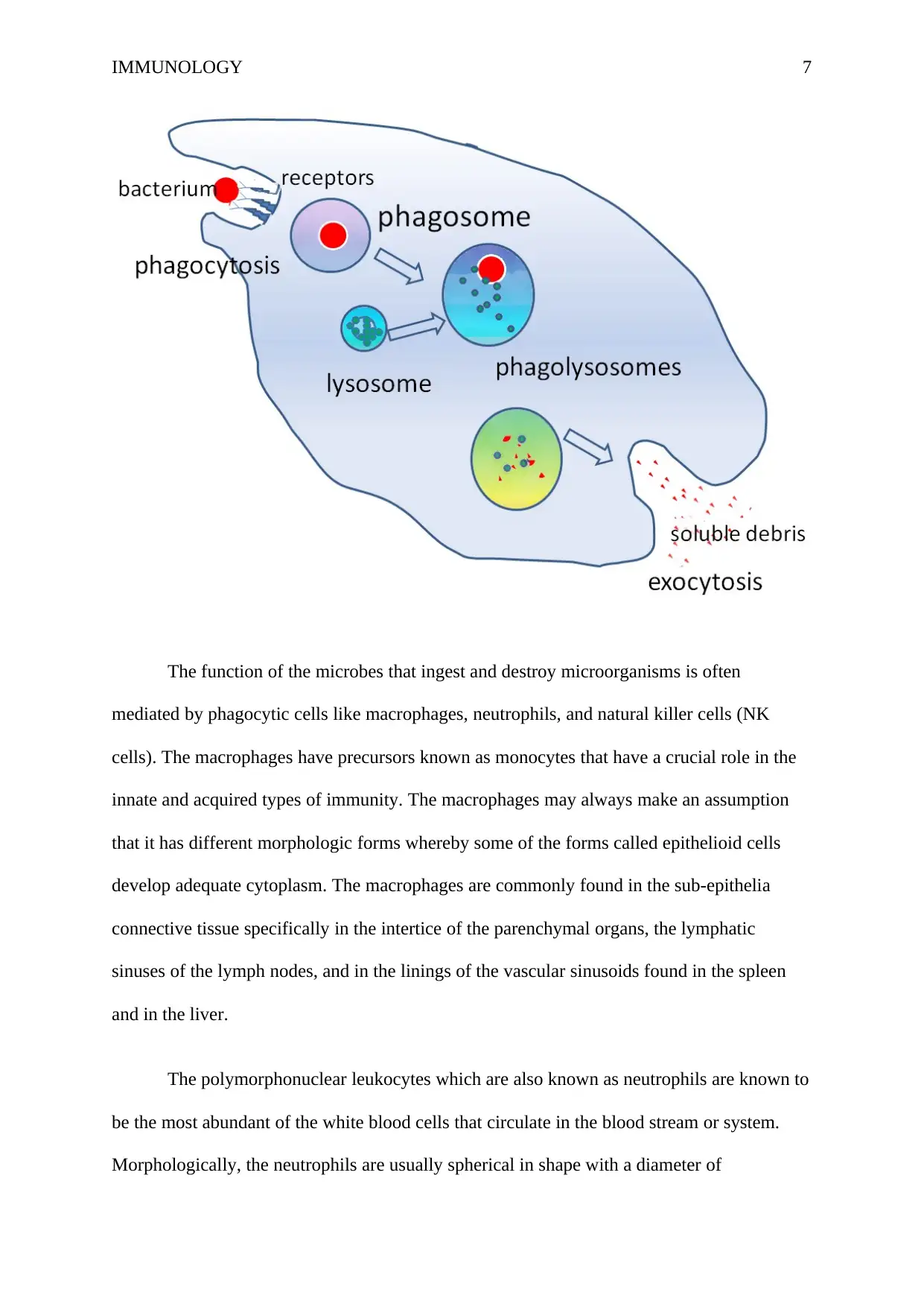
The function of the microbes that ingest and destroy microorganisms is often
mediated by phagocytic cells like macrophages, neutrophils, and natural killer cells (NK
cells). The macrophages have precursors known as monocytes that have a crucial role in the
innate and acquired types of immunity. The macrophages may always make an assumption
that it has different morphologic forms whereby some of the forms called epithelioid cells
develop adequate cytoplasm. The macrophages are commonly found in the sub-epithelia
connective tissue specifically in the intertice of the parenchymal organs, the lymphatic
sinuses of the lymph nodes, and in the linings of the vascular sinusoids found in the spleen
and in the liver.
The polymorphonuclear leukocytes which are also known as neutrophils are known to
be the most abundant of the white blood cells that circulate in the blood stream or system.
Morphologically, the neutrophils are usually spherical in shape with a diameter of
Paraphrase This Document
Need a fresh take? Get an instant paraphrase of this document with our AI Paraphraser
IMMUNOLOGY 8
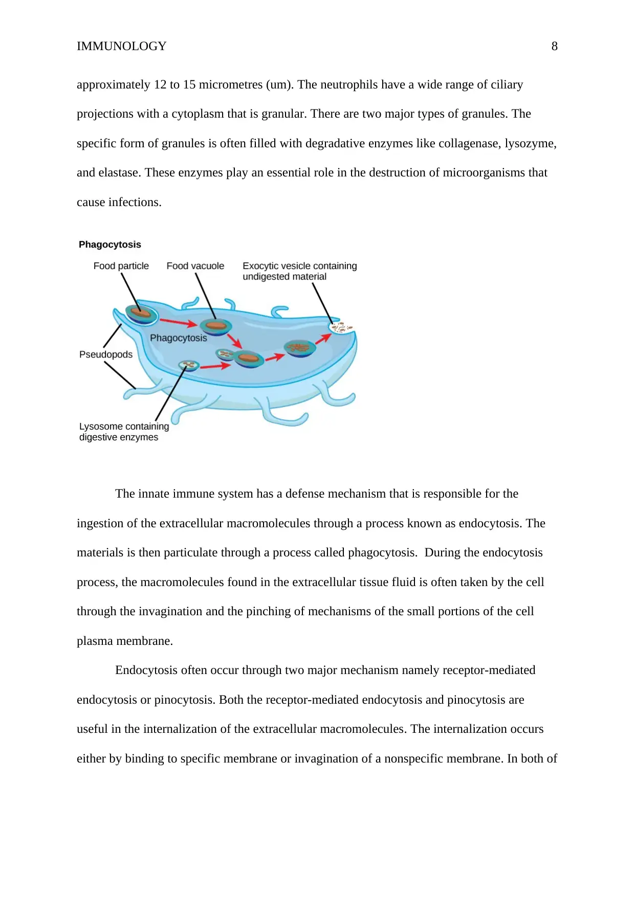
approximately 12 to 15 micrometres (um). The neutrophils have a wide range of ciliary
projections with a cytoplasm that is granular. There are two major types of granules. The
specific form of granules is often filled with degradative enzymes like collagenase, lysozyme,
and elastase. These enzymes play an essential role in the destruction of microorganisms that
cause infections.
The innate immune system has a defense mechanism that is responsible for the
ingestion of the extracellular macromolecules through a process known as endocytosis. The
materials is then particulate through a process called phagocytosis. During the endocytosis
process, the macromolecules found in the extracellular tissue fluid is often taken by the cell
through the invagination and the pinching of mechanisms of the small portions of the cell
plasma membrane.
Endocytosis often occur through two major mechanism namely receptor-mediated
endocytosis or pinocytosis. Both the receptor-mediated endocytosis and pinocytosis are
useful in the internalization of the extracellular macromolecules. The internalization occurs
either by binding to specific membrane or invagination of a nonspecific membrane. In both of
IMMUNOLOGY 9
these mechanisms, the ingested material, often a microbe, is often degraded through the
endocytic processing pathway.
After the fusion of the endocytic vesicles with each other and with endosomes, there
is a dissociation of the ligands of the macromolecules from the receptors. This enables the
macromolecules fuse with the primary lysosome hence leading to the formation of a
secondary lysosomes. The primary lysosomes often originate from the Golgi apparatus of the
cell. The primary lysosome encompasses the degrading enzymes together with nucleases,
proteases, hydrolytic enzymes, and lipases. These enzymes are responsible for the digestion
of macromolecules in the secondary lysosomes. The degrading enzymes also break down the
macromolecules into very small useless products.
The phagocytosis mechanism is often regarded as a cytoskeleton dependent process
that engulfs a large particulate material. The large particulate material may include the entire
pathogenic molecules. During the phagocytosis mechanisms, there is an expansion of the
plasma membrane to cover the whole particulate material resulting in a large vesicle known
as phagosome. Before the formation of a phagosome, the neutrophils and macrophages are
often attracted towards many substances that are generated following an immune response.
The phagosomes are known to be large than the endocytic vesicles by far.
The phagosome vesicle often contains the foreign particles which had been ingested.
The particles usually break off from the plasma membrane and gets into the endocytic
pathway where they are ingested and degraded. The phagosome then moves to the interior
aspect of the cell and fuses of lysomes or lysosomes forming a phagolysosome.
The natural killer cells also form a very essential aspect of the innate immune system.
The natural killer cells are regarded as the third lymphoid lineage of cells that are distinct
⊘ This is a preview!⊘
Do you want full access?
Subscribe today to unlock all pages.
Trusted by 1+ million students worldwide
IMMUNOLOGY 10
from T-cells and B cells and their progeny. The large, granular, and non-phagocytic
lymphocytes are named on the basis of their ability to destroy the abnormal cells of the host
organism. The abnormal cells could be malignant or infected with microbes. The natural
killer cells make 5 percent to 10 percent of the lymphocytes found in the blood circulation
system.
The NK cells as lymphocytes are large, non-T, granular and no-B. Considerably, the
NK cells are less prominent compared to the ones found in the granulocytes for example
eosinophils, basophils, and neutrophils despite the fact they contain a good number of
cytoplasmic granules. The natural killer cells are often obtained form the precursors of the
bone marrow and they usually appear to be large lymphocytes due to the numerous
cytoplasmic granules found on them. These types of cells have approximately five to twenty
percent of the mononuclear cells found in the spleen and in the blood.
Third component: The blood proteins.
This component primarily involved the complement system whereby numerous blood
plasma proteins called complement proteins are responsible for the linking the recognition of
microbes to the effector function. The complement system has a wide range of functions in
the immune system. The classical pathway and the alternative pathways of the complement
system have similar biological functions. They initiate acute inflammation by directly
activating the mast cells.
The two pathways of the complement system attract the neutrophils to the site of the
microbial attack through a process known as chemotaxis. They enhance the attachment of a
phagocyte to the microbe through a process known as opsonization. The two pathways also
Paraphrase This Document
Need a fresh take? Get an instant paraphrase of this document with our AI Paraphraser
IMMUNOLOGY 11
are responsible for the killing of the microbes through activation of the membrane attack
complex. This process is referred to as lysis.
The plasma proteins also promote development of an inflammatory response by the
“alternative pathway” and the “classical pathway”. The two pathways of the complement
system results to the creation of pores for induction of apoptosis of the target cells that are
infected with virus. Following these mechanisms, the granzymes then enter via the created
pores for induction of the apoptosis process. Therefore, the natural killer cells play a key role
in the elimination of the reservoir of the viral infection.
The complement system often makes use three different strategies for recognition of
microorganisms. Each of the strategies initiates the complement activation pathways leading
to the covalent bonding of the complement proteins to microbial surfaces. The classical
pathway of the complement activation is often triggered by antibodies which are bound to the
antigens on the surface of the viral microbes. In this instance, the complement proteins
collaborate with the antibodies to promote antigen clearance which are body’s antibody
complexes. On the other hand, the alternative pathways offer for the complement activation
without the presence of the antibodies. These extra pathways are also considered as important
parts of the innate immune defense system.
The alternative pathway of the complement system activation is directly triggered by
the components of the viral cell surfaces and the lectin-mediated pathway. The lectin-
mediated pathway is commonly activated when the mannose-binding protein binds on the
proteoglycans containing mannose on the surfaces of the bacteria or virus. The mannose-
binding protein is commonly found in the blood plasma.
IMMUNOLOGY 12
The deposition of the complement of the surface of particular pathogens such as virus
may directly lead to the breakdown of the coated pathogen via the assembly of the terminal
components of the complement system. The assembly of these components results in the
formation of a hold in the cell membrane hence destroying its integrity. Consequently, this
results to the impairment of the pathogenic mechanisms of the viral and death hence
preventing infection.
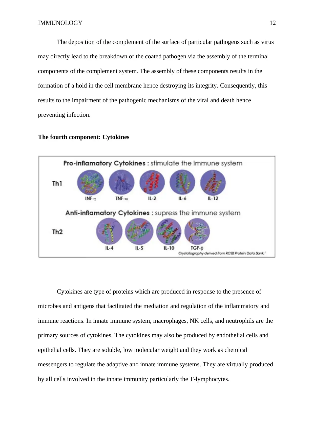
The fourth component: Cytokines
Cytokines are type of proteins which are produced in response to the presence of
microbes and antigens that facilitated the mediation and regulation of the inflammatory and
immune reactions. In innate immune system, macrophages, NK cells, and neutrophils are the
primary sources of cytokines. The cytokines may also be produced by endothelial cells and
epithelial cells. They are soluble, low molecular weight and they work as chemical
messengers to regulate the adaptive and innate immune systems. They are virtually produced
by all cells involved in the innate immunity particularly the T-lymphocytes.
The deposition of the complement of the surface of particular pathogens such as virus
may directly lead to the breakdown of the coated pathogen via the assembly of the terminal
components of the complement system. The assembly of these components results in the
formation of a hold in the cell membrane hence destroying its integrity. Consequently, this
results to the impairment of the pathogenic mechanisms of the viral and death hence
preventing infection.
The fourth component: Cytokines
Cytokines are type of proteins which are produced in response to the presence of
microbes and antigens that facilitated the mediation and regulation of the inflammatory and
immune reactions. In innate immune system, macrophages, NK cells, and neutrophils are the
primary sources of cytokines. The cytokines may also be produced by endothelial cells and
epithelial cells. They are soluble, low molecular weight and they work as chemical
messengers to regulate the adaptive and innate immune systems. They are virtually produced
by all cells involved in the innate immunity particularly the T-lymphocytes.
⊘ This is a preview!⊘
Do you want full access?
Subscribe today to unlock all pages.
Trusted by 1+ million students worldwide
1 out of 25
Related Documents


Your All-in-One AI-Powered Toolkit for Academic Success.
+13062052269
info@desklib.com
Available 24*7 on WhatsApp / Email
![[object Object]](/_next/static/media/star-bottom.7253800d.svg)
Unlock your academic potential
Copyright © 2020–2026 A2Z Services. All Rights Reserved. Developed and managed by ZUCOL.