Microbiology Report: Bacterial Structures, Growth, and Disease Control
VerifiedAdded on 2022/12/23
|11
|3433
|68
Report
AI Summary
This report provides a comprehensive overview of key topics in microbiology. It begins by detailing the structure and function of bacterial pili and flagella, comparing and contrasting their characteristics. The report then explores the life cycle of Leishmaniasis, emphasizing the challenges in controlling this pathogen due to its range of mammalian hosts. The use of Salmonella chromogenic agar plates to distinguish enteric pathogens is discussed, highlighting the methods used to identify and differentiate bacterial colonies. Finally, the report examines various methods for assessing microbial growth, including both direct techniques like plate counts, serial dilution, and filtration, and indirect techniques such as turbidity, metabolic activity, and dry weight analysis. The report provides insights into the methodologies used to measure and understand microbial populations.

Medical Questions
Paraphrase This Document
Need a fresh take? Get an instant paraphrase of this document with our AI Paraphraser
Table of Contents
1. The structure and function of bacterial pili and flagella..............................................................3
2. Life cycle of Leishmaniasis and its range of mammalian hosts make it a particularly difficult
pathogen to control. .......................................................................................................................5
3. Salmonella chromogenic agar plates used to distinguish enteric pathogens and normal floral...6
4. The growth of the microbes can be assessed in many ways ......................................................7
Metabolic Activity.......................................................................................................................8
5.. The life cycle of Schistosomiasis and ways they cause disease in the humans .........................9
REFERENCES..............................................................................................................................12
1. The structure and function of bacterial pili and flagella.
Pili
The cell surfaces of the prokaryotic cells have short, hair-like structures on them, these
structures are called pili. They are attached to the surface which cause infections and have the
ability to overcome host defences. Pili are commonly attached to Gram-negative bacteria.
(Jacobsen, Bardiaux and et.al, 2020).
Depending on the assembly pathway pili are divided into four groups in Garm-negative bacteria:
i) Usher pathway pili
ii) Type IV pili(Drame, Lafforgue and et.al, 2021)
iii) Curli pili
iv) CS1 pilus family
They are divided into two types of groups:
Short and Thin Pili – These type of pili are found in normal body of flora streptococcus
species.
Longer and Flexible Pili- These types of pili are found on Corynebacterium species and
pathogenic streptococci.
Functions
Pili help the bacterial cells to avoid the attack from the white blood cells.
Pili are called sex pilus or F pilus because they recognize the recipient cell to receive the donors
genetic material and stabilize the bacteria during DNA transfer which is done by conjugation.
Pili is used for the movement in Pseudomonas aeruginosa and Myxococcus xanthus.
Flagella
The word flagellum means whip. The bacterial flagella is a microscopic-hair like
structure and are used for the locomotion of a cell (Poghosyan, Iacovache and et.al, 2020). Types
of flagella:
• Monotrichous
• Peritrichous
• Lophotrichous
• Amphitrichous
Structure
⊘ This is a preview!⊘
Do you want full access?
Subscribe today to unlock all pages.
Trusted by 1+ million students worldwide
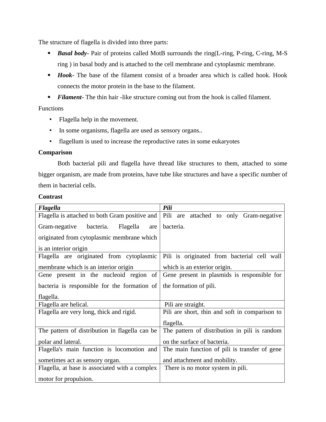
The structure of flagella is divided into three parts:
Basal body- Pair of proteins called MotB surrounds the ring(L-ring, P-ring, C-ring, M-S
ring ) in basal body and is attached to the cell membrane and cytoplasmic membrane.
Hook- The base of the filament consist of a broader area which is called hook. Hook
connects the motor protein in the base to the filament.
Filament- The thin hair -like structure coming out from the hook is called filament.
Functions
• Flagella help in the movement.
• In some organisms, flagella are used as sensory organs..
• flagellum is used to increase the reproductive rates in some eukaryotes
Comparison
Both bacterial pili and flagella have thread like structures to them, attached to some
bigger organism, are made from proteins, have tube like structures and have a specific number of
them in bacterial cells.
Contrast
Flagella Pili
Flagella is attached to both Gram positive and
Gram-negative bacteria. Flagella are
originated from cytoplasmic membrane which
is an interior origin
Pili are attached to only Gram-negative
bacteria.
Flagella are originated from cytoplasmic
membrane which is an interior origin
Pili is originated from bacterial cell wall
which is an exterior origin.
Gene present in the nucleoid region of
bacteria is responsible for the formation of
flagella.
Gene present in plasmids is responsible for
the formation of pili.
Flagella are helical. Pili are straight.
Flagella are very long, thick and rigid. Pili are short, thin and soft in comparison to
flagella.
The pattern of distribution in flagella can be
polar and lateral.
The pattern of distribution in pili is random
on the surface of bacteria.
Flagella's main function is locomotion and
sometimes act as sensory organ.
The main function of pili is transfer of gene
and attachment and mobility.
Flagella, at base is associated with a complex
motor for propulsion.
There is no motor system in pili.
Paraphrase This Document
Need a fresh take? Get an instant paraphrase of this document with our AI Paraphraser
Flagella do not act as receptors. Pili act as receptors in different viruses.
2. Life cycle of Leishmaniasis and its range of mammalian hosts make it a
particularly difficult pathogen to control.
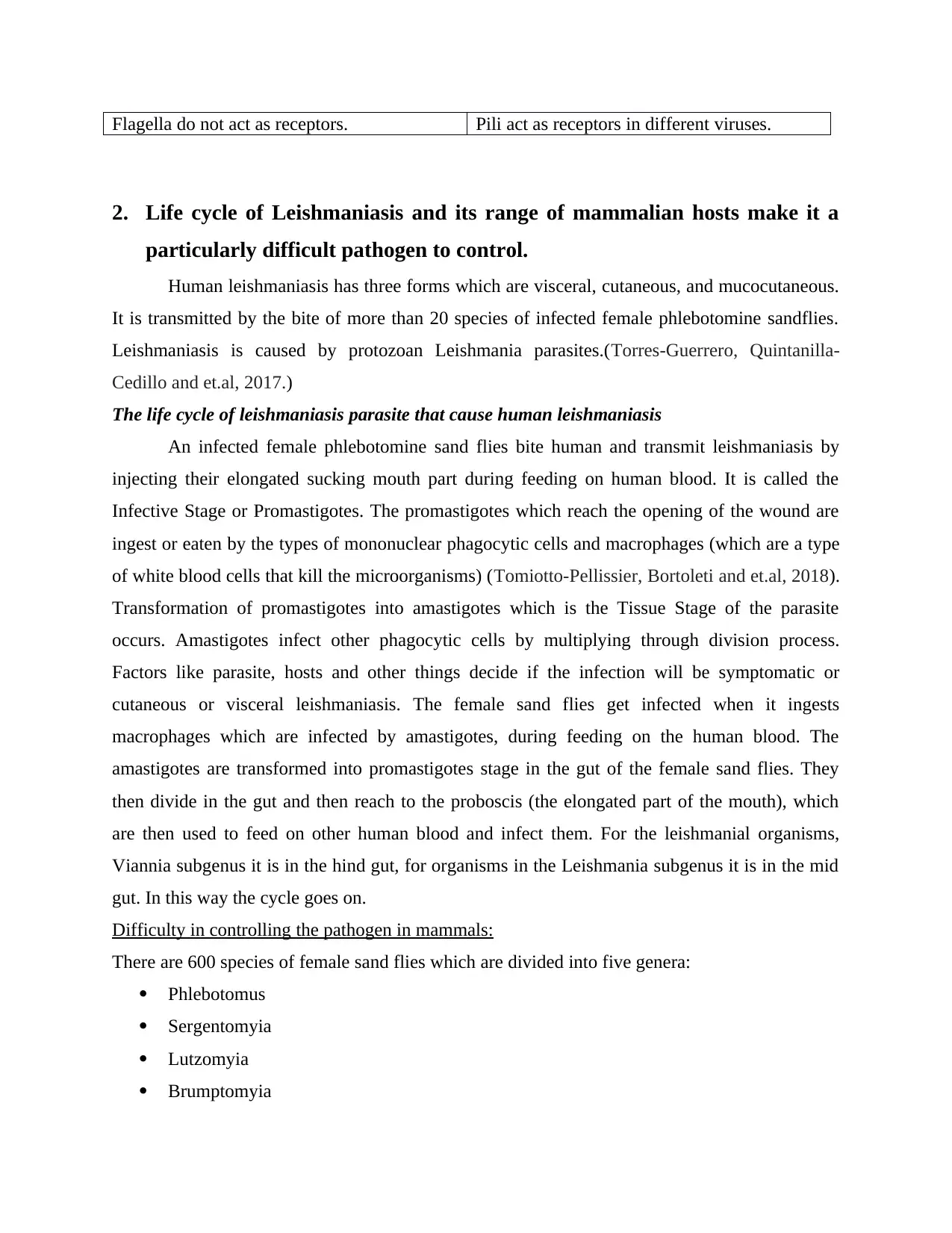
Human leishmaniasis has three forms which are visceral, cutaneous, and mucocutaneous.
It is transmitted by the bite of more than 20 species of infected female phlebotomine sandflies.
Leishmaniasis is caused by protozoan Leishmania parasites.(Torres-Guerrero, Quintanilla-
Cedillo and et.al, 2017.)
The life cycle of leishmaniasis parasite that cause human leishmaniasis
An infected female phlebotomine sand flies bite human and transmit leishmaniasis by
injecting their elongated sucking mouth part during feeding on human blood. It is called the
Infective Stage or Promastigotes. The promastigotes which reach the opening of the wound are
ingest or eaten by the types of mononuclear phagocytic cells and macrophages (which are a type
of white blood cells that kill the microorganisms) (Tomiotto-Pellissier, Bortoleti and et.al, 2018).
Transformation of promastigotes into amastigotes which is the Tissue Stage of the parasite
occurs. Amastigotes infect other phagocytic cells by multiplying through division process.
Factors like parasite, hosts and other things decide if the infection will be symptomatic or
cutaneous or visceral leishmaniasis. The female sand flies get infected when it ingests
macrophages which are infected by amastigotes, during feeding on the human blood. The
amastigotes are transformed into promastigotes stage in the gut of the female sand flies. They
then divide in the gut and then reach to the proboscis (the elongated part of the mouth), which
are then used to feed on other human blood and infect them. For the leishmanial organisms,
Viannia subgenus it is in the hind gut, for organisms in the Leishmania subgenus it is in the mid
gut. In this way the cycle goes on.
Difficulty in controlling the pathogen in mammals:
There are 600 species of female sand flies which are divided into five genera:
Phlebotomus
Sergentomyia
Lutzomyia
Brumptomyia
2. Life cycle of Leishmaniasis and its range of mammalian hosts make it a
particularly difficult pathogen to control.
Human leishmaniasis has three forms which are visceral, cutaneous, and mucocutaneous.
It is transmitted by the bite of more than 20 species of infected female phlebotomine sandflies.
Leishmaniasis is caused by protozoan Leishmania parasites.(Torres-Guerrero, Quintanilla-
Cedillo and et.al, 2017.)
The life cycle of leishmaniasis parasite that cause human leishmaniasis
An infected female phlebotomine sand flies bite human and transmit leishmaniasis by
injecting their elongated sucking mouth part during feeding on human blood. It is called the
Infective Stage or Promastigotes. The promastigotes which reach the opening of the wound are
ingest or eaten by the types of mononuclear phagocytic cells and macrophages (which are a type
of white blood cells that kill the microorganisms) (Tomiotto-Pellissier, Bortoleti and et.al, 2018).
Transformation of promastigotes into amastigotes which is the Tissue Stage of the parasite
occurs. Amastigotes infect other phagocytic cells by multiplying through division process.
Factors like parasite, hosts and other things decide if the infection will be symptomatic or
cutaneous or visceral leishmaniasis. The female sand flies get infected when it ingests
macrophages which are infected by amastigotes, during feeding on the human blood. The
amastigotes are transformed into promastigotes stage in the gut of the female sand flies. They
then divide in the gut and then reach to the proboscis (the elongated part of the mouth), which
are then used to feed on other human blood and infect them. For the leishmanial organisms,
Viannia subgenus it is in the hind gut, for organisms in the Leishmania subgenus it is in the mid
gut. In this way the cycle goes on.
Difficulty in controlling the pathogen in mammals:
There are 600 species of female sand flies which are divided into five genera:
Phlebotomus
Sergentomyia
Lutzomyia
Brumptomyia
Warileya
One species of parasite is transmitted by one female sand fly. A specific disease is caused by
each parasite. The Leishmania parasite develop in the female sand flies which is an unavoidable
stage and cause transmission of Leishmania in various mammalian.
Two main source of human leishmaniasis
Zoonotic leishmaniasis- The reservoir hosts in this type are wild animals, commensals or
domestic animals. A single species of parasite can survive in many hosts. Example, rock
hyrax, rodents, dogs, cats are the hosts that maintain the transmission of Leishmania in
different parts.
Anthroponotic Leishmania- The reservoir hosts in this type is human. Humans transmit
two types of Leishmania namely visceral leishmaniasis which is caused by L. donovani
and cutaneous leishmaniasis which is caused by L. tropica (Bennai, Tahir and et.al,
2018).
The behaviour of humans is also affecting the transmission like keeping the animals close
to their buildings and homes attract the female sand flies and increase the transmission. The
symptoms of this disease is different in different person. The disease have symptoms comparable
to other diseases also making it more difficult to diagnose.
3. Salmonella chromogenic agar plates used to distinguish enteric pathogens
and normal floral
The salmonella chromogenic agar plates are used as a selective chromogenic medium.
These chromogenic agar plates are used to detect and distinguish the enteric pathogen or the
salmonella species and the normal flora . The enteric pathogens are detected on the basis of the
amount of the hydrogen sulphide they produce and not fermenting lactose. Around 2000 or more
species of salmonella are there which do not show these characters (Kuijpers, Jacobs and et.al,
2018).
Amino acids, minerals, nitrogen as well as vitamins which are essential for growth are
provided by casein peptone along with beef extract. To prevent Gram- positive organisms,
Proteus as well as coliform, chromogenic mixture and sodium citrate are added. Bacteriological
agar are used as solidifying agent. The false positive results can be prevented by bacteriological
agar which prevent the occurrence of flora (Furukawa, Stirling and et.al, 2018).
⊘ This is a preview!⊘
Do you want full access?
Subscribe today to unlock all pages.
Trusted by 1+ million students worldwide
The quick identification and detection of the enteric pathogen from the normal flora can
be done by chromogenic agent which is based on two chromogenic substrates. The salmonella
species cause one chromogenic species to hydrolyse and as a result of this magenta coloured
colonies are generated. This is due the fact that the salmonella species cannot hydrolyse the other
chromogenic species. The magenta colour colonies can be clearly seen in the given picture of
salmonella chromogenic agar plate. The second chromogenic species is cleaved by the enzymes
produced by the normal flora. The cleavage of the chromogenic species gives blue- green colour
to the colonies of bacteria. The blue-green coloured colonies are clearly visible in the picture
provided and the two colonies can be distinguished easily.
The organisms present in the medium which are not salmonella will appear either blue-
green in colour or will not gain any colour by any of two chromogenic species present in the
medium. In case where more selectivity is required the amount of supplement (chromogenic
agent) is increased. The addition of more supplement prevent the flora, specifically the
pseudomonas or the pseudomonas can appear in the same colour as that for the detection of the
enteric pathogens.
Example; for the detection of staphylococcus aureus, S.aureus ID medium is used in
which S. aureus form green colonies with Staph because of α‐glucosidase and the other
staphylococci cause the white colour and sometimes pink coloured colonies with the hydrolysis
of substrate and form β‐glucosidase (Papadopoulos, Papadopoulos and et.al, 2018).
4. The growth of the microbes can be assessed in many ways
Direct methods of measuring microbial growth
Plate Counts
The number of variable cells are counted in this method. The method take quite a lot of
time for the formation of the colonies. The method is also called CFU(colony- forming units).
There is a risk of overcrowding in the formation of colonies as it can stop the development thus,
it is necessary to select the amount of colonies to be developed. Serial dilution can be used to
overcome this problem (Van Nevel, Koetzsch and et.al, 2017).
Serial Dilution
Paraphrase This Document
Need a fresh take? Get an instant paraphrase of this document with our AI Paraphraser
A substance is continuously diluted in the solution. It is logarithmic and also show
geometric progression, so, in the process the factor is always kept constant. (Irwin, Fisher and
et.al, 2017.)
Pour Plate and Spread plates
In pour plate method the bacterial suspension is poured into the Petri dish with nutrient
agar and mixed. They are then incubated. The colonies grow in the agar and the surface. In
spread plate method, inoculum is introduced to the surface of agar and is spread uniformly with a
rod. The rod is used to avoid the contact of melted agar and cells.
Filtration
In this method the bacteria can be counted when they are present in the lakes or pure
streams in small portion. The bacteria are filtered and the bacteria remain in the filter surface
which are then collected in the petri dish of nutrient. The bacteria are then allowed to grow
colonies there.
Indirect methods of measuring microbial growth:
Turbidity
It is a simple method. Spectrophotometer or calorimeter are used to measure the cell
mass. The light which pass through the cell suspension is measured by spectrophotometer to
determine the turbidity. The culture should be dense enough to be able to measure the turbidity
on spectrometer. (Farrell, Hassard and et.al, 2018)
Metabolic Activity
In this method the metabolic activity of the bacterial population is measured. The magnitude of
the population of bacteria is proportional to the amount of oxygen consumed or the acid
produced when the conditions are specific and the time is constant.
Dry Weight
This method is used only for the suspensions which are very dense and the cells should
be clean from all extra matter. The dry weight of the filter paper with the pellets of bacterial cells
are weighed and the dry weight of the filter paper is subtracted to get the dry weight of the
bacterial cells.(Soares, Rousk and et.al, 2019)
Cell Count (Manual)- the microbial growth can be measured by the counting the number of the
microbes present in the given sample. This can be done by the microscope glass slide also known
as Petroff- Hausser Counting Chamber.
The glass slide have grids marked on the chamber in squares whose area are known. The
number of cells in each square is counted per ml of the sample. Each grid have 25 squares in it.
Direct counting of the dilute colonies can be done easily but if the cells are not dilute the
colonies can be diluted and then counted. If the cell colonies are not diluted then clump of cells
will appear in the colonies and will create a problem in the counting.
5.. The life cycle of Schistosomiasis and ways they cause disease in the humans
The schistosomiasis disease is also called snail fever and bilharzia. It is caused by
schistosomes which is a parasitic flatworm. In total five species are known which infect the
human species. These are:
• S. Haematobium
• S. Intercalatum
• S. Japonicum
• S. Mansoni
• S. Mekongi
life cycle of schistosomes:
Cercariae containing water causes schistosomiasis. Depending upon the species the
schistosoma release eggs which swim and the parasite enters the human body by the skin. They
then leave their shedding tail inside the skin which form schistosomula. This schistosomula
travels in the body tissue by blood circulation and it develops to become schistosomes and adult
worms. These species have ZZ chromosomes and ZW chromosomes in males and females
respectively.
Adult worm locate in different places according to each species like S.haematobium is
present in the bladder and ureters and can be found sometimes in the rectal venules, S.japonicum
will be found in small intestine, S.mansoni will be present in both large or small intestine and
can change places. Eggs are laid in the small venules of the portal and perivesical systems and
are moved slowly to the intestine, bladder and ureters and are eliminated by excreting faeces and
urine.(Nelwan, 2019. )
The ways organisms cause diseases in humans
Animals like cats, dogs, cattle, pigs, horses act as host for the S. japonicum, dogs act as
host for S. mekongi. Humans act as a host for S. mansoni.
Schistosomiasis caused by trematode worms is an acute and chronic parasitic disease.
⊘ This is a preview!⊘
Do you want full access?
Subscribe today to unlock all pages.
Trusted by 1+ million students worldwide
The people get infected when the larva released by the fresh water snails comes in contact with
the human skin and penetrate through the skin. The infected person when release the excreta
which contain the parasite eggs, in the freshwater, they continue to hatch in the water bodies and
infect other people who come in contact with the infected water.
The organism causes certain diseases :
Intestinal Schistosomiasis
The eggs of the parasite gets collected in the intestinal wall which cause the immune
system to react. The reaction of the immune system is called a granulomatous reaction. This
reaction of immune system can cause obstruction of the colon and blood loss to the infected
person and a pot-belly is visible.
Eggs can also get collected in the liver and cause portal hypertension, splenomegaly, collection
of fluid in abdomen, the gastrointestinal tract and oesophagus can swell and burst and bleed .
Dermatitis
The penetration of parasite in the skin can result in papular rash, round bumps. These
rash can occur in first few hours of the hours .
Katayama Fever
It is known as acute schistosomiasis or katayama fever. Infection from the worm causes
fever, lethargy with severe urticarial rash, liver and spleen becomes large in sizes and
bronchospasm. This type of symptoms are seen after weeks or months after being infected. The
initial symptoms can be seen when the parasite move from the lungs to the liver. Usually the
symptoms leave their effect with time but leave some effects on people effected by the diseases
like weight loss, diarrhoea, diffuse abdominal pain, rash.
Chronic Disease
The eggs laid by the worms create inflammatory reactions. The eggs migrate to bladder
and intestines by secreting proteolytic enzymes. The enzymes released by the eggs also cause
inflammatory reaction when they are trapped in the tissues.
Diseases like anaemia and malnutrition show mild reactions.
Gastrointestinal Disease
Eggs can travel to the veins of gastrointestinal tract along with liver of S.haematobium
worms. The accumulation of the eggs in the gut wall cause pain and blood in the stool.
Paraphrase This Document
Need a fresh take? Get an instant paraphrase of this document with our AI Paraphraser
REFERENCES
Books and journals
Bennai, Tahir and et.al, 2018. Molecular detection of Leishmania infantum DNA and host blood
meal identification in Phlebotomus in a hypoendemic focus of human leishmaniasis in
northern Algeria. PLoS neglected tropical diseases. 12(6). p.e0006513.
Drame, Lafforgue and et.al, 2021. Pili and other surface proteins influence the structure and the
nanomechanical properties of Lactococcus lactis biofilms. Scientific reports.11.
Farrell, Hassard and et.al, 2018. Turbidity composition and the relationship with microbial
attachment and UV inactivation efficacy. Science of the total environment. 624. pp.638-
647.
Furukawa, Stirling and et.al, 2018. Furukawa Agar–A novel bacteriological agar designed to
inhibit fungal contamination when sampling organic compost. Journal of
microbiological methods. 144. pp.88-90.
Irwin, Fisher and et.al, 2017. Sulfites inhibit the growth of four species of beneficial gut bacteria
at concentrations regarded as safe for food. PLoS One. 12(10). p.e0186629.
Jacobsen, Bardiaux and et.al, 2020. Structure and function of minor pilins of type IV pili.
Medical microbiology and immunology. 209(3). pp.301-308.
Kuijpers, Jacobs and et.al, 2018. Chromogenic media for the detection of Salmonella enterica
serovar Paratyphi A in human stool samples: evaluation in a reference setting.
European Journal of Clinical Microbiology & Infectious Diseases. 37(11).pp.2181-
2190.
Nelwan, 2019. Schistosomiasis: life cycle, diagnosis, and control. Current Therapeutic Research,
91. pp.5-9.
Papadopoulos, Papadopoulos and et.al, 2018. Prevalence of Staphylococcus aureus and of
methicillin-resistant S. aureus (MRSA) along the production chain of dairy products in
north-western Greece. Food microbiology. 69.pp.43-50.
Poghosyan, Iacovache and et.al, 2020. The structure and symmetry of the radial spoke protein
complex in Chlamydomonas flagella. Journal of cell science. 133(16).
Soares, Rousk and et.al, 2019. Microbial growth and carbon use efficiency in soil: links to
fungal-bacterial dominance, SOC-quality and stoichiometry. Soil Biology and
Biochemistry.131.pp.195-205.
Tomiotto-Pellissier, Bortoleti and et.al, 2018. Macrophage polarization in leishmaniasis:
broadening horizons. Frontiers in immunology. 9. p.2529.
Torres-Guerrero, Quintanilla-Cedillo and et.al, 2017. Leishmaniasis: a review.
F1000Research .6.
Van Nevel, Koetzsch and et.al, 2017. Flow cytometric bacterial cell counts challenge
conventional heterotrophic plate counts for routine microbiological drinking water
monitoring. Water Research. 113. pp.191-206.
1 out of 11

Your All-in-One AI-Powered Toolkit for Academic Success.
+13062052269
info@desklib.com
Available 24*7 on WhatsApp / Email
![[object Object]](/_next/static/media/star-bottom.7253800d.svg)
Unlock your academic potential
Copyright © 2020–2026 A2Z Services. All Rights Reserved. Developed and managed by ZUCOL.