Life Sciences Workbook: RPL Portfolio, Mental Health Nursing
VerifiedAdded on 2021/07/09
|46
|5776
|317
Homework Assignment
AI Summary
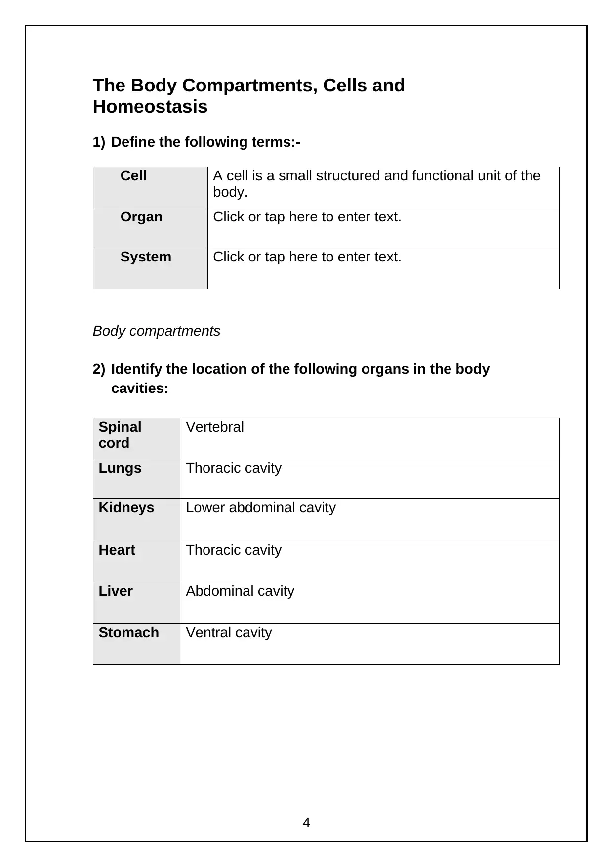
This life sciences workbook is designed for nursing students enrolled in a Pg Dip/MSc program, specifically focusing on mental health nursing. The workbook covers a wide range of topics related to human anatomy and physiology, including body compartments, cells, homeostasis, the integumentary, musculoskeletal, cardiovascular, respiratory, renal, gastrointestinal, nervous, endocrine, and reproductive systems. It also addresses wound healing and temperature regulation. The workbook serves as a learning resource to enhance the students' understanding of the body's structures and systems, providing a foundation for their nursing studies. The content includes definitions, identification of organ locations, descriptions of cell structures and functions, explanations of physiological processes like osmosis and diffusion, and outlines of key concepts like homeostasis, skin layers, and wound healing stages. The workbook aims to equip students with the necessary knowledge to succeed in their nursing program, and is to be submitted with their RPL portfolio as evidence of acquired knowledge. The workbook also includes learning resources such as a recommended reading list.
Contribute Materials
Your contribution can guide someone’s learning journey. Share your
documents today.

Life Sciences Workbook
To accompany RPL portfolio for
Pg. Dip/MSc nursing programme.
Candidate Name Jhanneal Whyte
Candidate Course Mental health Nursing
Date of completion 09/07/21
1
To accompany RPL portfolio for
Pg. Dip/MSc nursing programme.
Candidate Name Jhanneal Whyte
Candidate Course Mental health Nursing
Date of completion 09/07/21
1
Secure Best Marks with AI Grader
Need help grading? Try our AI Grader for instant feedback on your assignments.

Contents
Life Sciences Workbook.................................................................1
The Body Compartments, Cells and Homeostasis.........................4
The Skin (Integumentary System), Wound Healing and
Temperature Regulation.................................................................8
The Musculo Skeletal System.......................................................13
The Heart......................................................................................18
The Blood and Blood Vessels.......................................................21
The Respiratory System...............................................................25
The Renal System, Fluid and Electrolytes....................................29
The Gastro Intestinal System........................................................34
The Nervous System....................................................................38
The Endocrine System..................................................................41
The Reproductive System.............................................................43
Learning Resources......................................................................45
2

Workbook on Body Systems
The aim of this workbook is to enable our Pg Dip / MSc nursing
applicants, who have been offered a place on the programme and
who have not studied a health/science related degree before, to
develop and enhance their knowledge of the structures and
systems of the body.
It is essential to have a good understanding of the anatomy and
physiology of the body prior to commencing your programme of
study as one of your modules will build upon this knowledge.
The completed workbook must be submitted with your RPL
portfolio as evidence that you have acquired this knowledge.
You may use any resources you wish to help you with its
completion and at the back of this workbook you will find a
recommended reading list.
3

The Body Compartments, Cells and
Homeostasis
1) Define the following terms:-
Cell A cell is a small structured and functional unit of the
body.
Organ Click or tap here to enter text.
System Click or tap here to enter text.
Body compartments
2) Identify the location of the following organs in the body
cavities:
Spinal
cord
Vertebral
Lungs Thoracic cavity
Kidneys Lower abdominal cavity
Heart Thoracic cavity
Liver Abdominal cavity
Stomach Ventral cavity
4
Secure Best Marks with AI Grader
Need help grading? Try our AI Grader for instant feedback on your assignments.

Cells
3) Insert the appropriate words in the blank spaces against
the correct description below:-
Ribosomes
Mitochondria
Nucleus
Golgi apparatus
Centrioles
Mitochondria is where production of energy in the form of
ATP occurs
Centrioles are found near the nucleus and are
involved in cell division
Ribosomes is an intracellular factory that manufactures
protein
Golgi aaratus is the processing and packaging station for
the protein from the endoplasmic reticulum.
Nucleus is essential for all cellular activities and
contains genetic materials
4) Insert the following appropriate words in the blank
space against the correct description below:-
Osmosis
Active transport
Pinocytosis
Simple diffusion
Lysosomes
Osmosis is the movement of water through a semi
permeable membrane from a region of low
solute
Active transport is the movement of solutes/gas through a
semi permeable membrane from a region
of higher solute/gas
Pinocytosis is a mechanism by which cell membrane
indents and actually engulf the substance
5

to be absorbed into the cell
Simple diffusion requires energy derived from cellular
chemical reactions to move molecules or
ions against their concentration gradient
Lysosome contains digestive enzymes which digest
bacteria and cellular debris
5) Which word fully describes the cell structure; please enter
your response in the spaces provided under each heading?
Cell structure Location Function
Plasma membrane Forms the external
boundary of cells
Serves as a selectively
permeable membrane
which regulate material to
and from the cell
Lysome Found in the
cytoplasm
Contains digestive
enzymes
Mitochrondria Scattered
throughout the cells
Control release of energy
from food and forms ATP
Ribosomes Found in all cells Manufactures protein
Nucleolus Found in the
cytoplasm
Contains ribosomes and
is involved in protein
synthesis
6) Describe the structure of the cell membrane
Cell membrane consist mainly of double layers of phospholip
molecules that are align by chemical attractions. The phospholip
molecules are designed with polar hydrophilic water head and two
non polar hydrophobic water fearing tails. The membrane is
formed with the heads face out and the tails face in forms a bilayer
that acts like fence.
7) Explain the function of the cell membrane
Click or tap here to enter text.
6

8) Explain briefly what occurs during osmosis
Osmosis is the passage of water from a region of high water
concentration through a semi-permeable membrane to a region of
low water concentration. This is basically done to re establish the
equilibrium
9) Explain how diffusion across the cell membrane occurs
Click or tap here to enter text.
Homeostasis
10) What are ‘receptors’?
These are biological transducers that changes energy from both
external and internal environments into elecetrical impulses.
11) What are the receptors that detect body temperature
called?
Thermoreceptor
12) The ‘pathway’ that conveys messages towards the control
centre is known as the Click or tap here to enter text.
pathway.
13) The ‘pathway’ that conveys messages away from the
control centre is known as the Click or tap here to enter text.
pathway.
14) What are ‘effectors’?
Click or tap here to enter text.
.
15) The 2 most important systems involved in regulation and
the maintenance of homeostasis are:
1 Click or tap here to enter text.
2 Click or tap here to enter text.
7
Paraphrase This Document
Need a fresh take? Get an instant paraphrase of this document with our AI Paraphraser

16) Explain how the body strives to maintain homeostasis.
Click or tap here to enter text.
8
Click or tap here to enter text.
8

The Skin (Integumentary System), Wound
Healing and Temperature Regulation
The Skin
1) Select the correct term with the definition given.
Stratum basale Hypodermis
Stratum corneum Papillary layer
Stratum granulosum Epidermis (whole)
Stratum lucidum Dermis (whole)
Term Description
Dermis Layer of cells present only in thick skin
Stratum corneum Layer containing all dead cells
Stratum corneum Layer that sloughs off
Dermis Contains the blood vessels
Stratum basale Epidermal layer undergoing mitosis (cell
division)
Stratum lucidum Cells with most keratin in them
Epidermis Outer layer of skin
Hypodermis Deeper layer of skin
Epidermis Layer where accessory structures are
found
Stratum basale Epidermal layer where most melanocytes
are found
Stratum granulosum Layer that produces lipid rich secretion
Stratum basale Epidermal layer containing the oldest cells
Hypodermis Largely adipose tissue and anchors skin
to underlying tissue
9

2) Name the 5 layers of the epidermis starting with the
innermost layer
1 Stratum basale
2 Stratum spinosum
3 Stratum granulosum
4 Stratum lucidum
5 Stratum corneum
3) For the protective functions of skin, match the term
given with the definition of the role each part plays in
providing protection. Choose an answer from the
following:
Melanin, Sweat, Normal flora, Skin, Macrophages,
Lipids, Langerhans cells, Keratin
Answer Description
Click or tap
here to enter
text.
Involved in the sensation of touch
Macrophages Resist invasion by organisms by preventing harmful
bacteria from colonising
Click or tap
here to enter
text.
Prevent some evaporation on skin, protects hair and
skin from drying out, antibacterial due to low pH
Melanin Protects from UV light
Sweat Increases heat loss
Click or tap
here to enter
text.
Forms a protective barrier and protects underlying
tissues
Click or tap Phagocytose bacteria and viruses
10
Secure Best Marks with AI Grader
Need help grading? Try our AI Grader for instant feedback on your assignments.

here to enter
text.
Click or tap
here to enter
text.
A tough fibrous protein
4) What is an exocrine gland?
This is a cellur sub-structures, organs in a body that provide a
system to secrete substances out and external to the body.
5) List some of the structures found in the dermis
Click or tap here to enter text.
6) List the cells found in the epidermis and briefly describe
their function
Click or tap here to enter text.
Wound healing
7) If the epidermis is damaged during injury, which layer of
the epidermis enables new cells to grow?
Click or tap here to enter text.
8) Name the cells found in the skin that protect the skin from
pathogenic organisms
Click or tap here to enter text.
9) State what happens during each stage of wound healing.
Inflammation Click or tap here to enter text.
Regeneration Click or tap here to enter text.
11
text.
Click or tap
here to enter
text.
A tough fibrous protein
4) What is an exocrine gland?
This is a cellur sub-structures, organs in a body that provide a
system to secrete substances out and external to the body.
5) List some of the structures found in the dermis
Click or tap here to enter text.
6) List the cells found in the epidermis and briefly describe
their function
Click or tap here to enter text.
Wound healing
7) If the epidermis is damaged during injury, which layer of
the epidermis enables new cells to grow?
Click or tap here to enter text.
8) Name the cells found in the skin that protect the skin from
pathogenic organisms
Click or tap here to enter text.
9) State what happens during each stage of wound healing.
Inflammation Click or tap here to enter text.
Regeneration Click or tap here to enter text.
11

Maturation Click or tap here to enter text.
Homeostatic control of temperature
10) Using the key choices, select the terms that match the
following descriptions pertaining to body temperature
regulation.
Key choices
Hypothermia, Hyperthermia, Heat, Sensory (afferent pathway),
Thermoreceptors, Blood, Hypothalamus, Sweating, Shivering,
Temperature control centre, Motor (efferent pathway),
Vasoconstriction, Vasodilation
Answer Description
Click or tap here to
enter text.
Produced as a result of cell metabolism
Hypothalamus Thermoregulatory centre
Click or tap here to
enter text.
Medium that distributes heat to all the tissues
and carries it to the skin
Hypothalamus Site of the body’s temperature control centre
Click or tap here to
enter text.
Nerves that carry information about
temperature to the hypothalamus (control
centre)
Motor (efferent
pathway)
Nerves convey impulses away from the control
centre and activate the effectors
Shivering Means of liberating excess body heat
Hypothermia Extremely low body temperature
Hyperthermia Extremely high body temperature
Thermoreceptors Receptors that detect temperature
Sweating Generates heat when temperature falls too low
12

Vaasodilation Increases blood flow to the skin
Vasoconstriction Reduces blood flow to the skin
13
Vasoconstriction Reduces blood flow to the skin
13
Paraphrase This Document
Need a fresh take? Get an instant paraphrase of this document with our AI Paraphraser

The Musculo Skeletal System
1) How many bones does the adult human body have?
206
2) Name 3 substances that bone contains when fully
developed
1 Calcium
2 Click or tap here to enter text.
3 Click or tap here to enter text.
3) Name, and give examples of the four main types (shapes)
of bone
1 Flat
2 Click or tap here to enter text.
3 Click or tap here to enter text.
4 Click or tap here to enter text.
4) State the main function of a:
i) ligament
Ligaments are bones that are attached to other bones which
allows body movement without pain.
ii) tendon
Click or tap here to enter text.
14

5) List the main functions of the skeleton
Click or tap here to enter text.
6) What are the two types of bone tissue?
1 Compact bone
2 Cancellous bone
7) What are the functions of:
i) Osteoblasts
Click or tap here to enter text.
ii) Osteoclasts
Click or tap here to enter text.
8) Name the 5 main regions of the vertebral column and state
how many vertebrae there are in each region
1 Click or tap here to enter text.
2 Click or tap here to enter text.
3 Click or tap here to enter text.
4 Click or tap here to enter text.
5 Click or tap here to enter text.
15
Click or tap here to enter text.
6) What are the two types of bone tissue?
1 Compact bone
2 Cancellous bone
7) What are the functions of:
i) Osteoblasts
Click or tap here to enter text.
ii) Osteoclasts
Click or tap here to enter text.
8) Name the 5 main regions of the vertebral column and state
how many vertebrae there are in each region
1 Click or tap here to enter text.
2 Click or tap here to enter text.
3 Click or tap here to enter text.
4 Click or tap here to enter text.
5 Click or tap here to enter text.
15

Joints, Tendons and Ligaments
9) Name the three types of joint
Fibrous, Cartilaginous and Synovia.
10) Give two examples of where in the body each of the
three types of joint can be found
1 Click or tap here to enter text.
2 Click or tap here to enter text.
3 Click or tap here to enter text.
11) What type of joint allows most movement?
Synovial joints
12) What type of joint does not allow movement?
Fibrous joints
13) What attaches muscle to bone?
Tendon
14) Outline the function(s) of ‘tendons’
Click or tap here to enter text.
15) Outline the function(s) of ‘ligaments’
Click or tap here to enter text.
16
Secure Best Marks with AI Grader
Need help grading? Try our AI Grader for instant feedback on your assignments.

Muscles
16) Complete the following table, identifying the type of
muscle
Type of muscle Description
Click or tap here to
enter text.
Is under the control of the will (voluntary)
Click or tap here to
enter text.
Moves substances such as food through the
gastrointestinal tract
Click or tap here to
enter text.
Enables movement and ability to hold the
body upright
Click or tap here to
enter text.
Is activated by a specialised conducting
system in the heart
Click or tap here to
enter text.
Causes blood vessels to constrict
Click or tap here to
enter text.
Has short branching fibres
Click or tap here to
enter text.
2 types of muscle not under the control of
the will (involuntary)
17) The 3 types of muscle tissues exhibit certain similarities
and differences. Tick the appropriate one in the following
table to indicate the specific characteristic of each muscle
type
Skeletal Cardiac Smooth
Voluntary controlled ☐ ☐ ☐
Involuntary
Controlled
☐ ☐ ☐
Single nucleus in
each cell
☐ ☐ ☐
Found attached to
bone
☐ ☐ ☐
Allows the
movement of your
eyeball
☐ ☐ ☐
Found in walls of
stomach uterus and
arteries
☐ ☐ ☐
17

Contain cylindrical
cells with branching
end
☐ ☐ ☐
Concerned with
locomotion of the
body
☐ ☐ ☐
18
cells with branching
end
☐ ☐ ☐
Concerned with
locomotion of the
body
☐ ☐ ☐
18

The Heart
1) The site in the thoracic cavity that the heart occupies is
called Click or tap here to enter text.
2) The structure that separates the right side of the heart from
the left side is called the Click or tap here to enter text.
3) Oxygenated blood found is found on which side of the
heart?
Click or tap here to enter text.
4) Name the tiny muscle that anchors the chordae tendinae to
the inner wall of the heart.
Click or tap here to enter text.
5) What are Chordae Tendinae and what are their functions?
Click or tap here to enter text.
6) Explain the reason why the atria has thinner muscle
compared to that of the ventricles
Click or tap here to enter text.
7) Describe the function of the conducting system in the
heart.
Click or tap here to enter text.
19
Paraphrase This Document
Need a fresh take? Get an instant paraphrase of this document with our AI Paraphraser

8) Explain why one chamber of the heart has a much thicker
muscle wall compared to other chambers and give a
reason for this difference.
Click or tap here to enter text.
9) Describe the flow of blood through the heart starting from
the right atrium, ending with the left ventricle. (Name the
chambers, valves and blood vessels that enter and leave
each chamber).
Click or tap here to enter text.
10) Name the 3 layers of the heart and explain each of their
functions
1 Epicardium
2 Mycocardium
3 Endocardium
11) Describe pulmonary circulation; where does it start and
where does it finish (name the blood vessels, heart
chambers and heart valves)?
Click or tap here to enter text.
12) Name the blood vessel(s) that carry blood away from the
left ventricle
Aorta
13) Name the blood vessel(s) that carry blood away from the
right ventricle
Pulmonary artery
20

14) Name the blood vessel(s) that carry blood into the left
atrium
Click or tap here to enter text.
15) Name the blood vessel(s) that carry blood into the right
atrium.
Click or tap here to enter text.
16) Name the parts of the conducting system numbered 1-5
on the diagram below
1 Click or tap here to enter text.
2 Click or tap here to enter text.
3 Click or tap here to enter text.
4 Click or tap here to enter text.
5 Click or tap here to enter text.
21

The Blood and Blood Vessels
1) Match the description with the relevant term below:-
Tunica externa Tunica media Tunica intima
Term Description
Click or tap here to enter text. single thin layer of endothelium
Click or tap here to enter text. bulky middle coat, containing
smooth muscle and elastin
Click or tap here to enter text. anchors blood vessels to
surrounding structures
Click or tap here to enter text. the innermost layer
Click or tap here to enter text. also called the adventitia
Click or tap here to enter text. which is in contact with blood in the
lumen
Click or tap here to enter text. a supporting and protecting coat
2)Define the term ‘vein’
Click or tap here to enter text.
3)Define the term ’artery’
Click or tap here to enter text.
4)Define the term ‘ capillary’
Click or tap here to enter text.
22
Secure Best Marks with AI Grader
Need help grading? Try our AI Grader for instant feedback on your assignments.

5) Artery walls have three layers; name these layers
1 Tunica intima
2 Tunica media
3 Tunica adventitia
6) Vein walls have three layers; name these layers
1 Tunica intima
2 Tunica media
3 Tunica adventitia
7) Insert the appropriate words/letter against the description
below:-
Cardiac output, Blood pressure, Korotkoff sounds, pressure
points, diastolic blood pressure, Systolic blood pressure,
pulse
Term Description
Click or tap here to enter text. an expansion and recoil of an artery
following a heart beat
Click or tap here to enter text. pressure exerted by blood against
the blood vessel wall.
Click or tap here to enter text. maximum blood pressure following
left ventricular contraction
Click or tap here to enter text. minimum blood pressure following
cardiac relaxation.
23
1 Tunica intima
2 Tunica media
3 Tunica adventitia
6) Vein walls have three layers; name these layers
1 Tunica intima
2 Tunica media
3 Tunica adventitia
7) Insert the appropriate words/letter against the description
below:-
Cardiac output, Blood pressure, Korotkoff sounds, pressure
points, diastolic blood pressure, Systolic blood pressure,
pulse
Term Description
Click or tap here to enter text. an expansion and recoil of an artery
following a heart beat
Click or tap here to enter text. pressure exerted by blood against
the blood vessel wall.
Click or tap here to enter text. maximum blood pressure following
left ventricular contraction
Click or tap here to enter text. minimum blood pressure following
cardiac relaxation.
23

Click or tap here to enter text. point at body surfaces where a
pulse may be felt
Click or tap here to enter text. the sounds heard over an artery
when blood pressure is determined
by the auscultation.
Click or tap here to enter text. one of the factors contributing to the
maintenance of blood pressure.
8) List the characteristics of blood
Click or tap here to enter text.
9) Name the two main components in whole blood and the
percentage of each one
Plasma
Formed elements – blood cells
10) List the functions of a red blood cell.
This cell carries oxygen from the lungs to the rest of the body then
take carbon dioxide back to the lungs.
11) What is the blood volume of an average size adult?
Click or tap here to enter text.
12) List the main blood groups.
-
13) Which cells are more numerous in the blood?
Red blood cells
14) What is/are the general function(s) of white blood cells?
24

The general function of the white blood cell is to fight against
infection by producing antibodies.
15) What is/are the general function(s) of red blood cells?
Click or tap here to enter text.
16) Describe the formation, structure, shape and life span of a
red blood cell
Click or tap here to enter text.
25
infection by producing antibodies.
15) What is/are the general function(s) of red blood cells?
Click or tap here to enter text.
16) Describe the formation, structure, shape and life span of a
red blood cell
Click or tap here to enter text.
25
Paraphrase This Document
Need a fresh take? Get an instant paraphrase of this document with our AI Paraphraser

The Respiratory System
1) In the correct order, list the structures that air passes
through before it reaches the lungs.
Click or tap here to enter text.
2) List the structures that help to change the characteristic of
inspired air before it reaches the lungs.
Click or tap here to enter text.
3) Explain the changes that occur to the characteristics of
inspired air before it enters the lungs
Click or tap here to enter text.
4) List the structures that air passes through as it passes
from the trachea to the alveoli
Click or tap here to enter text.
5) Where are the cilia found in the respiratory tract?
Click or tap here to enter text.
6) Explain the functions of the:
Cillia This is when water is mo
Goblet
cells
Click or tap here to enter text.
Mucus Click or tap here to enter text.
26

7) What is an alveolus?
Click or tap here to enter text.
8) What is the ‘respiratory membrane’?
Click or tap here to enter text.
9) How many cells thick are capillary walls?
Click or tap here to enter text.
10) What type of cells form the walls of the capillary?
Click or tap here to enter text.
11) What type of cells form the walls of the alveolus?
Click or tap here to enter text.
12) How many cells thick are alveoli walls?
Click or tap here to enter text.
13) Explain the changes that occur to the chest cavity
during
Inspiration Click or tap here to enter text.
Expiration Click or tap here to enter text.
27
Click or tap here to enter text.
8) What is the ‘respiratory membrane’?
Click or tap here to enter text.
9) How many cells thick are capillary walls?
Click or tap here to enter text.
10) What type of cells form the walls of the capillary?
Click or tap here to enter text.
11) What type of cells form the walls of the alveolus?
Click or tap here to enter text.
12) How many cells thick are alveoli walls?
Click or tap here to enter text.
13) Explain the changes that occur to the chest cavity
during
Inspiration Click or tap here to enter text.
Expiration Click or tap here to enter text.
27

14) Explain the mechanism which causes air to move in and
out of the lungs (chest movement)
Click or tap here to enter text.
15) Describe the location and functions of the pleural
membrane.
Click or tap here to enter text.
16) What is the function of pulmonary fluid?
Click or tap here to enter text.
17) Name the layers of the pleural membrane and state
where each layer is situated
Click or tap here to enter text.
18) Explain how the pleural membrane enables the lungs to
expand as the thoracic cavity expands
Click or tap here to enter text.
19) Explain how gases move across the respiratory
membrane
Click or tap here to enter text.
20) Explain how oxygen is transported in the blood stream
(give percentages)
Click or tap here to enter text.
28
Secure Best Marks with AI Grader
Need help grading? Try our AI Grader for instant feedback on your assignments.

21) Explain the thee ways by which carbon dioxide is
transported in the blood stream (give percentages)
Click or tap here to enter text.
22) Describe diffusion
Click or tap here to enter text.
23) Explain why diffusion is important for the movement of
gases
Click or tap here to enter text.
29
transported in the blood stream (give percentages)
Click or tap here to enter text.
22) Describe diffusion
Click or tap here to enter text.
23) Explain why diffusion is important for the movement of
gases
Click or tap here to enter text.
29

The Renal System, Fluid and Electrolytes
1) Label the diagram below.
A Renal vein
B Left kidney
C Renal artery
D Ureter
E Bladder
F Urethra
A
B
C
D
E
F
30
1) Label the diagram below.
A Renal vein
B Left kidney
C Renal artery
D Ureter
E Bladder
F Urethra
A
B
C
D
E
F
30

2) List the functions of the renal system
The function of the renal system is to exertion of waste and toxins
3) List the three processes by which urine is formed
1 filteration
2 reabsorption
3 secretion
4) Describe what occurs during each of the three process of
urine formation
1 Click or tap here to enter text.
2 Click or tap here to enter text.
3 Click or tap here to enter text.
5) List the contents of urine
Click or tap here to enter text.
6) List the parts of a nephron
Click or tap here to enter text.
7) What helps urine flow down the ureters?
Click or tap here to enter text.
8) Name the blood vessels that take blood to and away from
the glomerulus
31
Paraphrase This Document
Need a fresh take? Get an instant paraphrase of this document with our AI Paraphraser

Click or tap here to enter text.
9) What is the function of the bladder?
Click or tap here to enter text.
10)List the structures urine flows through after it leaves the
collecting duct until it leaves the body
Click or tap here to enter text.
11)Describe six different functions of the kidney
1 Click or tap here to enter text.
2 Click or tap here to enter text.
3 Click or tap here to enter text.
4 Click or tap here to enter text.
5 Click or tap here to enter text.
6 Click or tap here to enter text.
Fluid Balance
1) State the 3 ways by which we acquire water
1 Click or tap here to enter text.
2 Click or tap here to enter text.
3 Click or tap here to enter text.
2) How much should the average sized adult drink per day?
32

Click or tap here to enter text.
3) How much water do we lose per day?
Click or tap here to enter text.
4) State the 3 main ways fluid body fluid is lost (in health) and
state how much is lost by each way
1 Click or tap here to enter text.
2 Click or tap here to enter text.
3 Click or tap here to enter text.
5) Explain what electrolytes are
Click or tap here to enter text.
6) What are the 2 fluid compartments of the body?
1 Click or tap here to enter text.
2 Click or tap here to enter text.
7) Explain where each fluid compartment is located
Click or tap here to enter text.
8) What is a cation?
Click or tap here to enter text.
9) What is an anion?
Click or tap here to enter text.
33

10) List some of the cations and anions found in the body
Click or tap here to enter text.
11) Which ion is involved in maintaining the ‘acid balance’
(pH)?
Click or tap here to enter text.
12) What is the normal pH of blood?
Click or tap here to enter text.
34
Click or tap here to enter text.
11) Which ion is involved in maintaining the ‘acid balance’
(pH)?
Click or tap here to enter text.
12) What is the normal pH of blood?
Click or tap here to enter text.
34
Secure Best Marks with AI Grader
Need help grading? Try our AI Grader for instant feedback on your assignments.

The Gastro Intestinal System
1) What is an enzyme?
Click or tap here to enter text.
2) Name the enzyme in saliva that starts the breakdown of
carbohydrates.
Click or tap here to enter text.
3) Name the salivary glands and list their position in the
mouth
Click or tap here to enter text.
4) Name the constituents of saliva.
Click or tap here to enter text.
5) Define a sphincter
Click or tap here to enter text.
6) The Sphincter found between the stomach and the small
intestine is called:
Click or tap here to enter text.
35

7) Name the numbered structures in the diagram below:
1 Click or tap here to enter text.
2 Click or tap here to enter text.
3 Click or tap here to enter text.
4 Click or tap here to enter text.
5 Anus
6 Click or tap here to enter text.
7 Click or tap here to enter text.
36
1 Click or tap here to enter text.
2 Click or tap here to enter text.
3 Click or tap here to enter text.
4 Click or tap here to enter text.
5 Anus
6 Click or tap here to enter text.
7 Click or tap here to enter text.
36

8) What type of muscle contraction enables the movement of
food through the digestive tract?
Click or tap here to enter text.
9) Describe the following :-
The length of the
small intestine
Click or tap here to enter text.
The different
sections of the
small intestine
Click or tap here to enter text.
The functions of
the small
intestine.
Click or tap here to enter text.
10) Describe the following: -
The length,
position and
different parts of
the large intestine
Click or tap here to enter text.
The functions of
the large intestine
Click or tap here to enter text.
11) Explain the chemical digestion of the following
foodstuffs. Name the enzymes, where they are released
from and how they break down each of the following:
(i) Protein
Click or tap here to enter text.
37
Paraphrase This Document
Need a fresh take? Get an instant paraphrase of this document with our AI Paraphraser

(i) Fats
Click or tap here to enter text.
(iii) Carbohydrates
Click or tap here to enter text.
13) Before each nutrient can be absorbed it is broken down
into its smallest component. What are the following
absorbed as:
Nutrient Absorbed as:
Protein Click or tap here to enter text.
Fats Click or tap here to enter text.
Carbohydrates Click or tap here to enter text.
14) Define the following terms :-
Ingestion Click or tap here to enter text.
Digestion Click or tap here to enter text.
Absorption Click or tap here to enter text.
Excretion Click or tap here to enter text.
15) Explain the processes that occur during the act of
defecation.
Click or tap here to enter text.
38
Click or tap here to enter text.
(iii) Carbohydrates
Click or tap here to enter text.
13) Before each nutrient can be absorbed it is broken down
into its smallest component. What are the following
absorbed as:
Nutrient Absorbed as:
Protein Click or tap here to enter text.
Fats Click or tap here to enter text.
Carbohydrates Click or tap here to enter text.
14) Define the following terms :-
Ingestion Click or tap here to enter text.
Digestion Click or tap here to enter text.
Absorption Click or tap here to enter text.
Excretion Click or tap here to enter text.
15) Explain the processes that occur during the act of
defecation.
Click or tap here to enter text.
38

The Nervous System
1) Draw and label a typical neurone
2) Name the 2 parts that form the central nervous system?
1 Click or tap here to enter text.
2 Click or tap here to enter text.
3) What are the functions of dendrites?
Click or tap here to enter text.
4) What are the functions of axons?
Click or tap here to enter text.
5) Explain the function(s) of sensory neurones
Click or tap here to enter text.
6) Explain the function(s) of motor neurones
Click or tap here to enter text.
39

7) What is a synapse?
Click or tap here to enter text.
8) What are neuro transmitters? Where are they released?
What are their functions?
Click or tap here to enter text.
9) Name the two branches of the autonomic nervous system.
1 Click or tap here to enter text.
2 Click or tap here to enter text.
10) What are the coverings of the brain and spinal cord
collectively known as.?
Click or tap here to enter text.
11) What does CSF stand for and where is it produced?
Click or tap here to enter text.
12) List the functions of CSF.
Click or tap here to enter text.
13) The outer part of the brain is the Click or tap here to enter
text.
40
Click or tap here to enter text.
8) What are neuro transmitters? Where are they released?
What are their functions?
Click or tap here to enter text.
9) Name the two branches of the autonomic nervous system.
1 Click or tap here to enter text.
2 Click or tap here to enter text.
10) What are the coverings of the brain and spinal cord
collectively known as.?
Click or tap here to enter text.
11) What does CSF stand for and where is it produced?
Click or tap here to enter text.
12) List the functions of CSF.
Click or tap here to enter text.
13) The outer part of the brain is the Click or tap here to enter
text.
40
Secure Best Marks with AI Grader
Need help grading? Try our AI Grader for instant feedback on your assignments.

This is subdivided into 4 different lobes. These lobes have
the same names as the portion of the skull that covers that
lobe. The 4 different lobes are called:
1 Click or tap here to enter text.
2 Click or tap here to enter text.
3 Click or tap here to enter text.
4 Click or tap here to enter text.
14) The functions of each lobe are:
1 Click or tap here to enter text.
2 Click or tap here to enter text.
3 Click or tap here to enter text.
4 Click or tap here to enter text.
15) What is the function of the nervous system?
Click or tap here to enter text.
16) Describe the functional effects of the sympathetic
nervous system
Click or tap here to enter text.
17) Describe the functional effects of the parasympathetic
nervous system
Click or tap here to enter text.
41

The Endocrine System
1) Explain the difference between an endocrine and an
exocrine gland.
Click or tap here to enter text.
2) Give examples of some exocrine and endocrine glands.
Click or tap here to enter text.
3) What is a hormone?
Click or tap here to enter text.
4) Explain why hormones only influence specific cells and
tissues of the body.
Click or tap here to enter text.
5) Fill in the names of the hormones produced by the
following endocrine glands and briefly outline their action
Gland Hormone Name and Action
Posterior
Pituitary
1 Click or tap here to enter text.
2 Click or tap here to enter text.
Anterior
Pituitary
1 Click or tap here to enter text.
2 Click or tap here to enter text.
42

3 Click or tap here to enter text.
4 Click or tap here to enter text.
5 Click or tap here to enter text.
6 Click or tap here to enter text.
The
Thyroid
Gland
1 Click or tap here to enter text.
2 Click or tap here to enter text.
3 Click or tap here to enter text.
Parathyroid
Glands
1 Click or tap here to enter text.
2 Click or tap here to enter text.
Adrenal
Medulla
1 Click or tap here to enter text.
2 Click or tap here to enter text.
Adrenal
Cortex
1 Click or tap here to enter text.
2 Click or tap here to enter text.
Pancreas 1 Click or tap here to enter text.
2 Click or tap here to enter text.
43
Paraphrase This Document
Need a fresh take? Get an instant paraphrase of this document with our AI Paraphraser

The Reproductive System
1) Name the hormones released by the anterior pituitary that
act on the ovaries
Click or tap here to enter text.
2) On which day of the menstrual cycle is the mature egg
(ovum) released?
Click or tap here to enter text.
3) Into what area is the ovulated cell released?
Click or tap here to enter text.
4) Describe the structure and explain the function of Fimbriae
Click or tap here to enter text.
5) List the three layers of the uterus, starting with the outer
layer.
1 Click or tap here to enter text.
2 Click or tap here to enter text.
3 Click or tap here to enter text.
6) Explain how the ovum moves along the fallopian tube
Click or tap here to enter text.
7) Where does fertilization usually take place?
Click or tap here to enter text.
8) On what days of the menstrual cycle can fertilisation
occur? Explain why it can only occur on these days.
Click or tap here to enter text.
44

9) Which part of the uterus is broken down and lost during
menstruation?
Click or tap here to enter text.
10) Name the organ that delivers semen to the female
reproductive tract
Click or tap here to enter text.
11) Name the site of sperm and testosterone production
Click or tap here to enter text.
12) Name the external sac that houses the testes
Click or tap here to enter text.
13) Name the gland that surrounds the urethra at the base of
the bladder and produces a milky fluid
Click or tap here to enter text.
14) Approximately how much fluid is released per
ejaculation?
Click or tap here to enter text.
15) Approximately how many sperm are there in each 1ml of
ejaculatory fluid?
Click or tap here to enter text.
45

Learning Resources
Marieb, E.N. (2010) Essentials of Human Anatomy and
Physiology (10th Ed) Pearson, Benjamin Cummings San
Francisco.
McKissock, C. (2014) Great Ways to Learn Anatomy and
Physiology (2nd ed). Palgrave. London.
Tortora, G. & Derrickson, B. (2013) Essentials of Anatomy
and Physiology (9th Ed) John Wiley and sons, (Asia)
Thibodeau, G., Patton, K. (2013) The Human Body in Health
and Disease. (6th Ed) Mosby, Inc. USA.
Marieb, E.N., Hoehn, K. (2014) Human Anatomy and
Physiology (9th Ed) Pearson, Benjamin Cummings San
Francisco.
Tortora G.J., Derrickson B (2012) Principles of Anatomy and
Physiology: Organisation, Support and Movement and Control
Systems of the Human Body (13th Ed) John Wiley
46
1 out of 46

Your All-in-One AI-Powered Toolkit for Academic Success.
+13062052269
info@desklib.com
Available 24*7 on WhatsApp / Email
Unlock your academic potential
© 2024 | Zucol Services PVT LTD | All rights reserved.