CNA511 Oncology Nursing: Liver Cancer Case Study Analysis
VerifiedAdded on 2023/04/19
|12
|939
|145
Report
AI Summary
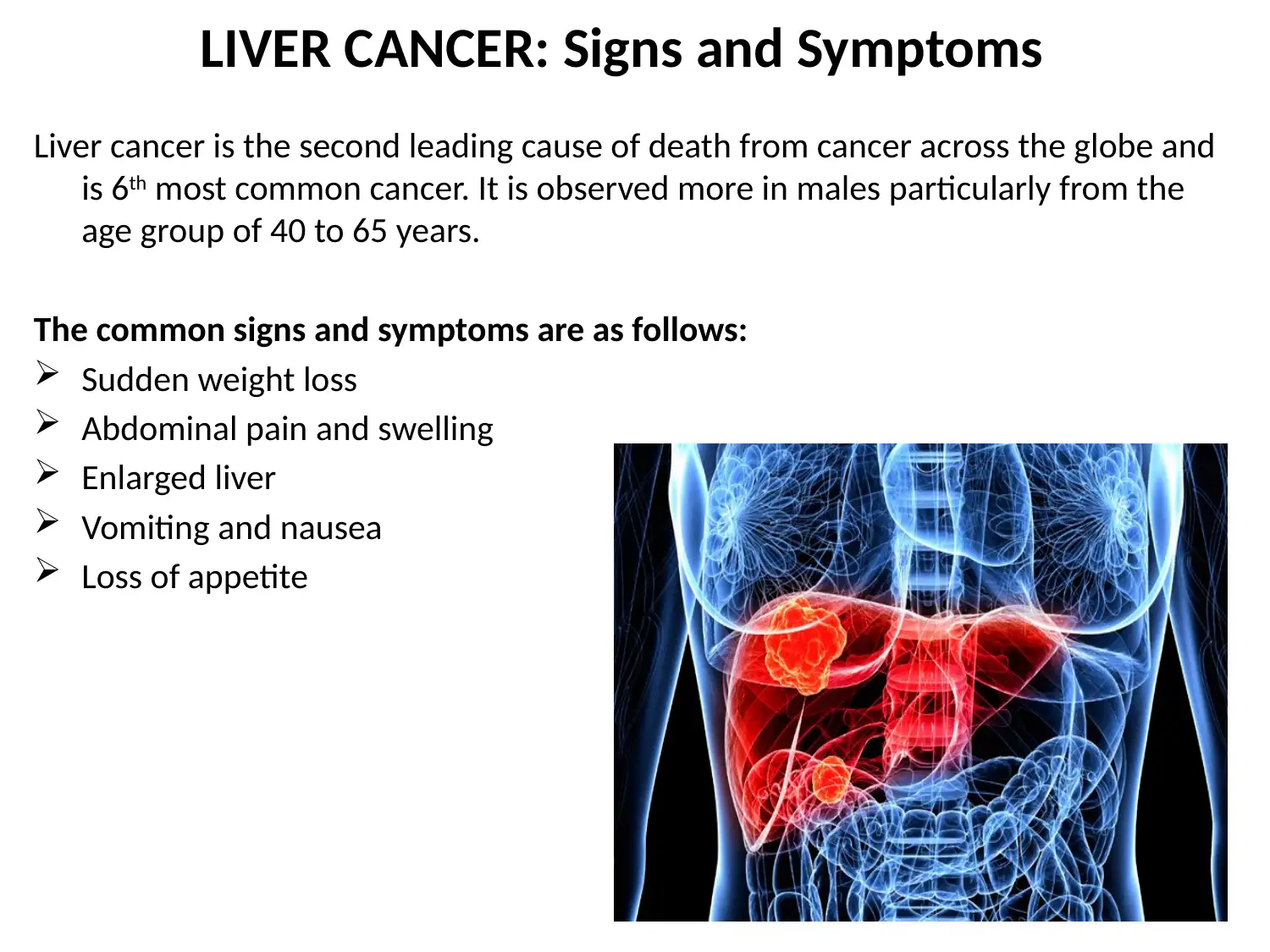
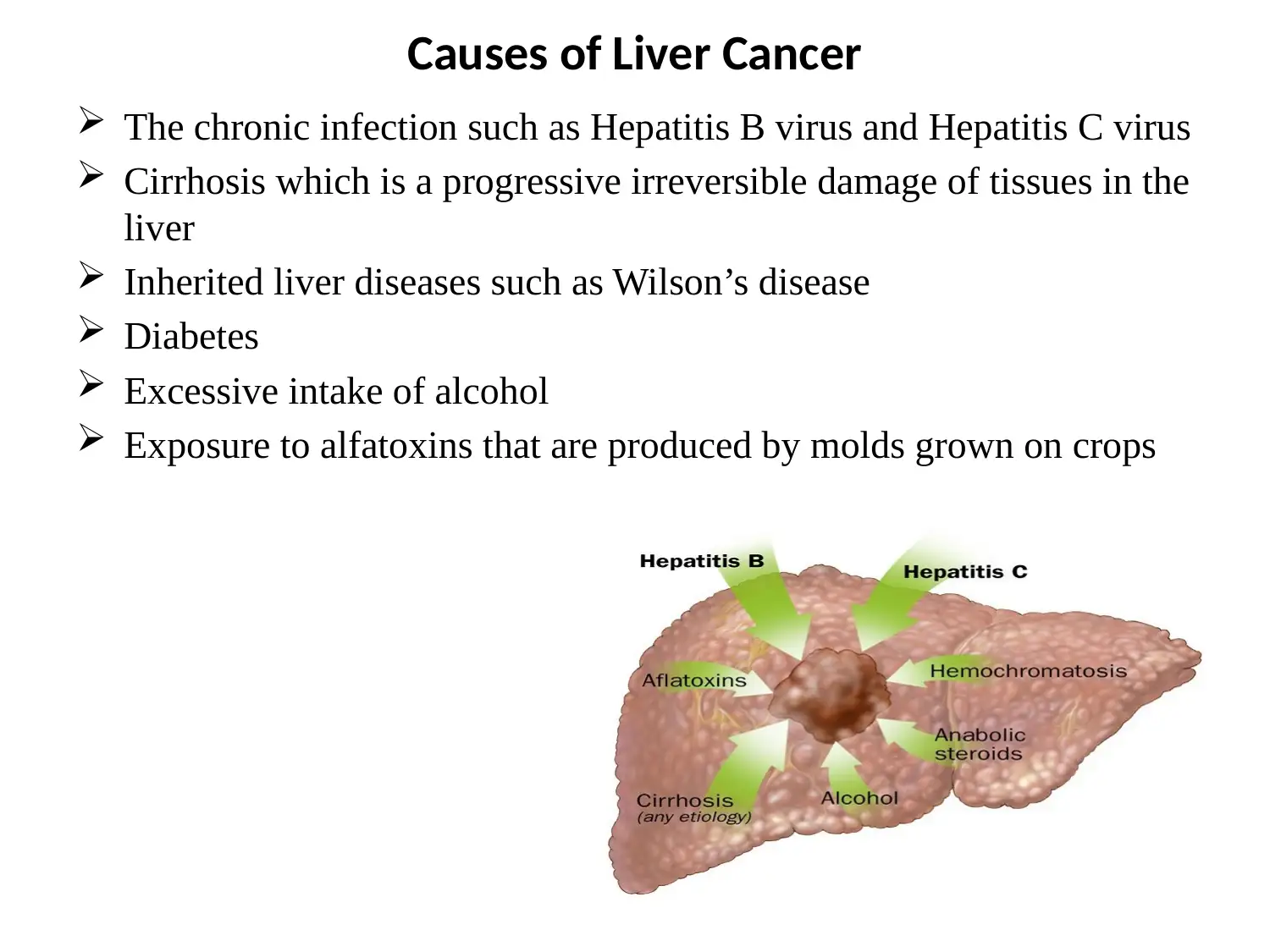
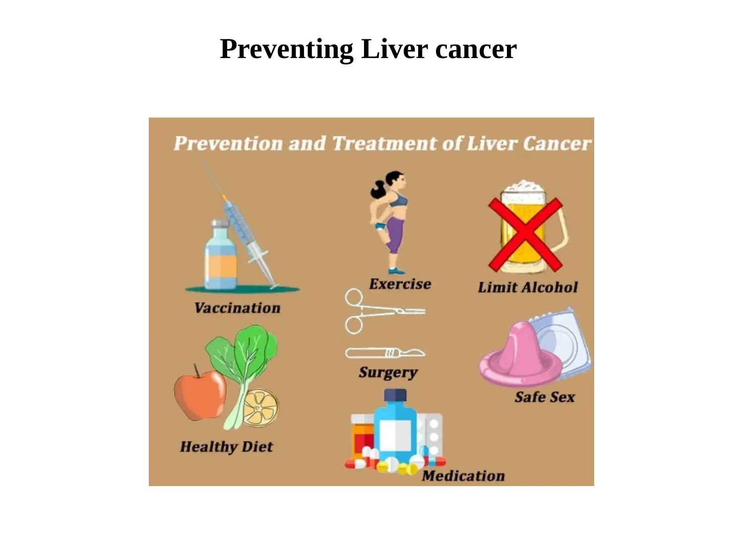
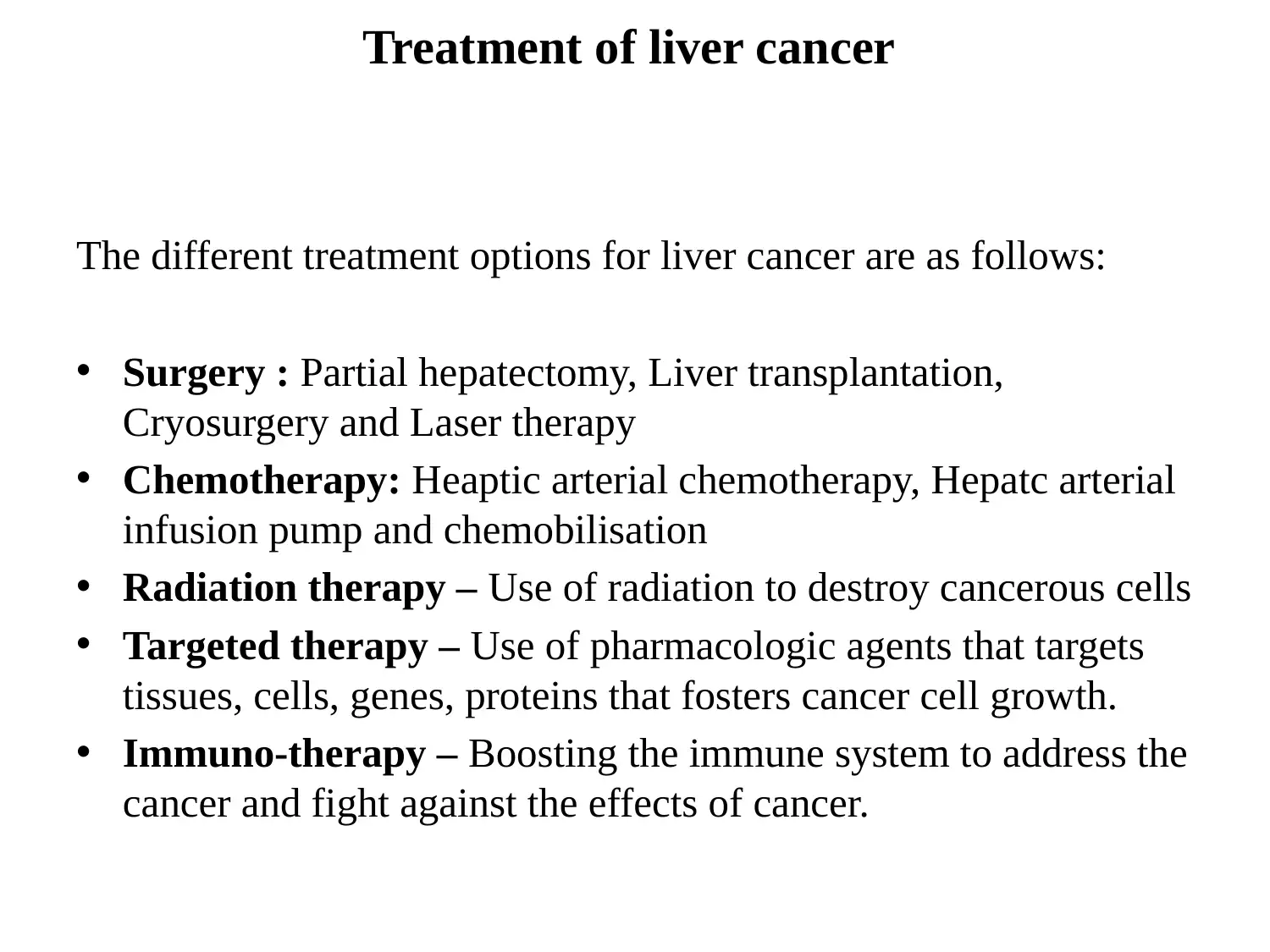
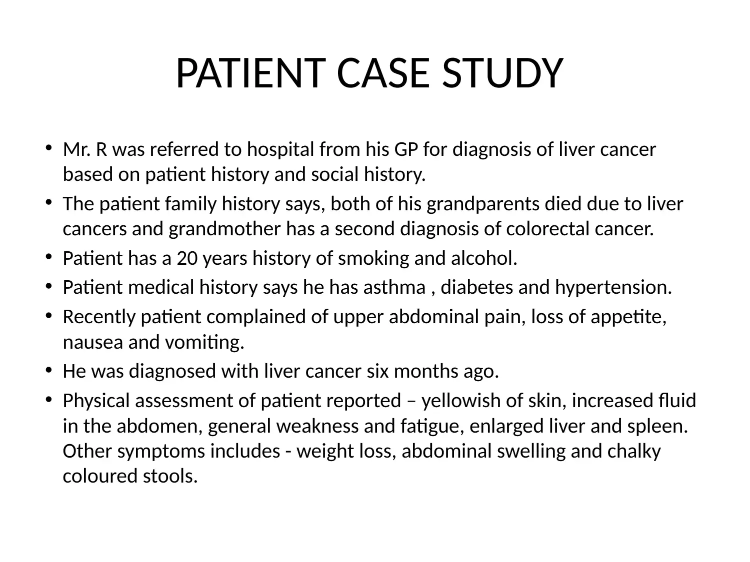
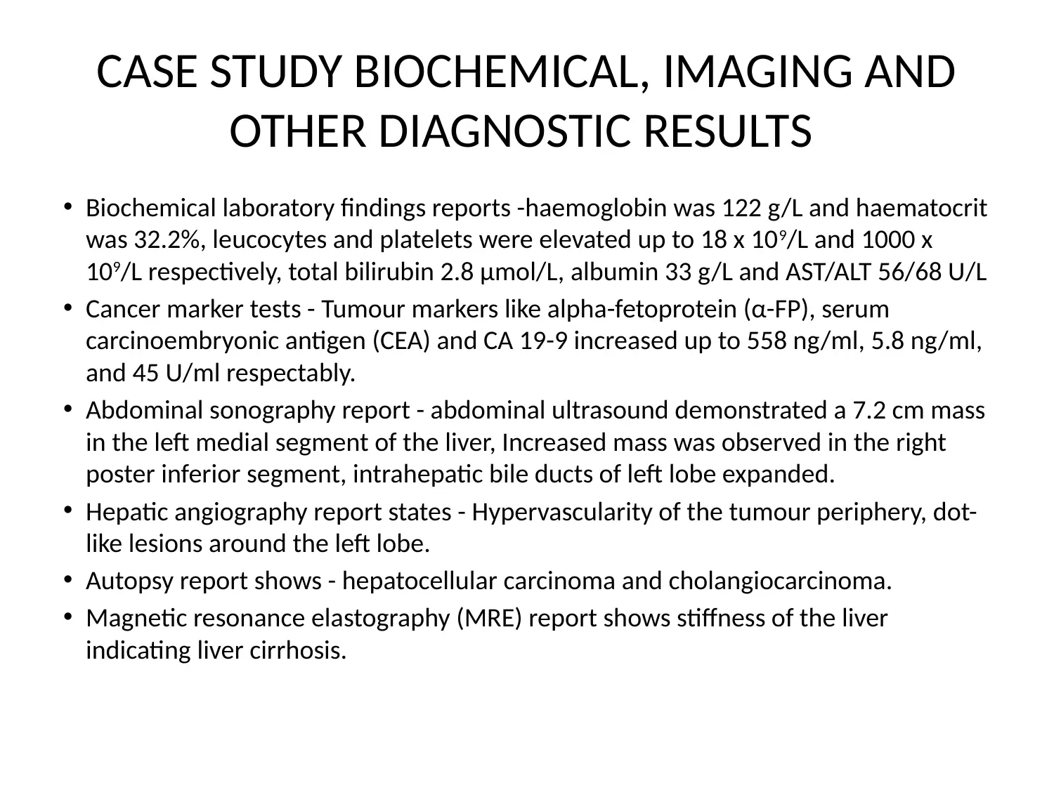
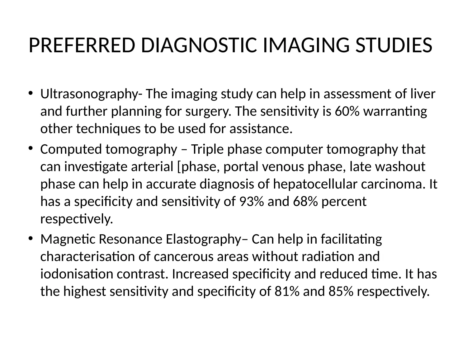
This report presents a comprehensive case study on liver cancer, detailing the signs, symptoms, causes, and various treatment options. The case study focuses on a 42-year-old male patient, Mr. R, with a family history of liver cancer, smoking, alcohol consumption, and pre-existing conditions such as asthma, diabetes, and hypertension. The report includes a review of the patient's history, physical assessment, and diagnostic results, including biochemical laboratory findings, cancer marker tests, and imaging reports such as abdominal sonography, hepatic angiography, and magnetic resonance elastography. The report also covers staging of liver cancer and nursing interventions. The references include research papers and medical journals on liver cancer, diagnosis, and treatment.

LIVER CANCER
Paraphrase This Document
Need a fresh take? Get an instant paraphrase of this document with our AI Paraphraser

LIVER CANCER: Signs and Symptoms
Liver cancer is the second leading cause of death from cancer across the globe and
is 6th most common cancer. It is observed more in males particularly from the
age group of 40 to 65 years.
The common signs and symptoms are as follows:
Sudden weight loss
Abdominal pain and swelling
Enlarged liver
Vomiting and nausea
Loss of appetite

Causes of Liver Cancer
The chronic infection such as Hepatitis B virus and Hepatitis C virus
Cirrhosis which is a progressive irreversible damage of tissues in the
liver
Inherited liver diseases such as Wilson’s disease
Diabetes
Excessive intake of alcohol
Exposure to alfatoxins that are produced by molds grown on crops
The chronic infection such as Hepatitis B virus and Hepatitis C virus
Cirrhosis which is a progressive irreversible damage of tissues in the
liver
Inherited liver diseases such as Wilson’s disease
Diabetes
Excessive intake of alcohol
Exposure to alfatoxins that are produced by molds grown on crops
⊘ This is a preview!⊘
Do you want full access?
Subscribe today to unlock all pages.

Trusted by 1+ million students worldwide

Preventing Liver cancer
Paraphrase This Document
Need a fresh take? Get an instant paraphrase of this document with our AI Paraphraser

Treatment of liver cancer
The different treatment options for liver cancer are as follows:
• Surgery : Partial hepatectomy, Liver transplantation,
Cryosurgery and Laser therapy
• Chemotherapy: Heaptic arterial chemotherapy, Hepatc arterial
infusion pump and chemobilisation
• Radiation therapy – Use of radiation to destroy cancerous cells
• Targeted therapy – Use of pharmacologic agents that targets
tissues, cells, genes, proteins that fosters cancer cell growth.
• Immuno-therapy – Boosting the immune system to address the
cancer and fight against the effects of cancer.

PATIENT CASE STUDY
• Mr. R was referred to hospital from his GP for diagnosis of liver cancer
based on patient history and social history.
• The patient family history says, both of his grandparents died due to liver
cancers and grandmother has a second diagnosis of colorectal cancer.
• Patient has a 20 years history of smoking and alcohol.
• Patient medical history says he has asthma , diabetes and hypertension.
• Recently patient complained of upper abdominal pain, loss of appetite,
nausea and vomiting.
• He was diagnosed with liver cancer six months ago.
• Physical assessment of patient reported – yellowish of skin, increased fluid
in the abdomen, general weakness and fatigue, enlarged liver and spleen.
Other symptoms includes - weight loss, abdominal swelling and chalky
coloured stools.
⊘ This is a preview!⊘
Do you want full access?
Subscribe today to unlock all pages.

Trusted by 1+ million students worldwide

CASE STUDY BIOCHEMICAL, IMAGING AND
OTHER DIAGNOSTIC RESULTS
• Biochemical laboratory findings reports -haemoglobin was 122 g/L and haematocrit
was 32.2%, leucocytes and platelets were elevated up to 18 x 109/L and 1000 x
109/L respectively, total bilirubin 2.8 μmol/L, albumin 33 g/L and AST/ALT 56/68 U/L
• Cancer marker tests - Tumour markers like alpha-fetoprotein (α-FP), serum
carcinoembryonic antigen (CEA) and CA 19-9 increased up to 558 ng/ml, 5.8 ng/ml,
and 45 U/ml respectably.
• Abdominal sonography report - abdominal ultrasound demonstrated a 7.2 cm mass
in the left medial segment of the liver, Increased mass was observed in the right
poster inferior segment, intrahepatic bile ducts of left lobe expanded.
• Hepatic angiography report states - Hypervascularity of the tumour periphery, dot-
like lesions around the left lobe.
• Autopsy report shows - hepatocellular carcinoma and cholangiocarcinoma.
• Magnetic resonance elastography (MRE) report shows stiffness of the liver
indicating liver cirrhosis.
Paraphrase This Document
Need a fresh take? Get an instant paraphrase of this document with our AI Paraphraser

PREFERRED DIAGNOSTIC IMAGING STUDIES
• Ultrasonography- The imaging study can help in assessment of liver
and further planning for surgery. The sensitivity is 60% warranting
other techniques to be used for assistance.
• Computed tomography – Triple phase computer tomography that
can investigate arterial [phase, portal venous phase, late washout
phase can help in accurate diagnosis of hepatocellular carcinoma. It
has a specificity and sensitivity of 93% and 68% percent
respectively.
• Magnetic Resonance Elastography– Can help in facilitating
characterisation of cancerous areas without radiation and
iodonisation contrast. Increased specificity and reduced time. It has
the highest sensitivity and specificity of 81% and 85% respectively.

LIVER BIOPSY PICTORIAL PRESENTATION
⊘ This is a preview!⊘
Do you want full access?
Subscribe today to unlock all pages.

Trusted by 1+ million students worldwide

STAGING OF LIVER CANCER
Paraphrase This Document
Need a fresh take? Get an instant paraphrase of this document with our AI Paraphraser

NURSING INTERVENTION- LIVER CANCER
• Nursing intervention depends on in which
stage the cancer is currently present
• Nursing intervention depends on in which
stage the cancer is currently present

REFERENCES
• Muriel, P. (2017). The liver: General aspects and epidemiology. In Liver
Pathophysiology (pp. 3-22). Academic Press.
• Kudo, M. (2015). Surveillance, diagnosis, treatment, and outcome of liver cancer in
Japan. Liver Cancer, 4(1), 39-50.
• Bruix, J., Reig, M., & Sherman, M. (2016). Evidence-based diagnosis, staging, and
treatment of patients with hepatocellular carcinoma. Gastroenterology, 150(4), 835-
853.
• Pepin, K. M., Ehman, R. L., & McGee, K. P. (2015). Magnetic resonance elastography
(MRE) in cancer: technique, analysis, and applications. Progress in nuclear magnetic
resonance spectroscopy, 90, 32-48.
• Oliveira, I. S., Kilcoyne, A., Everett, J. M., Mino-Kenudson, M., Harisinghani, M. G., &
Ganesan, K. (2017). Cholangiocarcinoma: classification, diagnosis, staging, imaging
features, and management. Abdominal radiology, 42(6), 1637-1649.
• Tapper, E. B., & Lok, A. S. F. (2017). Use of liver imaging and biopsy in clinical
practice. New England Journal of Medicine, 377(8), 756-768.
⊘ This is a preview!⊘
Do you want full access?
Subscribe today to unlock all pages.

Trusted by 1+ million students worldwide
1 out of 12
Your All-in-One AI-Powered Toolkit for Academic Success.
+13062052269
info@desklib.com
Available 24*7 on WhatsApp / Email
Unlock your academic potential
Copyright © 2020–2025 A2Z Services. All Rights Reserved. Developed and managed by ZUCOL.

