A Comprehensive Report on Lower Limb Deep Vein Thrombosis (DVT)
VerifiedAdded on  2022/10/17
|9
|1474
|14
Report
AI Summary
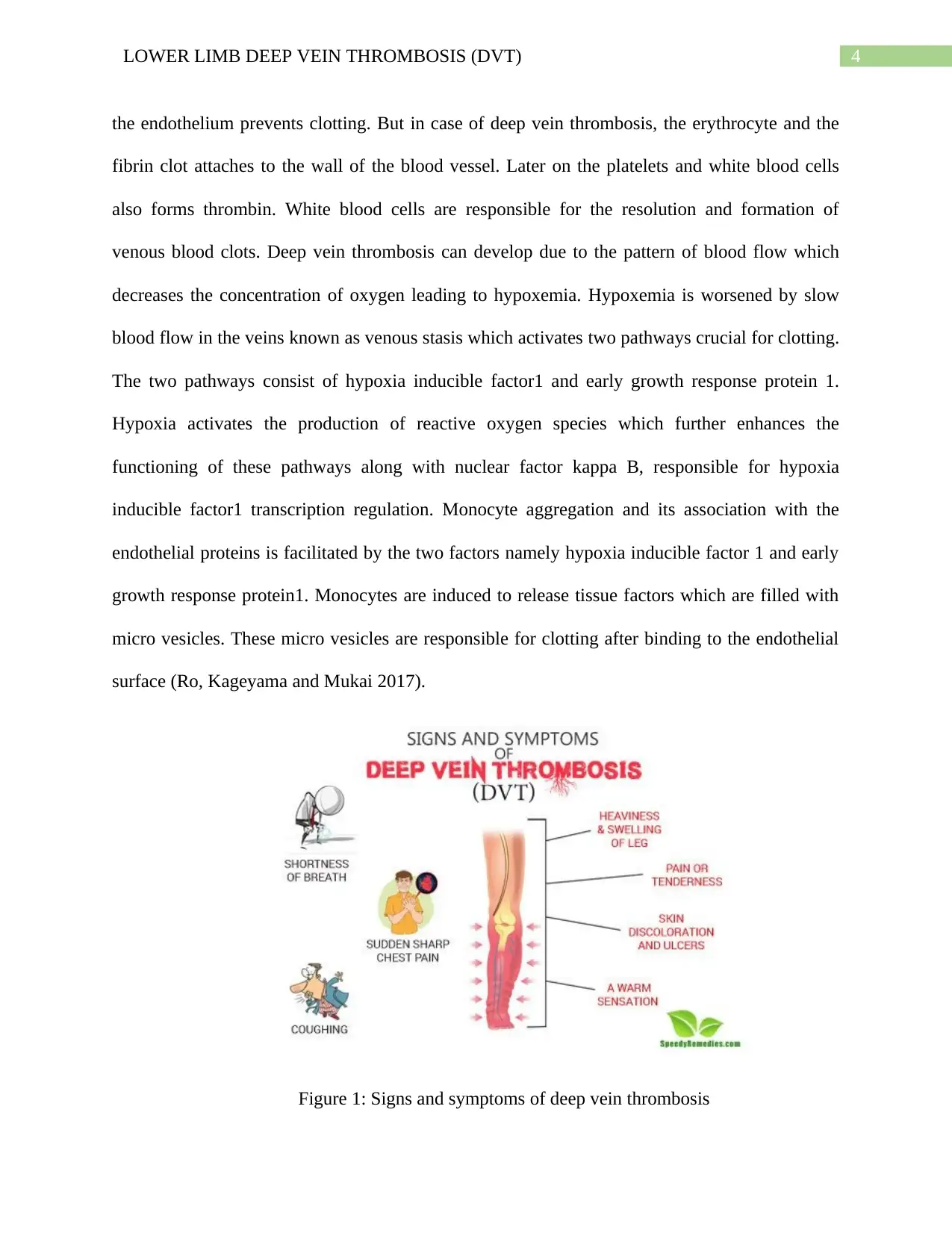
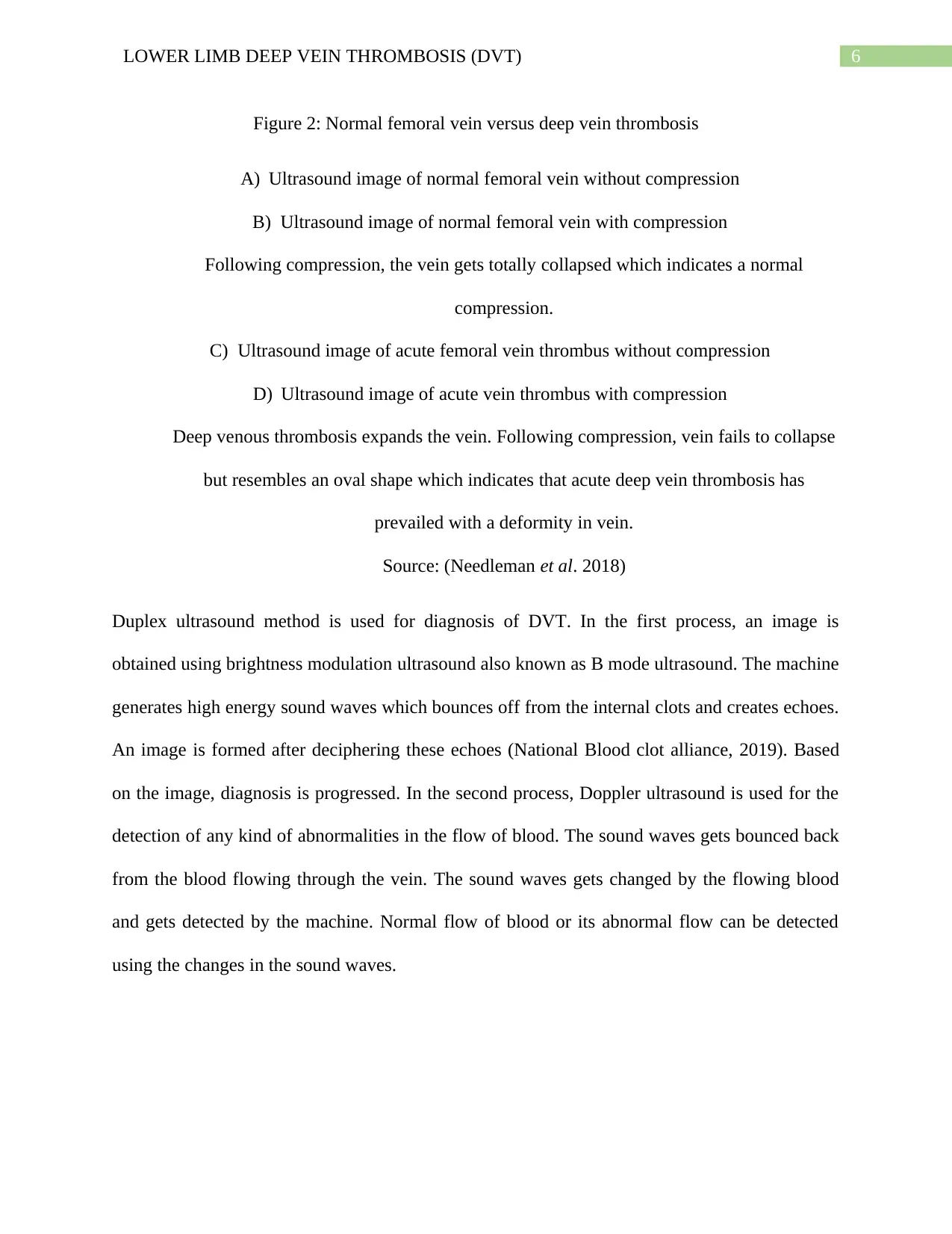
This report offers an in-depth analysis of Lower Limb Deep Vein Thrombosis (DVT), a clinical condition characterized by blood clots in the deep veins of the lower extremities. The executive summary highlights the report's focus on DVT, its pathogenesis, and sonographic appearance. The introduction defines DVT, outlining its symptoms, risk factors, and common treatment methods such as anticoagulation therapy. The discussion section delves into the pathophysiology, explaining how DVT develops and the role of fibrinolysis, the affected veins, and the mechanisms of blood clotting involving tissue factors, thrombin, and the impact of venous stasis and hypoxia. The report also describes the sonographic appearance of DVT, explaining how ultrasound imaging is used for diagnosis, differentiating between normal and thrombosed veins, and the use of duplex ultrasound. The conclusion emphasizes the treatability of DVT with early detection and the importance of preventive measures. References to relevant research papers are included to support the information presented.

Running Head: LOWER LIMB DEEP VEIN THROMBOSIS (DVT)
LOWER LIMB DEEP VEIN THROMBOSIS
Name of the Student
Name of the University
Authors Note
LOWER LIMB DEEP VEIN THROMBOSIS
Name of the Student
Name of the University
Authors Note
Secure Best Marks with AI Grader
Need help grading? Try our AI Grader for instant feedback on your assignments.

1LOWER LIMB DEEP VEIN THROMBOSIS (DVT)
Executive Summary:
The report deals with Lower Vein Deep Vein thrombosis (DVT). It is a clinical disease which
can occur in any type of individual. Thrombosis means a blood clot which gets accumulated in
the deep veins of lower extremities in our body. The report aims to give a brief description on
deep vein thrombosis followed by its pathogenesis. Ultrasound imaging appearance is also
discussed through an easy explanation from an image. The report finally concludes by addressing
certain measures of preventing the disease.

2LOWER LIMB DEEP VEIN THROMBOSIS (DVT)
Table of Contents
Introduction:....................................................................................................................................3
Discussion:.......................................................................................................................................3
Pathophysiology of the disease:...................................................................................................3
Sonographic appearance description:..........................................................................................5
Conclusion:......................................................................................................................................6
References:......................................................................................................................................7

3LOWER LIMB DEEP VEIN THROMBOSIS (DVT)
Introduction:
Deep vein thrombosis (DVT) is a disease where blood gets clotted in a deep vein, mostly
legs. The signs and symptoms of the disease are pain, swelling, redness, increase in temperature
of the affected area (Watson, Broderick and Armon 2016). Most critical case may include the
detachment of the clot from the leg vein and its transport to the lungs leading to difficulty in
breathing which is known as pulmonary embolism and in most severe cases, post-thrombotic
syndrome may arise. The factors which increases the risk of developing deep vein thrombosis
comprises of obesity, smoking, cancer, trauma, lack of locomotion, pregnancy, and certain
genetic disorders. The most common treatment procedure for thrombosis is anticoagulation
therapy which are known as blood thinners. The treatment includes drugs comprising of low
molecular weight heparin or direct oral anticoagulant. The report aims to address the
pathophysiology of the disease along with certain sonographic description.
Discussion:
Pathophysiology of the disease:
Deep vein thrombosis develops in the direction of venous blood flow that is towards
heart. DVT in initial stages can be cured naturally by the process of fibrinolysis where the clot
dissolves into blood (Riva, Donadini and Ageno 2015). The femoral vein, popliteal vein and the
iliofemoral vein is affected the most. Extreme cases can affect the inferior vena cava also.
Clotting in veins occurs without any damage to the vein wall unlike arterial thrombosis where
the blood vessel wall is damaged. The blood clotting in vein starts by the action of tissue factor
which converts prothrombin to thrombin leading to fibrin deposition. The thrombin in the vein
comprises of mainly red blood cells and fibrin. Usually the lining of the blood vessel wall that is
Secure Best Marks with AI Grader
Need help grading? Try our AI Grader for instant feedback on your assignments.

4LOWER LIMB DEEP VEIN THROMBOSIS (DVT)
the endothelium prevents clotting. But in case of deep vein thrombosis, the erythrocyte and the
fibrin clot attaches to the wall of the blood vessel. Later on the platelets and white blood cells
also forms thrombin. White blood cells are responsible for the resolution and formation of
venous blood clots. Deep vein thrombosis can develop due to the pattern of blood flow which
decreases the concentration of oxygen leading to hypoxemia. Hypoxemia is worsened by slow
blood flow in the veins known as venous stasis which activates two pathways crucial for clotting.
The two pathways consist of hypoxia inducible factor1 and early growth response protein 1.
Hypoxia activates the production of reactive oxygen species which further enhances the
functioning of these pathways along with nuclear factor kappa B, responsible for hypoxia
inducible factor1 transcription regulation. Monocyte aggregation and its association with the
endothelial proteins is facilitated by the two factors namely hypoxia inducible factor 1 and early
growth response protein1. Monocytes are induced to release tissue factors which are filled with
micro vesicles. These micro vesicles are responsible for clotting after binding to the endothelial
surface (Ro, Kageyama and Mukai 2017).
Figure 1: Signs and symptoms of deep vein thrombosis

5LOWER LIMB DEEP VEIN THROMBOSIS (DVT)
Source: (Tips, 2019)
Sonographic appearance description:
The symptoms of deep venous thrombosis are often non specific or absent. It is difficult
to detect the formation or presence of a blood clot in the veins which is deeply situated in the
lower extremities of the body. Diagnosis of few symptoms like pain or increase in temperature
leads to further confirmatory tests like ultrasonography (Govindarajan et al. 2018). It is a
technique where the echoes of ultrasound pulses are used to detect objects or areas of altered
density within a body. Venous ultrasound is the confirmatory imaging test done for the patients
who are suspected to suffer from deep venous thrombosis. Accumulation of clots in veins results
in expansion of the vein and dysfunction eventually. Increase in size of the clot leads to
displacement and its transport to lungs and heart through the bloodstream. This leads to
breathing problems and heart attack.

6LOWER LIMB DEEP VEIN THROMBOSIS (DVT)
Figure 2: Normal femoral vein versus deep vein thrombosis
A) Ultrasound image of normal femoral vein without compression
B) Ultrasound image of normal femoral vein with compression
Following compression, the vein gets totally collapsed which indicates a normal
compression.
C) Ultrasound image of acute femoral vein thrombus without compression
D) Ultrasound image of acute vein thrombus with compression
Deep venous thrombosis expands the vein. Following compression, vein fails to collapse
but resembles an oval shape which indicates that acute deep vein thrombosis has
prevailed with a deformity in vein.
Source: (Needleman et al. 2018)
Duplex ultrasound method is used for diagnosis of DVT. In the first process, an image is
obtained using brightness modulation ultrasound also known as B mode ultrasound. The machine
generates high energy sound waves which bounces off from the internal clots and creates echoes.
An image is formed after deciphering these echoes (National Blood clot alliance, 2019). Based
on the image, diagnosis is progressed. In the second process, Doppler ultrasound is used for the
detection of any kind of abnormalities in the flow of blood. The sound waves gets bounced back
from the blood flowing through the vein. The sound waves gets changed by the flowing blood
and gets detected by the machine. Normal flow of blood or its abnormal flow can be detected
using the changes in the sound waves.
Paraphrase This Document
Need a fresh take? Get an instant paraphrase of this document with our AI Paraphraser

7LOWER LIMB DEEP VEIN THROMBOSIS (DVT)
Conclusion:
Thus it can be concluded that deep vein thrombosis is an acute disease and can be easily
treated if detected early. Physical exercise, healthy eating and avoiding long sittings can prevent
the occurrence of such disease. Better treatment strategies have been developed so that the
disease can be treated faster without any patient sufferings.

8LOWER LIMB DEEP VEIN THROMBOSIS (DVT)
References:
Govindarajan, V., Zhu, S., Li, R., Lu, Y., Diamond, S.L., Reifman, J. and Mitrophanov, A.Y.,
2018. Impact of tissue factor localization on blood clot structure and resistance under venous
shear. Biophysical journal, 114(4), pp.978-991.
National Blood clot alliance (2019). How is DVT diagnosed? | Blood Clots. [online] Blood Clots.
Available at: https://www.stoptheclot.org/learn_more/signs-and-symptoms-of-blood-clots/
how_dvt_is_diagnosed/ [Accessed 25 Jul. 2019].
Needleman, L., Cronan, J., Lilly, M., Merli, G., Adhikari, S., Hertzberg, B., DeJong, M., Streiff,
M. and Meissner, M. (2018). Ultrasound for Lower Extremity Deep Venous
Thrombosis. Circulation, 137(14), pp.1505-1515.
Riva, N., Donadini, M.P. and Ageno, W., 2015. Epidemiology and pathophysiology of venous
thromboembolism: similarities with atherothrombosis and the role of inflammation. Thrombosis
and haemostasis, 113(06), pp.1176-1183.
Ro, A., Kageyama, N. and Mukai, T., 2017. Pathophysiology of venous thromboembolism with
respect to the anatomical features of the deep veins of lower limbs: a review. Annals of vascular
diseases, pp.ra-17.
Tips, A. (2019). Signs and Symptoms of Deep Vein Thrombosis | Speedy Remedies. [online]
Speedyremedies.com. Available at: https://www.speedyremedies.com/signs-and-symptoms-of-
deep-vein-thrombosis.html [Accessed 25 Jul. 2019].
Watson, L., Broderick, C. and Armon, M.P., 2016. Thrombolysis for acute deep vein
thrombosis. Cochrane Database of Systematic Reviews, (11).
1 out of 9
Related Documents

Your All-in-One AI-Powered Toolkit for Academic Success.
 +13062052269
info@desklib.com
Available 24*7 on WhatsApp / Email
Unlock your academic potential
© 2024  |  Zucol Services PVT LTD  |  All rights reserved.