Angiography in Diabetic Foot: Pathological Process and Imaging
VerifiedAdded on 2023/06/07
|11
|2897
|424
Report
AI Summary
This report delves into the critical role of angiography in diagnosing and managing macrovascular and microvascular diseases associated with diabetic foot. It begins by outlining the pathological processes underlying diabetic neuropathy, including the impact of hyperglycemia, advanced glycation, and oxidative stress on nerve and blood vessel integrity. The report then details the medical significance of these conditions, emphasizing the risk of amputations and premature deaths. Commonly used imaging procedures such as X-rays, CT scans, and MRI are discussed, with a focus on why angiography is the preferred method for evaluating the extent of atherosclerosis and planning interventions like angioplasty. The report outlines the angiography protocol, including the importance of renal function tests, and highlights medically significant findings such as stenosis, occlusion, and the presence of collateral circulation. The report also includes diagrams illustrating typical angiographic findings and discusses treatment options, including angioplasty, and expected patient outcomes. The report concludes by underscoring the crucial role of radiology in accurate diagnosis and intervention for diabetic neuropathy.
Running Head: NEUROPATHY 1
Macrovascular and Microvascular Disease in Diabetic Foot In Angiography
Student’s Name
Institution of Affiliation
Course Name
Date
Macrovascular and Microvascular Disease in Diabetic Foot In Angiography
Student’s Name
Institution of Affiliation
Course Name
Date
Paraphrase This Document
Need a fresh take? Get an instant paraphrase of this document with our AI Paraphraser
NEUROPATHY 2
Introduction
Diabetes Mellitus is a global public health concern that affects children, teenagers, adults
and the elderly. The World Health Organization currently approximates that more than 200
million people globally have either type I DM or type 2 DM and this number is expected to
double by the year 2030. In 2010, mortalities from diabetes associated complications accounted
for 8% of the global deaths most of which were as a result of macrovascular and microvascular
complications (Cade, 2013). These complications result to blindness, premature deaths, foot
ulceration and even amputations. Besides, their management incurs significant social care and
healthcare costs. For proper health outcomes, it is mandatory that healthcare providers
understand the pathological processes of microvascular and microvascular complications. This
knowledge can be integrated with new imaging approaches such as angiograph which increase
prevention possibilities and early treatment.
Discussion
This paper explains the pathological process of foot neuropathy in angiography. A critical
analysis of the radiological appearances of diabetic foot in angiography and the best practice
imaging protocols that evaluates diabetic foot will be discussed. Besides, a description of the
treatment options of diabetic foot and expected outcomes for a patient will also be provided. An
understanding of the pathological process of microvascular and macrovascular disease in
diabetic foot has proven to improve treatment options and health outcomes (Donnelly & Horton,
2015). With this knowledge, nurses can identify and order for the most appropriate imaging
procedure and interpret findings.
Pathological Process and Medical Significance
NEUROPATHY 3
Neuropathy and diabetic foot is as a result of diabetic neuropathy. It is one of the most
significant complications of diabetes that leads to reduced functionality, amputations and
premature deaths. Diabetic foot often occurs as a consequence of several risk factors which
include: a longstanding hyperglycemia, smoking, high blood pressure, elevated lipids, and
exposure to neurotoxic agents (Noha & Doupis, 2016). The pathology occurs in a cascade of
series which include: polyol pathway, advanced glycation and oxidative stress.
In the polyol pathway, longstanding hyperglycemia contributes to an increase in the
intracellular glucose levels in nerves which results to saturation of the normal glucose pathway.
In advanced glycation, there is a non-enzymatic reaction of lipids, nucleotides and proteins with
excess glucose which disrupts the mechanisms of repair and integrity of nerves by interfering
with metabolism and transport. This leads to an increase in the production of free radicals which
directly damage small and huge blood vessels resulting to ischemia in nerves (Donnelly &
Horton, 2015). With nerve ischemia, susceptibility to minor and major injuries increases and
most injuries tend to develop into wounds that take long to heal, ending up with a diabetic foot.
Diabetes mellitus type two is highly devastating and one that causes many complications
in its progression (Vora & Buse 2012). Some of the chronic reported complications include
retinopathy, ketoacidosis, nephropathy, heart diseases, kidney failure, stroke, and amputations.
Most patients with diabetes present with symptoms of numbness, polyuria, malaise, excessive
thirst, poorly healing wound and nausea and vomiting (Fonseca, 2012). The assessment question
that should be asked include signs of blurring of vision that is normally caused by retinopathy,
nutritional practices and caloric intake as they could affect glycemic control of the patient, signs
of numbness that usually signify neuropathy, as well activity patterns to assess the level of
fatigue.
⊘ This is a preview!⊘
Do you want full access?
Subscribe today to unlock all pages.
Trusted by 1+ million students worldwide
NEUROPATHY 4
Commonly Used Imaging procedures to evaluate Macrovascular and Microvascular
Disease in Diabetic Foot
Currently, the imaging procedures that are most commonly used to evaluate
macrovascular and microvascular disease in diabetic foot include; plain radiographs including X-
rays since they are readily available and inexpensive. A plain x-rays will show the key structural
and anatomic distribution of changes with osteomyelitis as the earliest sign (Reekers, 2016).
This can be noted through focal demineralization alongside the obscuring of the fat planes and
swelling of the soft tissues. A Computed Tomography (CT scan) is minimally used since it rarely
detects edema in the bone marrow and ischemia in nerves. Besides, patients are often exposed to
ionizing radiation (Reekers, 2016). A CT scan is only preferred in cases where the classical
features of involucre, sequestrate and cloacae for chronic osteomyelitis are visible.
Magnetic Resonance Imaging (MRI) is the widely used imaging modality to investigate
macrovascular and microvascular disease in diabetic foot. It provides a high resolution contrast
for tissues and is able to manipulate the images obtained for high sensitivity (Reekers, 2016).
MRI is able to diagnose the absence of osteomyelitis when there is infection of the soft tissue,
articular deformities, and calcifications in the arteries and gas in the soft tissue. Conventional
angiography is another imaging procedure that is done in especially in cases where surgical
endovascular and vascular treatment is considered (Reekers, 2016). Angiography helps to
describe the extent of atherosclerosis of small and big blood vessels.
Why Angiography Is Used to Evaluate Macrovascular and Microvascular Disease in
Diabetic Foot
Angiography is the recommended imaging procedure in evaluating macrovascular and
microvascular disease in diabetic foot since healing of most diabetic foot is often delayed or not
Paraphrase This Document
Need a fresh take? Get an instant paraphrase of this document with our AI Paraphraser
NEUROPATHY 5
achieved. Therefore, angiography helps to define the extent of damage of macrovascular and
microvascular blood vessels before considering angioplasty as an alternative treatment option
(Eyes & MacFarlane, 2016). Angiography is used in the stage of severe ischemia of the foot
which is a sign of advanced atherosclerosis with stenosis of blood vessels, plaques and
occlusions. The protocol observed in conducting an angiography is to ensure that a patient has no
underlying renal insufficiency especially patients with diabetes since the contrast is nephrotoxic
and many exacerbate renal failure (Eyes & MacFarlane, 2016). Therefore, a kidney function test
must be done to determine an acceptable level of serum creatinine. In case of severe renal
impairment, angiography is avoided.
The main tests that are usually used to monitor the blood glucose levels are random blood
glucose levels, and the HbA1c blood tests. Fasting blood glucose is monitored in the morning
before the patient eats anything. The patient should have fasted for at least 8 hours (Ignatavicious
et al., 2015). A diabetic patient is likely to have fasting blood glucose of 7.0 mmol/L or more.
The oral glucose tolerance test is conducted to a patient who has fasted overnight and served
with 75g of glucose then the blood glucose levels determined after 2 hours. The HbA1c test
analyses the concentration of the molecules of red blood cells thus showing the level of glucose
concentration in blood. The test provides an accurate strategy unlike the other tests that monitor
blood glucose levels for just a few hours.
Individuals with type 2 diabetes are more susceptible to infection because an elevated
blood glucose levels weaken the immune system of patients (Bernstein, 2011). Moreover, some
of the health issues related to diabetes such as reduced blood flow to the extremities and
neuropathy increase a patient’s vulnerability to infections. Peripheral neuropathy is caused by the
damage to the blood vessels supplying the nerves by the high blood glucose levels. This can lead
NEUROPATHY 6
to gradual loss of sensation in the affected area. Neuropathy caused by diabetes can lead to foot
wound as is seen in the client’s case (Lewis et al., 2014). The blood supply to the lower
extremities is compromised leading to foot ulcers or diabetic foot. If the blood glucose is poorly
controlled, bacteria are able to multiply rapidly in the affected area and gain access into the
bloodstream causing sepsis. Neuropathy causes the ulcers not to be painful; therefore, they
frequently become infected resulting to sepsis.
Medically significant findings in Angiography
In a patient with macrovascular and microvascular disease in diabetic foot, an
angiography should reveal peripheral artery disease with stenosis or occlusion of small blood
vessels. The nerves may have a deviated course, a fusiform enlargement with an abnormal
fascicular pattern. An angiogram also shows involution and atrophy of fat tissues of the soft
tissue of the plantar surface. Callus may appear to be hypotense and ulcerations may appear with
headed-up edges with the granular tissue at the base.
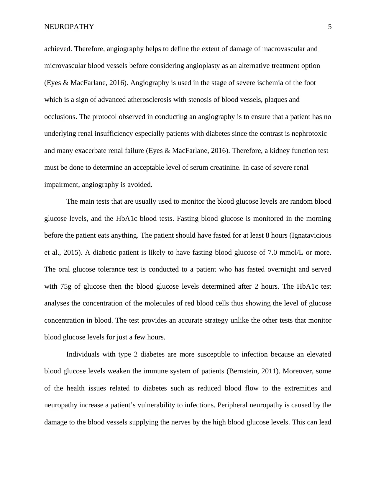
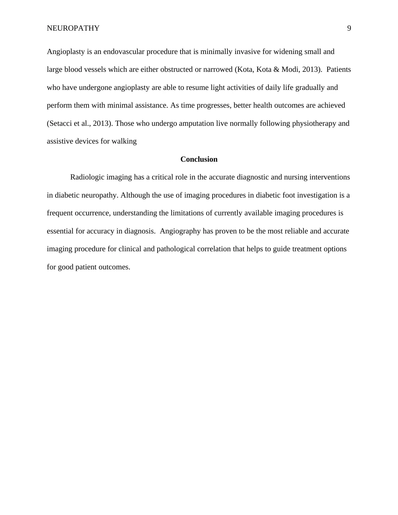
Diagram 1: Source: Manzi et al. 2012
(a) (b)
Figure 1 :( a) photograph of a 65 year old male diabetic patient that shows infected wounds and
various ischemic points. There is a large lesion on the front foot, the medial plantar surface and
the posterior aspects of the ankle. (b) Baseline angiogram of both the distal and proximal aspects
to gradual loss of sensation in the affected area. Neuropathy caused by diabetes can lead to foot
wound as is seen in the client’s case (Lewis et al., 2014). The blood supply to the lower
extremities is compromised leading to foot ulcers or diabetic foot. If the blood glucose is poorly
controlled, bacteria are able to multiply rapidly in the affected area and gain access into the
bloodstream causing sepsis. Neuropathy causes the ulcers not to be painful; therefore, they
frequently become infected resulting to sepsis.
Medically significant findings in Angiography
In a patient with macrovascular and microvascular disease in diabetic foot, an
angiography should reveal peripheral artery disease with stenosis or occlusion of small blood
vessels. The nerves may have a deviated course, a fusiform enlargement with an abnormal
fascicular pattern. An angiogram also shows involution and atrophy of fat tissues of the soft
tissue of the plantar surface. Callus may appear to be hypotense and ulcerations may appear with
headed-up edges with the granular tissue at the base.
Diagram 1: Source: Manzi et al. 2012
(a) (b)
Figure 1 :( a) photograph of a 65 year old male diabetic patient that shows infected wounds and
various ischemic points. There is a large lesion on the front foot, the medial plantar surface and
the posterior aspects of the ankle. (b) Baseline angiogram of both the distal and proximal aspects
⊘ This is a preview!⊘
Do you want full access?
Subscribe today to unlock all pages.
Trusted by 1+ million students worldwide
NEUROPATHY 7
of the blood vessels in the leg (left antero-posterior projection and right lateral projection)
showing total blockage of the posterior and anterior tibial arteries and complete blockage of the
perineal artery with distal ad middle segments which are open as indicated by the arrows.
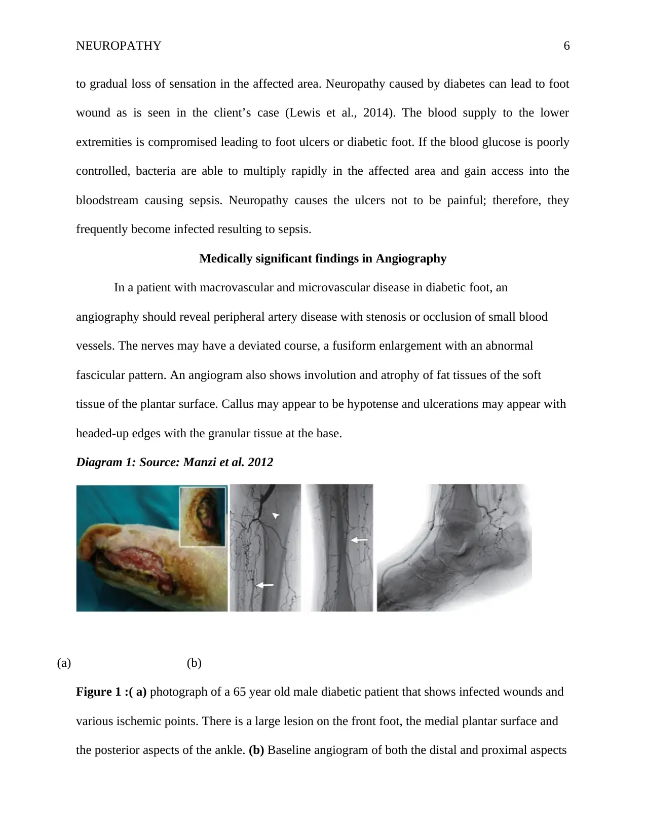
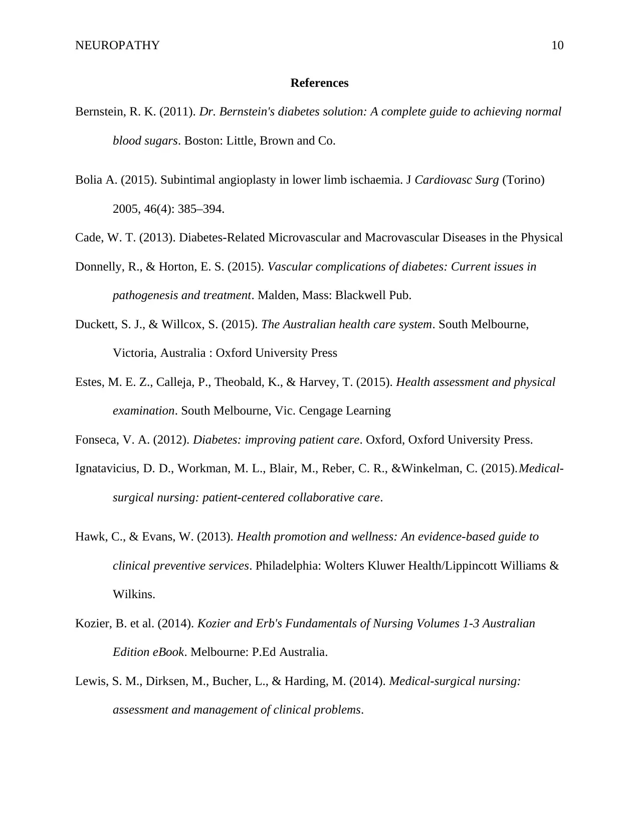
Diagram 2: Source: Manzi et al. 2012
Figure 2: Image of a 50 year old male with critical limb ischemia that shows lack of a linkage
between the anterior-posterior pedal circulation following the absence of a pedal-plantar loop
and a plantar arch. The left and right anteroposterior projections reveal that the medial plantar
and dorsalis pedis arteries supply blood to the first and second toes while the third and fourth
toes are supplied by the lateral plantar and arcuate arteries (Patel et al. 2018). The fifth toe is
only supplied with the lateral plantar artery.
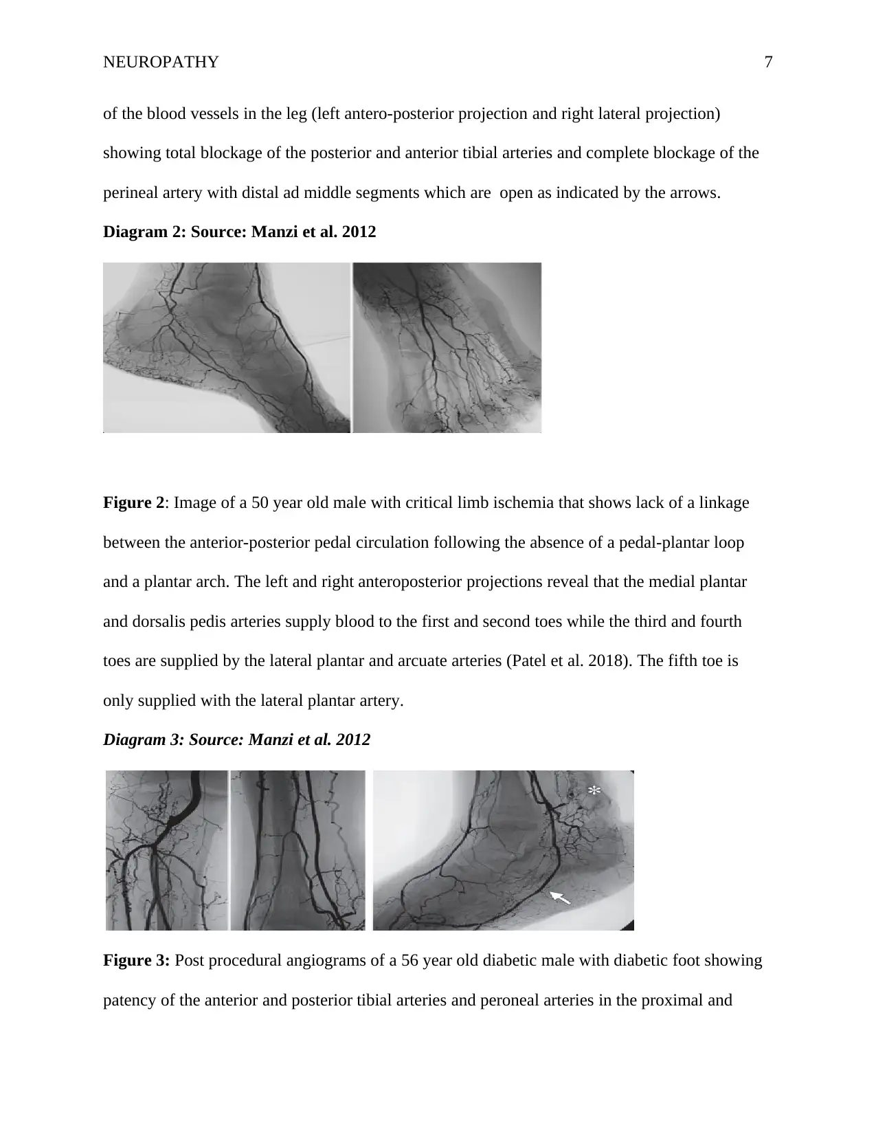
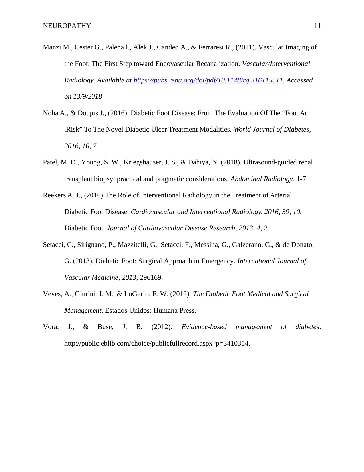
Diagram 3: Source: Manzi et al. 2012
Figure 3: Post procedural angiograms of a 56 year old diabetic male with diabetic foot showing
patency of the anterior and posterior tibial arteries and peroneal arteries in the proximal and
of the blood vessels in the leg (left antero-posterior projection and right lateral projection)
showing total blockage of the posterior and anterior tibial arteries and complete blockage of the
perineal artery with distal ad middle segments which are open as indicated by the arrows.
Diagram 2: Source: Manzi et al. 2012
Figure 2: Image of a 50 year old male with critical limb ischemia that shows lack of a linkage
between the anterior-posterior pedal circulation following the absence of a pedal-plantar loop
and a plantar arch. The left and right anteroposterior projections reveal that the medial plantar
and dorsalis pedis arteries supply blood to the first and second toes while the third and fourth
toes are supplied by the lateral plantar and arcuate arteries (Patel et al. 2018). The fifth toe is
only supplied with the lateral plantar artery.
Diagram 3: Source: Manzi et al. 2012
Figure 3: Post procedural angiograms of a 56 year old diabetic male with diabetic foot showing
patency of the anterior and posterior tibial arteries and peroneal arteries in the proximal and
Paraphrase This Document
Need a fresh take? Get an instant paraphrase of this document with our AI Paraphraser
NEUROPATHY 8
distal regions (Kozier, 2014). Angiographic projections reveal the patency of the dorsalis pedis
and lateral plantar arteries following an arterial revascularization.
Brief Discussion
Critical limb ischemia is one of the most prevalent form of chronic complicatiosn in
clients with microvascular and macrovascular disease in diabetic foot as illustrated by the
angiography in Figure 1 and Figure 2. That reveals atherosclerosis disease in peripheral arteries.
In critical limb ischemia, blood vessels are severely damaged and ischemia gradually develops in
cases where the flow of blood fails to meet the demands of tissues at rest (Bolia, 2015). The
resultant manifestation is severe pain. Damage to blood vessels will also manifest with non-
healing leg ulcers and gangrene that are commonly located in the leg, the heel or the toes of the
affected limb as shown in Figure 1.
This is the major non-traumatic indication for the amputation of lower limbs in patients
with chronic leg ulcers or gangrene (Hawk and Evans, 2013). Despite the fact that
pharmacologic therapy remains to be widely used in most patients with diabetic foot, critical
limb ischemia is best managed with arterial revascularization (angioplasty) as observed in Figure
3. Angioplasty guarantees the replacement of sufficient flow of blood to the leg in critical limb
ischemia and this is vital for the provision of relief from pain, to promote healing of the wound
and to prevent amputation (Estes et al, 2015). Angioplasty is not only visible but also safe in
settings with adequate resources and helps to regain full functionality of the limb.
Treatment options and Outcomes for the patient
Various treatment options have been attempted for the vascular illnesses with considerable
success (Ducket and Willcox, 2015). The most effective treatment option for microvascular and
macrovascular disease in diabetic foot following and angiography is amputation or angioplasty.
NEUROPATHY 9
Angioplasty is an endovascular procedure that is minimally invasive for widening small and
large blood vessels which are either obstructed or narrowed (Kota, Kota & Modi, 2013). Patients
who have undergone angioplasty are able to resume light activities of daily life gradually and
perform them with minimal assistance. As time progresses, better health outcomes are achieved
(Setacci et al., 2013). Those who undergo amputation live normally following physiotherapy and
assistive devices for walking
Conclusion
Radiologic imaging has a critical role in the accurate diagnostic and nursing interventions
in diabetic neuropathy. Although the use of imaging procedures in diabetic foot investigation is a
frequent occurrence, understanding the limitations of currently available imaging procedures is
essential for accuracy in diagnosis. Angiography has proven to be the most reliable and accurate
imaging procedure for clinical and pathological correlation that helps to guide treatment options
for good patient outcomes.
⊘ This is a preview!⊘
Do you want full access?
Subscribe today to unlock all pages.
Trusted by 1+ million students worldwide
NEUROPATHY 10
References
Bernstein, R. K. (2011). Dr. Bernstein's diabetes solution: A complete guide to achieving normal
blood sugars. Boston: Little, Brown and Co.
Bolia A. (2015). Subintimal angioplasty in lower limb ischaemia. J Cardiovasc Surg (Torino)
2005, 46(4): 385–394.
Cade, W. T. (2013). Diabetes-Related Microvascular and Macrovascular Diseases in the Physical
Donnelly, R., & Horton, E. S. (2015). Vascular complications of diabetes: Current issues in
pathogenesis and treatment. Malden, Mass: Blackwell Pub.
Duckett, S. J., & Willcox, S. (2015). The Australian health care system. South Melbourne,
Victoria, Australia : Oxford University Press
Estes, M. E. Z., Calleja, P., Theobald, K., & Harvey, T. (2015). Health assessment and physical
examination. South Melbourne, Vic. Cengage Learning
Fonseca, V. A. (2012). Diabetes: improving patient care. Oxford, Oxford University Press.
Ignatavicius, D. D., Workman, M. L., Blair, M., Reber, C. R., &Winkelman, C. (2015).Medical-
surgical nursing: patient-centered collaborative care.
Hawk, C., & Evans, W. (2013). Health promotion and wellness: An evidence-based guide to
clinical preventive services. Philadelphia: Wolters Kluwer Health/Lippincott Williams &
Wilkins.
Kozier, B. et al. (2014). Kozier and Erb's Fundamentals of Nursing Volumes 1-3 Australian
Edition eBook. Melbourne: P.Ed Australia.
Lewis, S. M., Dirksen, M., Bucher, L., & Harding, M. (2014). Medical-surgical nursing:
assessment and management of clinical problems.
Paraphrase This Document
Need a fresh take? Get an instant paraphrase of this document with our AI Paraphraser
NEUROPATHY 11
Manzi M., Cester G., Palena l., Alek J., Candeo A., & Ferraresi R., (2011). Vascular Imaging of
the Foot: The First Step toward Endovascular Recanalization. Vascular/Interventional
Radiology. Available at https://pubs.rsna.org/doi/pdf/10.1148/rg.316115511. Accessed
on 13/9/2018
Noha A., & Doupis J., (2016). Diabetic Foot Disease: From The Evaluation Of The “Foot At
,Risk” To The Novel Diabetic Ulcer Treatment Modalities. World Journal of Diabetes,
2016, 10, 7
Patel, M. D., Young, S. W., Kriegshauser, J. S., & Dahiya, N. (2018). Ultrasound-guided renal
transplant biopsy: practical and pragmatic considerations. Abdominal Radiology, 1-7.
Reekers A. J., (2016).The Role of Interventional Radiology in the Treatment of Arterial
Diabetic Foot Disease. Cardiovascular and Interventional Radiology, 2016, 39, 10.
Diabetic Foot. Journal of Cardiovascular Disease Research, 2013, 4, 2.
Setacci, C., Sirignano, P., Mazzitelli, G., Setacci, F., Messina, G., Galzerano, G., & de Donato,
G. (2013). Diabetic Foot: Surgical Approach in Emergency. International Journal of
Vascular Medicine, 2013, 296169.
Veves, A., Giurini, J. M., & LoGerfo, F. W. (2012). The Diabetic Foot Medical and Surgical
Management. Estados Unidos: Humana Press.
Vora, J., & Buse, J. B. (2012). Evidence-based management of diabetes.
http://public.eblib.com/choice/publicfullrecord.aspx?p=3410354.
1 out of 11


Your All-in-One AI-Powered Toolkit for Academic Success.
+13062052269
info@desklib.com
Available 24*7 on WhatsApp / Email
![[object Object]](/_next/static/media/star-bottom.7253800d.svg)
Unlock your academic potential
Copyright © 2020–2026 A2Z Services. All Rights Reserved. Developed and managed by ZUCOL.