Magnetic Resonance Imaging Critique: Stroke Protocol Analysis
VerifiedAdded on 2023/01/19
|12
|2688
|76
Report
AI Summary
This report provides a comprehensive critique of Magnetic Resonance Imaging (MRI) and its application in healthcare, particularly in stroke diagnosis and treatment. It explores the advantages of MRI as a non-invasive imaging technique, detailing its clinical indications and the anatomical protocols utilized, including DWI/ADC, GREWI, T2WI, TOF MRA, T2 FLAIR, and T1FSWI sequences. The report discusses the ischemic cascade, alternative imaging techniques like CT perfusion scans, and the hardware equipment used, such as the Magnetom Sola scanner and radiofrequency coils. Advantages and disadvantages of the MRI stroke protocol are analyzed, along with pulse sequence parameters and diagrams. The conclusion emphasizes the significance of MRI in modern healthcare, highlighting its role in disease detection, diagnosis, and treatment, especially during the acute stroke stage. The report references various studies and publications to support the analysis.
Running head: MAGNETIC RESONANCE IMAGING 1
Magnetic Resonance Imaging Critique
Name
Institutional Affiliation
Magnetic Resonance Imaging Critique
Name
Institutional Affiliation
Paraphrase This Document
Need a fresh take? Get an instant paraphrase of this document with our AI Paraphraser
MAGNETIN RESONANCE IMAGING CRITIQUE 2
Executive Summary
Today, advancement in technology is engulfing every industry. Healthcare is not left
behind with corporation of sophisticated technological equipment like Magnetic Resonance
Imaging being useful in patient care. Notably, MRI is considered a non-invasive technology of
imaging that produces comprehensive anatomical images that are three dimensional. Besides,
MRI is useful in diagnosis, disease detection as well as treatment monitoring (Hashemi, Bradley,
& Lisanti, 2012). Categorically, MRI can be very useful not only in detecting blood vessel as
well as brain abnormalities but also in determination of the region of brain affected by stroke.
Notably, stroke can be defined as a neurological dysfunction which occurs when there is absence
in flow of blood to a given brain region. In clinical setting, according Zhao, Ma, Greiser,
Zhang, An, and Fan (2016), a Magnetic Resonance Imaging scan be useful in the diagnosis as
well as aid in the pathology management through the use of sequences which rely on
pathological changes which occurs post ictus with sensitivity as well as specificity. However, it
is worth noting that, for effective management as well as treatment of a patient suffering from
stroke; diagnosis is best and effectively done within therapeutic window through the use of
DWI/ADC sequence which allows the demonstration of stroke within some onset minutes
(Weishaupt, Froehlich, Nanz, Koechli, Pruessmann, & Marincek, 2013). Moreover, immediate
and effective treatment of stroke can be helpful in reducing disability as well as saving lives by
reducing pressure and controlling bleeding in hemorrhagic stroke case or restoring blood flow
especially in ischemic stroke.
Clinical Indication and Anatomical Protocol
The necessity of patients requiring brain imaging is rising since stroke is regarded as the
3rd leading cause of death across the world with a considerable disabilities among survivors.
MAGNETIN RESONANCE IMAGING CRITIQUE 3
According to Fiebach and Schellinger (2013), in the quest to help solve complexity of stroke,
imaging attempts to optimise therapeutic window phase of patients. Therefore, it is worth noting
that, MRI medical imaging is not only considered as a sensitive modality in the detection of
stroke but also in stroke diagnosis in patients that shows an acute neurological deficit as well as
consciousness alternation. Furthermore, the techniques involved include DWI/ADC (diffusion-
weighted imaging/apparent diffusion co-efficient), GREWI (gradient-recalled echo weighted
imaging), T2WI (weight image), TOF MRA (time of flight magnetic resonance angiography), T2
FLAIR (Fluid-attenuated inversion recovery imaging), and T1FSWI (T1 fat suppression
weighted image).
Sequentially, MR imaging visualized can be ischemic cascade which happens after
vascular occlusion. According to Caplan (2016), ischemic cascade include apoptosis and
inflammation, depolarization, K+/Na+ pump failure, free radicals generation, intracellular Ca2+
increase, and blood brain barrier disruption. Besides, it worth noting that DWI/ADC especially
during therapeutic window stage is a stroke indicator mostly as a result of the sequence. The
result of intracellular cytotoxic oedema is the prolongation of T1 and T2 relaxation with
T2weighed images showing six to eight hours post stroke, subcortical hypointensityan and loss
of normal vessel signal.
Moreover, T2 Flair has higher detection rates for small infarctions due to its ability to
null the signal from cerebrospinal fluid despite having a hypersensitivity to oedema (González et
al., 2010). Furthermore, The Oxygination content in blood changes the magnetic susceptibility
hence breaking down red blood cells. In addition, this degradation process is sensitive to GRE
T2 sequence, thus, providing images of acute or chronic haemorrhages, with hypointense regions
of interest makes GRE T2 a sequence of choice for detection (Bushong & Clarke, 2014).
⊘ This is a preview!⊘
Do you want full access?
Subscribe today to unlock all pages.
Trusted by 1+ million students worldwide
MAGNETIN RESONANCE IMAGING CRITIQUE 4
Consequently, MRI sequences show sensitivity to pathophysiological changes that follow a
stroke thus providing prognostic and diagnostic approach in assessing brain infarction in a none
invasive setting (Haaga et al., 2008).
Alternative Techniques and Other Imaging Modalities
Detection as well as management of a cute stroke requires improved MRA as well as
perfusion weighted imaging sequences as alternative techniques. First, A CT perfusion scan is an
alternative to MR stroke imaging, offering a current view of cerebralvascular pathology. Also,
Gadolinium contrast media administered intravenously causing T2 weighted images a non-linear
signal loss. It is worth noting that, gadolinium administered to the patient intravenously, CE-
MRA is used (Baraee, Faeghi, Shokoohi, & Saeedi, 2016). Also, usage of T1 properties of the
contrast to reduce T1 relaxation time instead of flow dynamics creates a difference in appearance
between the intravascular lumen and adjacent tissue.
Furthermore, creating semi-quantitative perfusion maps as a CE technique estimates
cerebral blood volumes mean transit time of the bolus to transverse the network of capillaries as
well as cerebral blood flow. However, a T2 weighted images appear normal during an acute
stroke window thereby PWI can show cerebral ischemia (Bare, Faeghi, Shokoohi, & Saeedi,
2016). Notably, the integrity of the intra and extracranial vessels are evaluated using CE-MRA,
most usefully in the subacute as well as chronic stroke stages especially when pathophysiology is
not established.
Paraphrase This Document
Need a fresh take? Get an instant paraphrase of this document with our AI Paraphraser
MAGNETIN RESONANCE IMAGING CRITIQUE 5
Hardware Equipment
Production of images with high resolution requires high field strengths. Thus, the patient
is placed in a resilient magnetic field also known as Bo which is produced by super conductor
magnet thereby aligning the body of the patient in a hydrogen nuclei.
Scanner
For this protocol, the scanner used is Magnetom Sola which is the first 1.5T MRI
system considered to have BioMatrix Technology. Besides, the scanner automatically overcome
unwarranted variations especially during examination by adjusting to patient biovariability.
Additionally, it does not only embrace efficiency at Simultaneous Multi-Slice as well as with GO
technologies but also embrace improved clinical capabilities at 1.5T with Inline Compressed
Sensing. Furthermore, the scanner’s capabilities results to outcomes like predictable consistent
and scheduling, fewer scans, and high-quality personalized examinations which ultimately
results to increased productivity.
Coil
For the brain MRI scan, a radiofrequency trans-receiver coil is used for receiving as well as
creating radiofrequencies (Haacke, & Reichenbach, 2014). The two ends of the coil that are
circular rings are joined by Z direction running conductive wires. The equal amount of Z
direction conductive wires arranged to provide a cosine current variation thereby surrounding the
coil periphery reference. Also, RF pulses create an oscillating magnetic field perpendicular to the
resilient magnetic field in which the filed oscillation coincides with the hydrogen nuclei at the
Larmor frequency thereby causing the protons to not only to press oscillating magnetic field
phase but also lean way from magnetic field Bo (Berkhemer, Fransen, Beumer, van den Berg,
MAGNETIN RESONANCE IMAGING CRITIQUE 6
Lingsma, Yoo, & van Walderveen,. (2015). Notably, RF pulses removal settle nuclei to
equilibrium. Besides, field the gradient coil creates distorts Bo in an anticipated sequence
causing the resonance frequency of the nuclei to alter as a function of spatial location.
Furthermore, according to Rajan (2012), MRA and diffusion imaging of the brain for a stroke
require gradients.
Protocol Advantages and Disadvantages
MRI stroke protocol has its advantages and disadvantages. First, MRI stroke protocol
are non-invasive. Therefore, can be used for patients that pass the MRI screen. Secondly, T2WI
sensitivity characterise a stroke that increases in advance of 6 hours of ictus. However, it is
worth noting that, T2 FLAIR indicates within the first 4-6 shours hypersensitivity of the
ischemic brain (Haacke, & Reichenbach, 2014). On the other hand, T2 FLAIR will not establish
before a strokes actue stage since the sequence most sensitive for demonstrating sub arachnoid
haemorrhages as it is able to hypersensitise extra-axial blood (Burgener & Meyers, 2011).
Furthermore, a combination of DWI sequencing as well as ADC mapping allows for
stage and age of the stroke to be identified. According to Fiebach and Schellinger (2013), T1 fat
suppression weighted images is beneficial in detecting intramural blood as a result of vascular
dissection. Also, T1 sequence can also suppress signals from tissue or fluid that contains a
similar T1 to fat (Weishaupt et al., 2008). Additionally, patients that have contraindications for
contrast medial TOF-MRA is useful as it doesn’t require intravenous contrast to be administered
(Westbrook, 2013). Although, TOF-MRA has several advantages, its major disadvantage is that
it can overestimate length/degree of a stenosis due to the blood flow in the case of distal
vasculature. This is because the vascular signal depends on the velocity and flow direction of
blood flow.
⊘ This is a preview!⊘
Do you want full access?
Subscribe today to unlock all pages.
Trusted by 1+ million students worldwide
MAGNETIN RESONANCE IMAGING CRITIQUE 7
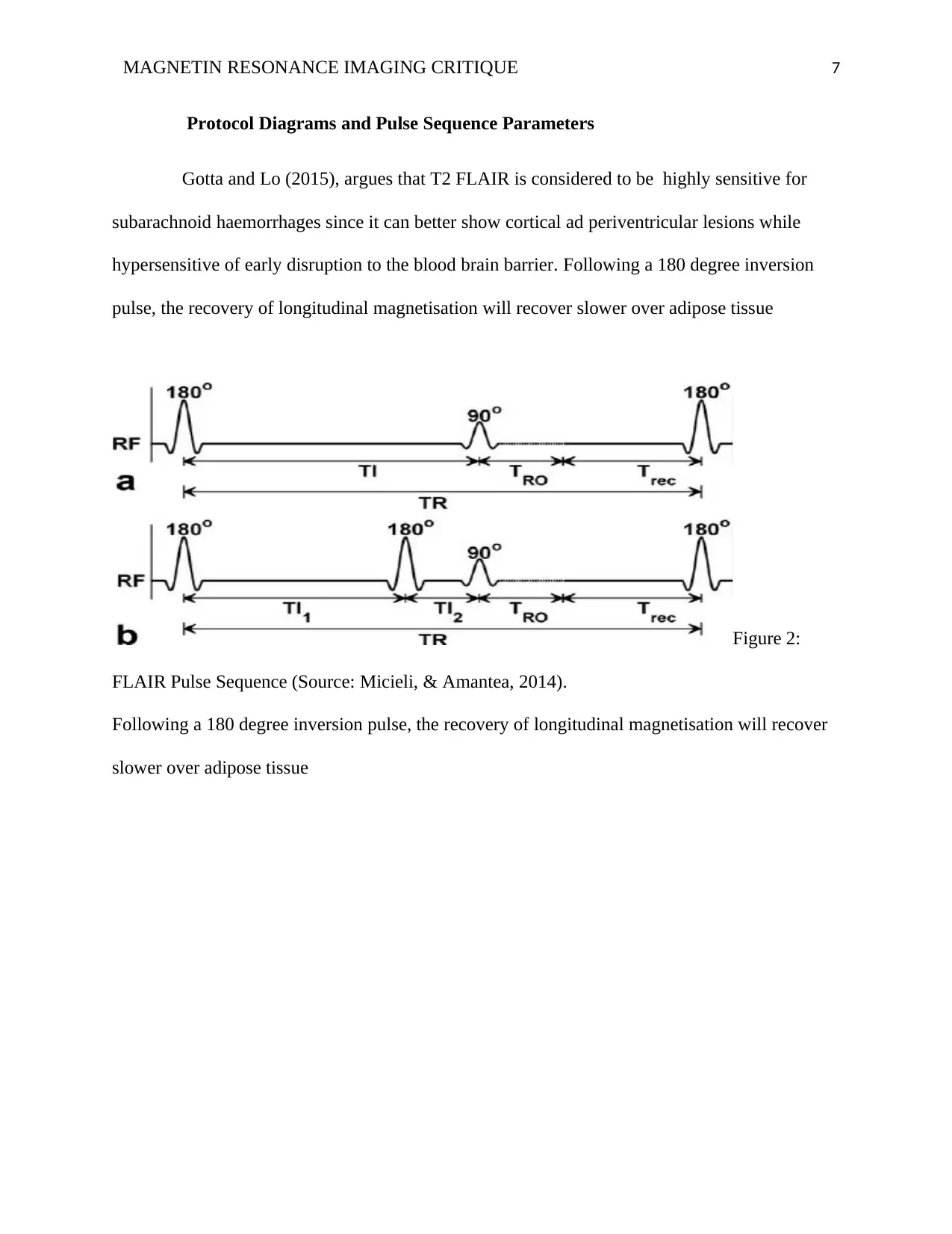
Protocol Diagrams and Pulse Sequence Parameters
Gotta and Lo (2015), argues that T2 FLAIR is considered to be highly sensitive for
subarachnoid haemorrhages since it can better show cortical ad periventricular lesions while
hypersensitive of early disruption to the blood brain barrier. Following a 180 degree inversion
pulse, the recovery of longitudinal magnetisation will recover slower over adipose tissue
Figure 2:
FLAIR Pulse Sequence (Source: Micieli, & Amantea, 2014).
Following a 180 degree inversion pulse, the recovery of longitudinal magnetisation will recover
slower over adipose tissue
Paraphrase This Document
Need a fresh take? Get an instant paraphrase of this document with our AI Paraphraser
MAGNETIN RESONANCE IMAGING CRITIQUE 8
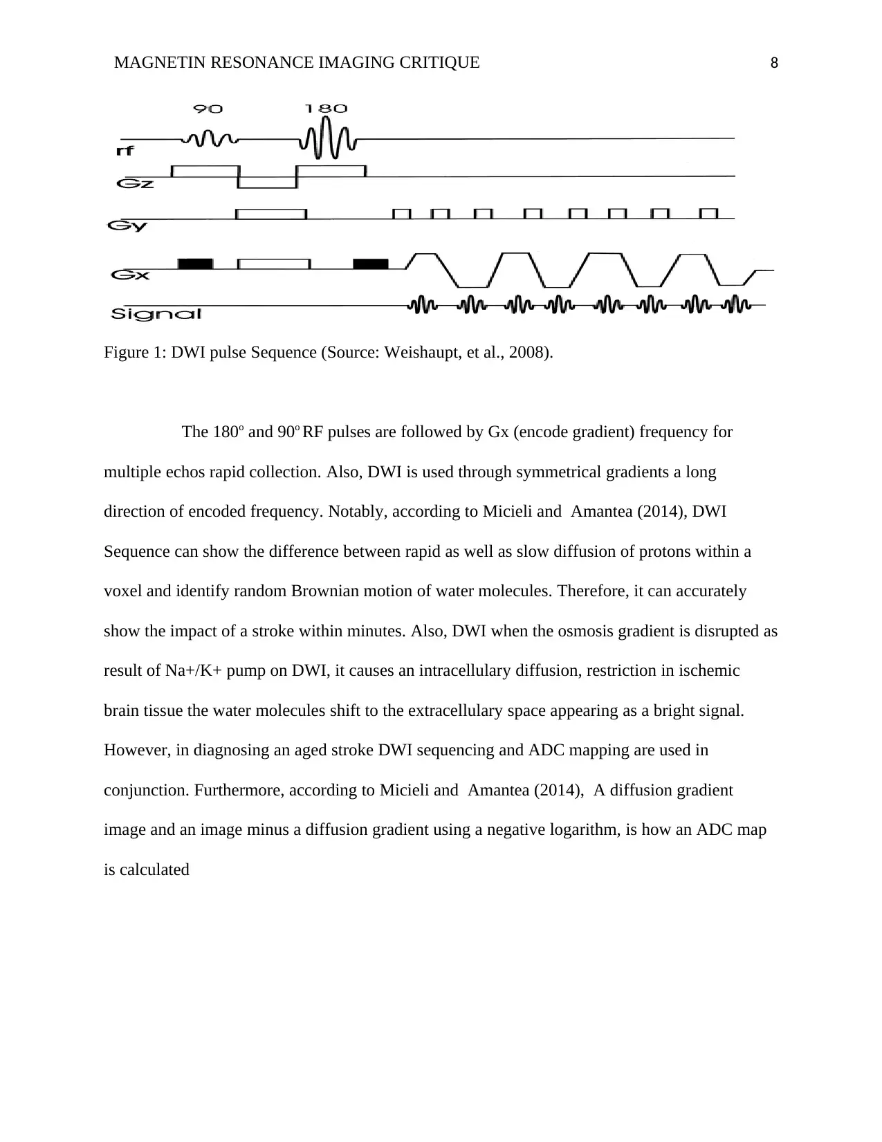
Figure 1: DWI pulse Sequence (Source: Weishaupt, et al., 2008).
The 180o and 90o RF pulses are followed by Gx (encode gradient) frequency for
multiple echos rapid collection. Also, DWI is used through symmetrical gradients a long
direction of encoded frequency. Notably, according to Micieli and Amantea (2014), DWI
Sequence can show the difference between rapid as well as slow diffusion of protons within a
voxel and identify random Brownian motion of water molecules. Therefore, it can accurately
show the impact of a stroke within minutes. Also, DWI when the osmosis gradient is disrupted as
result of Na+/K+ pump on DWI, it causes an intracellulary diffusion, restriction in ischemic
brain tissue the water molecules shift to the extracellulary space appearing as a bright signal.
However, in diagnosing an aged stroke DWI sequencing and ADC mapping are used in
conjunction. Furthermore, according to Micieli and Amantea (2014), A diffusion gradient
image and an image minus a diffusion gradient using a negative logarithm, is how an ADC map
is calculated
MAGNETIN RESONANCE IMAGING CRITIQUE 9
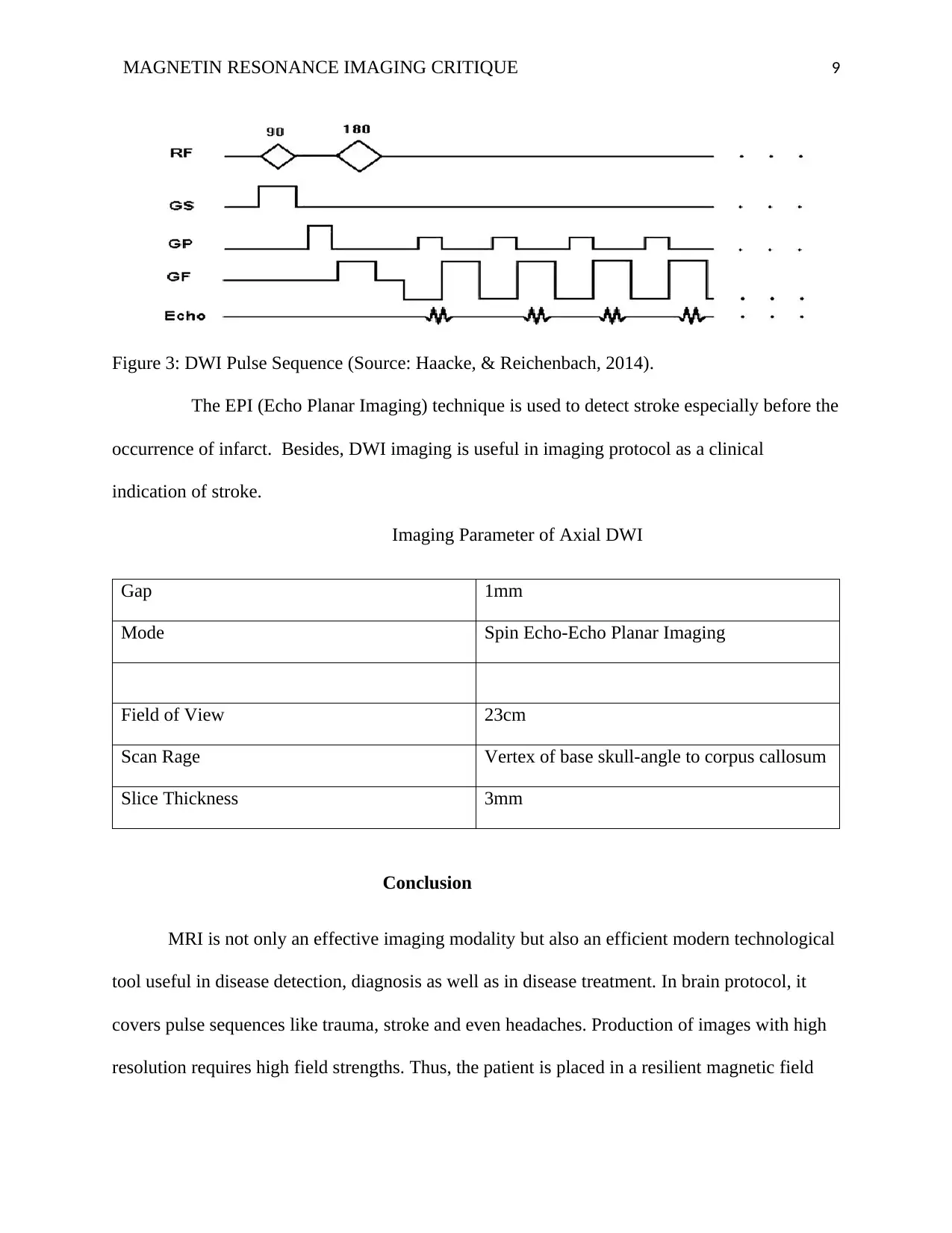
Figure 3: DWI Pulse Sequence (Source: Haacke, & Reichenbach, 2014).
The EPI (Echo Planar Imaging) technique is used to detect stroke especially before the
occurrence of infarct. Besides, DWI imaging is useful in imaging protocol as a clinical
indication of stroke.
Imaging Parameter of Axial DWI
Gap 1mm
Mode Spin Echo-Echo Planar Imaging
Field of View 23cm
Scan Rage Vertex of base skull-angle to corpus callosum
Slice Thickness 3mm
Conclusion
MRI is not only an effective imaging modality but also an efficient modern technological
tool useful in disease detection, diagnosis as well as in disease treatment. In brain protocol, it
covers pulse sequences like trauma, stroke and even headaches. Production of images with high
resolution requires high field strengths. Thus, the patient is placed in a resilient magnetic field
⊘ This is a preview!⊘
Do you want full access?
Subscribe today to unlock all pages.
Trusted by 1+ million students worldwide
MAGNETIN RESONANCE IMAGING CRITIQUE 10
also known as Bo which is produced by super conductor magnet thereby aligning the body of the
patient in a hydrogen nuclei. Notably, MRI has its advantages as well as its disadvantages. For
instance, diagnosis as well the non-invasive assessment useful especially during acute stroke
stage where T2, TOF MRA, and DWI/ADC are incorporated. Therefore, MRI is great
significance in the modern healthcare setting.
Paraphrase This Document
Need a fresh take? Get an instant paraphrase of this document with our AI Paraphraser
MAGNETIN RESONANCE IMAGING CRITIQUE 11
Reference
Baraee, K. R. Z., Faeghi, F., Shokoohi, J. J., & Saeedi, A. (2016). Comparison of Maximum
Signal Intensity of Magnevist Contrast Agent in Modified T1 Weighted Spin Echo, T1
Weighted Fast Spin Echo and T1 Weighted Gradient Echo Sequences. Journal of
Biomedical Physics and Engineering. doi.org/10.22086/jbpe.v0i0.512
Bammer, R. (2016). MR and CT Perfusion and Pharmacokinetic Imaging: Clinical Applications
and Theoretical Principles: Wolters Kluwer Health.
Burgener, F. A., & Meyers, S. P. (2011). Differential Diagnosis in Magnetic Resonance
Imaging: Thieme.
Berkhemer, O. A., Fransen, P. S., Beumer, D., van den Berg, L. A., Lingsma, H. F., Yoo, A. J.,
& van Walderveen, M. A. (2015). A randomized trial of intraarterial treatment for acute
ischemic stroke. New England Journal of Medicine, 372(1), 11-20:
doi:10.1056/NEJMoa1411587
Caplan, L. R. (2016). Caplan's Stroke: A Clinical Approach: Cambridge University Press.
González, R. G., Hirsch, J. A., Lev, M. H., Schaefer, P. W., & Schwamm, L. H. (2010). Acute
Ischemic Stroke: Imaging and Intervention: Springer Berlin Heidelberg.
Grotta, J. C., & Lo, E. H. (2015). Stroke: Pathophysiology, Diagnosis, and Management:
Elsevier.
Haacke, E. M., & Reichenbach, J. R. (2014). Susceptibility Weighted Imaging in MRI: Basic
Concepts and Clinical Applications: Wiley.
Hashemi, R. H., Bradley, W. G., & Lisanti, C. J. (2012). MRI: The Basics: The Basics: Wolters
Kluwer Health.
MAGNETIN RESONANCE IMAGING CRITIQUE 12
Micieli, G., & Amantea, D. (2014). Rational Basis for Clinical Translation in Stroke Therapy:
CRC Press.
Rajan, S. S. (2012). MRI: A Conceptual Overview: Springer New York.
Weishaupt, D., Froehlich, J. M., Nanz, D., Koechli, V. D., Pruessmann, K. P., & Marincek, B.
(2008). How does MRI work?: An Introduction to the Physics and Function of Magnetic
Resonance Imaging: Springer Berlin Heidelberg.
Weishaupt, D., Froehlich, J. M., Nanz, D., Koechli, V. D., Pruessmann, K. P., & Marincek, B.
(2013). How does MRI work?: An Introduction to the Physics and Function of Magnetic
Resonance Imaging: Springer Berlin Heidelberg.
Zhao, L., Li, S., Ma, X., Greiser, A., Zhang, T., An, J., ... & Fan, Z. (2016). Systolic MOLLI T1
mapping with heart-rate-dependent pulse sequence sampling scheme is feasible in
patients with atrial fibrillation. Journal of Cardiovascular Magnetic Resonance, 18(1),
13: doi.org/10.1186/s12968-016-0232-7
⊘ This is a preview!⊘
Do you want full access?
Subscribe today to unlock all pages.
Trusted by 1+ million students worldwide
1 out of 12
Related Documents


Your All-in-One AI-Powered Toolkit for Academic Success.
+13062052269
info@desklib.com
Available 24*7 on WhatsApp / Email
![[object Object]](/_next/static/media/star-bottom.7253800d.svg)
Unlock your academic potential
Copyright © 2020–2026 A2Z Services. All Rights Reserved. Developed and managed by ZUCOL.