Cell Division: Meiosis, Mitosis Diagrams & Genetic Cross Analysis

Verified

Added on  2023/06/15

|7
|659
|127
Homework Assignment
AI Summary
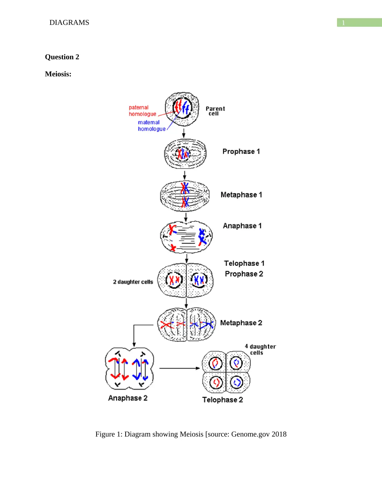
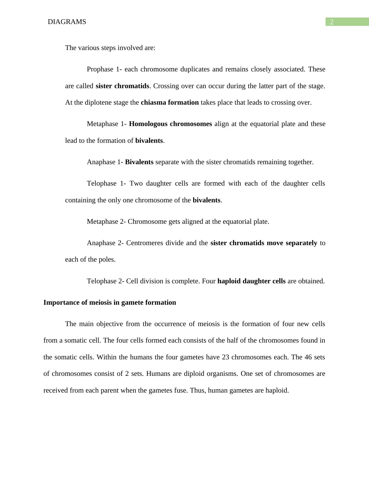
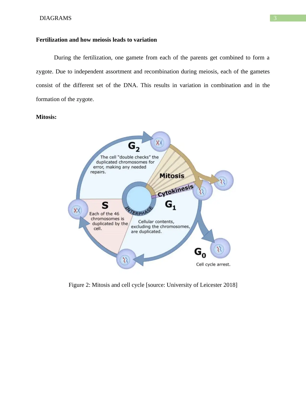
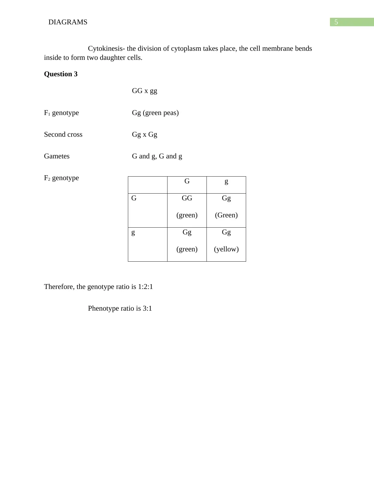
This assignment provides a detailed overview of meiosis and mitosis, including illustrative diagrams that highlight the various stages of each process. Meiosis is explained with a focus on gamete formation, emphasizing the creation of four haploid daughter cells from a somatic cell and the importance of independent assortment and recombination in generating genetic variation during fertilization. The stages of meiosis, including Prophase 1, Metaphase 1, Anaphase 1, Telophase 1, Metaphase 2, Anaphase 2, and Telophase 2, are thoroughly described. Mitosis is presented as a cell division process resulting in two identical daughter cells, with detailed explanations of Interphase, Prophase, Anaphase, Telophase, and Cytokinesis. Additionally, the assignment includes a genetic cross problem involving green and yellow peas, demonstrating the calculation of genotype and phenotype ratios in the F2 generation. The solution illustrates the principles of Mendelian genetics and the expected outcomes of a dihybrid cross.
tabler-icon-diamond-filled.svg

Contribute Materials

Your contribution can guide someone’s learning journey. Share your documents today.
Document Page
Running head: DIAGRAMS
Diagram
Name of the Student:
Name of the University:
Author Note:
tabler-icon-diamond-filled.svg

Secure Best Marks with AI Grader

Need help grading? Try our AI Grader for instant feedback on your assignments.
Document Page
1DIAGRAMS
Question 2
Meiosis:
Figure 1: Diagram showing Meiosis [source: Genome.gov 2018
Document Page
2DIAGRAMS
The various steps involved are:
Prophase 1- each chromosome duplicates and remains closely associated. These
are called sister chromatids. Crossing over can occur during the latter part of the stage.
At the diplotene stage the chiasma formation takes place that leads to crossing over.
Metaphase 1- Homologous chromosomes align at the equatorial plate and these
lead to the formation of bivalents.
Anaphase 1- Bivalents separate with the sister chromatids remaining together.
Telophase 1- Two daughter cells are formed with each of the daughter cells
containing the only one chromosome of the bivalents.
Metaphase 2- Chromosome gets aligned at the equatorial plate.
Anaphase 2- Centromeres divide and the sister chromatids move separately to
each of the poles.
Telophase 2- Cell division is complete. Four haploid daughter cells are obtained.
Importance of meiosis in gamete formation
The main objective from the occurrence of meiosis is the formation of four new cells
from a somatic cell. The four cells formed each consists of the half of the chromosomes found in
the somatic cells. Within the humans the four gametes have 23 chromosomes each. The 46 sets
of chromosomes consist of 2 sets. Humans are diploid organisms. One set of chromosomes are
received from each parent when the gametes fuse. Thus, human gametes are haploid.
Document Page
3DIAGRAMS
Fertilization and how meiosis leads to variation
During the fertilization, one gamete from each of the parents get combined to form a
zygote. Due to independent assortment and recombination during meiosis, each of the gametes
consist of the different set of the DNA. This results in variation in combination and in the
formation of the zygote.
Mitosis:
Figure 2: Mitosis and cell cycle [source: University of Leicester 2018]
tabler-icon-diamond-filled.svg

Secure Best Marks with AI Grader

Need help grading? Try our AI Grader for instant feedback on your assignments.
Document Page
4DIAGRAMS
Figure 3: Mitosis in detail in relation to cell cycle [source: Genome.gov 2018]
Mitosis is a process of cell division which results in the production of two daughter cells
from one parent cell. The daughter cells are similar to one another and to the parent cell.
The steps involved in cell division include
Interphase- this phase is also known as Gap 1 phase or growth phase, synthesis of
DNA takes place, and the Gap 2 phase include the production of organelle.
Prophase- in the interphase, chromatin gets diffused and condenses into the
chromosomes. Each of the chromatids gets duplicated and consists of 2 sister chromatids. When
the prophase ends, the nuclear envelop gets broken down to form vesicles.
Anaphase- centromeres gets divided, the sister chromatids move to the different
poles.
Telophase- the microtubules disappear when the daughter chromosomes move to
the poles. The nuclear envelop reappears and the chromatin material expands.
Document Page
5DIAGRAMS
Cytokinesis- the division of cytoplasm takes place, the cell membrane bends
inside to form two daughter cells.
Question 3
GG x gg
F1 genotype Gg (green peas)
Second cross Gg x Gg
Gametes G and g, G and g
F2 genotype
Therefore, the genotype ratio is 1:2:1
Phenotype ratio is 3:1
G g
G GG
(green)
Gg
(Green)
g Gg
(green)
Gg
(yellow)
Document Page
6DIAGRAMS
Reference
Genome.gov, 2018. [online] Genome.gov. Available at:
https://www.genome.gov/edkit/pdfs/1902b2.pdf [Accessed 5 Feb. 2018].
University of Leicester, 2018. The cell cycle, mitosis and meiosis — University of Leicester.
[online] Www2.le.ac.uk. Available at:
https://www2.le.ac.uk/projects/vgec/schoolsandcolleges/topics/cellcycle-mitosis-meiosis
[Accessed 5 Feb. 2018].
chevron_up_icon
1 out of 7
circle_padding
hide_on_mobile
zoom_out_icon
logo.png

Your All-in-One AI-Powered Toolkit for Academic Success.

Available 24*7 on WhatsApp / Email

[object Object]