Melanoma Immunotherapy for Melanoma: Pharmacology and Pathophysiology
VerifiedAdded on 2022/08/26
|10
|2347
|11
Report
AI Summary
This report provides a comprehensive overview of melanoma, a skin disorder characterized by the uncontrolled growth of melanocytes, and its treatment via immunotherapy. It details the pathophysiology of melanoma, including the role of UV radiation and genetic factors, as well as the cellular and molecular dynamics involved. The report focuses on Nivolumab (Opdivo®), an immunotherapeutic drug that targets the PD-1 pathway, outlining its pharmacodynamics, pharmacokinetics, side effects, and potential interactions. The analysis includes the drug's mode of action in restoring the patient's immune response against tumor cells, and the clinical findings from studies. It concludes by highlighting the importance of understanding both the disease and the treatment's implications for effective management, while also acknowledging the associated side effects of the treatment. The assignment is a report for NSC2500, focusing on the disease/disorder plus pharmacology that the student selected.
Running head = MELANOMA 0
Melanoma
Immunotherapy for Melanoma
Student detail
[Pick the date]
Melanoma
Immunotherapy for Melanoma
Student detail
[Pick the date]
Paraphrase This Document
Need a fresh take? Get an instant paraphrase of this document with our AI Paraphraser
Melanoma 1
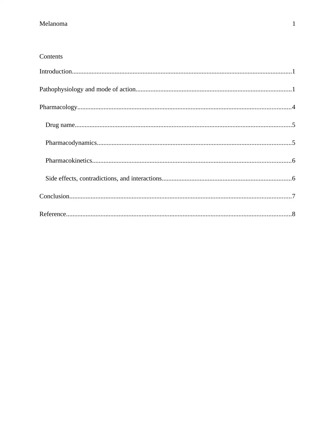
Contents
Introduction......................................................................................................................................1
Pathophysiology and mode of action...............................................................................................1
Pharmacology..................................................................................................................................4
Drug name....................................................................................................................................5
Pharmacodynamics......................................................................................................................5
Pharmacokinetics.........................................................................................................................6
Side effects, contradictions, and interactions...............................................................................6
Conclusion.......................................................................................................................................7
Reference.........................................................................................................................................8
Melanoma 2
Introduction
The normal homeostasis of melanocyte involves division, differentiation from neural crest
cells, which is distinctive of these cells and extension of dendrites to make contacts with other
cells such as keratinocytes. The growth and expression of melanocyte are controlled by the
keratinocytes. Melanoma is a skin disorder that involves the uncontrolled growth of melanocyte
cells causing cancer, the primary function of melanocyte is to produce melanin pigment
(Kawakami & Fisher, 2017). The primary reason for the development of melanoma is not
known, however, Ultraviolet (UV) radiations and tanning lamps have been associated as the
intrinsic factor that triggers skin melanoma. The occurrence of melanoma has been associated
with loss of control of keratinocytes over melanocyte functioning, which results in cancerous
growth (Heistein & Acharya, 2019). The spreading of cancer in the body could be stopped by the
application of immunotherapy, which triggers the patient's own immune system to identify and
cancer cells (Lugowska, Teterycz, & Rutkowski, 2018).
Pathophysiology and mode of action
Melanoma is one of the fastest and frequently found cancerous disorders throughout the
world. In terms of epidemiology, melanoma accounts for 5th in men and 7th most common
malignancy, attributing to 5% and 4% cases of cancer in men and women respectively. The
pathophysiology of melanoma could be studied, on an already remaining precursor lesion or
fresh skin sample that would appear healthy. There are studies which show some individual are
more susceptible to melanoma due to UV radiation and could be attributed to genetics and they
are 8% to 12% of total melanoma cases (Liu & Sheikh, 2014). If a malignant melanoma has been
developed over a healthy skin it is called arise de novo, as there is no involvement of precursor
⊘ This is a preview!⊘
Do you want full access?
Subscribe today to unlock all pages.

Trusted by 1+ million students worldwide
Melanoma 3
lesion. The primary cause of this is radiation from the sun; however, melanoma has been
reported in the unexposed area body (Kanavy & Gerstenblith, 2011).
There are some lesions that have been associated with melanoma, such as
I. Common acquired nevus
II. Dysplastic nevus
III. Congenital nevus
IV. Cellular blue nevus
The growth phase of melanoma could be divided into two phases radial and vertical
growth phase. Generally, at the start of melanoma the growth occurs in the radial way around
and over the epidermis, over a period of time the melanoma shifts in a vertical direction resulting
in the invasion of the dermis layer along with it, melanoma advances acquire the capability to
metastasize (Betti, Agape, Vergani, Moneghini, & Cerri, 2016). Lesion classification could be
established on their depths: thin (1 mm), moderate (1mm to 4 mm) and thick more than (4 mm)
(Heistein & Acharya, 2019).
The classification of melanoma is generally done based on varying in its growth pattern
that could be done as follows (Shain & Bastian, 2016):
I. Superficial: This kind of melanoma contributes to 70 % of the total melanomas, generally
appears flat, however, it could develop in irregular shape and elevated form in the
advanced stages if not treated.
II. Nodular: This kind of melanoma accounts for roughly 15% to 30% of the melanoma
cases, the general appearance of the tumor is blue or black in color, however, it could
appear as a lack of pigmentation also.
Paraphrase This Document
Need a fresh take? Get an instant paraphrase of this document with our AI Paraphraser
Melanoma 4
III. Lentigo maligna: This type of melanoma contributes to 4% to 10% of the total occurrence
of melanoma.
IV. Acral lentiginous: This type of melanoma accounts for a total of 2% to 8% of the total
melanoma cases in whites and 35% to 60% in the skin of people of color.
The advancement of melanoma in metastasizes results in spreading to other tissues, a 5%
to 20% dip in response towards the treatment along with the drastic 10% decrease in survival rate
is observed (Heistein & Acharya, 2019). The molecular dynamics of the area affected by
melanoma could be used as an identifier of the stage of melanoma and also help in deciding the
proper treatment model. The pathophysiology of molecular changes has been reported in the
literature. The cellular dynamics of cancer are very different from a normal cell and therefore the
molecular dynamics study of the cancer cell is very important, which would help in developing
an effective targeted therapy (Force & Salama, 2017).
BRAF gene belongs to the category of oncogenes, in the dysfunctional stage, they are
capable of mutating a normal cell into a mutated cancerous cell. Recent studies have shown that
about 40% to 60% have an active BRAF mutation. A mutation in BRAF leads to the activation
of a monomer that is continually stimulated, which results in uninhibited cell division and
proliferation that could attribute to tumor growth and development. However, over 60% to 70%
of developed melanoma have BRAF mutations, which indicate the involvement of the BRAF
gene in cancer progression (Stadler, Weina, Gebhardt, & Utikal, 2015). NRAS is a gene which
has been associated with the initiation of melanoma, although both the genes NRAS and BRAF
are found on the same pathway, the simultaneous mutation in a single individual is a very rare
occurrence (Colombino, et al., 2012). The patient suffering due to NRAS mutation has a vertical
tumor growth which has been attributed to the high cell multiplying rate prompted by NRAS
Melanoma 5
mutation. Some other genetic pathways that have been associated with the progression of cancer
are, P13K-ATK/PTEN associated with 3% melanoma cases, p53 found in 11% melanoma cases,
CDK4/CDKN24 present in 5% to 10% melanoma cases and c-KIT has been associated with 2%
to 5% melanoma cases (Liu & Sheikh, 2014).
Pharmacology
Surgeries have been used for the removal of melanoma, the advanced stage of melanoma
known as metastatic melanoma is not treatable using a surgical pathway, hence knew and
innovative methods need to be developed for the treatment. Among the various treatments
available for cancer, immunotherapy is one of them and certainly most promising in terms of
treatment of melanoma (Lugowska, Teterycz, & Rutkowski, 2018).
Drug name
Nivolumab (Opdivo®) is a new one of kind immunotherapeutic drug that is used in the
treatment of advanced melanoma; the drug obstructs the programmed death -1 (PD-L) 1
signaling mediated by the tumor. It is human immunoglobulin belonging to the G4 group of a
monoclonal antibody and has a high affinity for PD-1.
Pharmacodynamics
The tumor cells preserve themselves by the normal detection methods through there
modified pathways that help them in developing tolerance towards normal immunogenic
response in the tissues. The pharmacodynamics of drugs has been studied in vitro, which
presented that patients suffering from melanoma have an overall survivor rate of 63%. It was
observed that in the condition of the tumor, the ligand PD-L1 and PD-L2 attaches to the PD-1
and creates a condition of inhibition for the signals of T cells that are responsible for cell death in
⊘ This is a preview!⊘
Do you want full access?
Subscribe today to unlock all pages.

Trusted by 1+ million students worldwide
Melanoma 6
normal circumstances. The drug is used as an inhibitor of these ligands, working as a blockage
for PD-1, further reduction in the hindrance of immune inhibitory signals, that helps in restoring
patient natural immune response through the action of tumor definite T-cells.
Pharmacokinetics
The pharmacokinetics of the drug was studied by (Force & Salama, 2017), provided the
results for the treatment of melanoma using nivolumab and intravenous mode of administration
was selected for the introduction of the drug in the body. The study included data from a total of
35 patients, and analysis of anti-PD-1 concentration in serum was carried out using an
immunosorbent assay. The drug was infused intravenously over a period of 1 to 4 hours with a
steady increase in the median to peak concentration. It was observed that the pharmacokinetics
of the drug followed a linear trend, the dose was increased as per the peak concentration, the
measurement was carried out through calculating area under the curve for day 1 to day 14 for
dosage ranging from 0.1 mg/kg to 10. 0 mg/kg. The mean elimination half-life was calculated
and it was observed at 26.7 days. The steady-state of the drug in terms of concentration was
achieved at 12th week for the nivolumab dosage of 3mg/kg for a time interval of 2 weeks.
Side effects, contradictions, and interactions
As most of the drugs following side effects should be considered, the following side
effects have been associated with nivolumab (Lainez, Tissot, Cottier, & Vergnon, 2017):
I. Unresponsiveness of extremities
II. Other immune disorders
III. Inflammation in the oral region
IV. Pain in the joints
Paraphrase This Document
Need a fresh take? Get an instant paraphrase of this document with our AI Paraphraser
Melanoma 7
V. Lowering of blood serum level
In terms of interaction, there is no known interaction of nivolumab with any other drugs,
however, a patient should consider with the medical professional regarding various drugs that
they are using in the duration of immunotherapy. The major contradiction associated with
nivolumab is a systemic fungal infection, slowing of the wound healing process (Campitelli,
Zeineddine, Samaha, & Maslak, 2017), (Loh, Davis, Martin, & Siva, 2014).
Conclusion
Melanoma is a skin disorder that involves the uncontrolled growth of melanocyte cells
causing cancer, the primary function of melanocyte is to produce melanin pigment. The
manifestation melanoma is started by the regulation in normal homeostasis of keratinocyte and
melanocytes. The chief cause for the growth of melanoma is not known, however, Ultraviolet
(UV) radiations and tanning lamps have been associated as the intrinsic factor that triggers skin
melanoma. Studies have shown some individual is more susceptible to melanoma due to UV
radiation could be attributed to the genetics and they are 8 % to 12 % of total melanoma cases
Immunotherapy has been applied as an innovative treatment method. The studies of molecular
dynamics of melanoma have helped to identify various treatment approaches. Nivolumab is
being used for the treatment of melanoma with an overall survival rate of 63 %. However, some
side effects have been associated with nivolumab that needs to be considered before the
treatment.
Melanoma 8
Reference
Betti, R., Agape, E., Vergani, R., Moneghini, L., & Cerri, A. (2016). An observational study
regarding the rate of growth in vertical and radial growth phase superficial spreading
melanomas. Oncology letters, 2099-2102.
Campitelli, M., Zeineddine, N., Samaha, G., & Maslak, S. (2017). Combination antifungal
therapy: a review of current data. Journal of clinical medicine research, 451.
Colombino, M., Capone, M., Lissia, A., Antonio, C., Rubino, C., De Giorgi, V., et al. (2012).
BRAF/NRAS mutation frequencies among primary tumors and metastases in patients
with melanoma. J Clin Oncol, 2522-2529.
Force, J., & Salama, A. K. (2017). First-line treatment of metastatic melanoma: role of
nivolumab. ImmunoTargets and therapy, 1.
Heistein, J. B., & Acharya, U. (2019, December 20). Cancer, Malignant Melanoma. Retrieved
March 29, 2020, from StatPearls: https://www.ncbi.nlm.nih.gov/books/NBK470409/
Kanavy, H. E., & Gerstenblith, M. R. (2011). Ultraviolet radiation and melanoma. Seminars in
cutaneous medicine and surger, 222-228.
Kawakami, A., & Fisher, D. E. (2017). The master role of microphthalmia-associated
transcription factor in melanocyte and melanoma biology. Laboratory investigation, 649-
656.
⊘ This is a preview!⊘
Do you want full access?
Subscribe today to unlock all pages.

Trusted by 1+ million students worldwide
Melanoma 9
Lainez, S., Tissot, C., Cottier, M., & Vergnon, J.-M. (2017). EBUS-TBNA can distinguish
sarcoid-like side effect of nivolumab treatment from tumor progression in non-small cell
lung cancer. Respiration, 518-521.
Liu, Y., & Sheikh, S. M. (2014). Melanoma: molecular pathogenesis and therapeutic
management. Molecular and cellular pharmacology, 228.
Loh, J., Davis, I. D., Martin, J. M., & Siva, S. (2014). Extracranial oligometastatic renal cell
carcinoma: current management and future directions. Future oncology, 761-774.
Lugowska, I., Teterycz, P., & Rutkowski, P. (2018). Immunotherapy of melanoma.
Contemporary Oncology, 61.
Shain, H. A., & Bastian, B. C. (2016). From melanocytes to melanomas. nature reviews Cancer,
345.
Stadler, S., Weina, K., Gebhardt, C., & Utikal, J. (2015). New therapeutic options for advanced
non-resectable malignant melanoma. Advances in medical sciences, 83-88.
1 out of 10
Your All-in-One AI-Powered Toolkit for Academic Success.
+13062052269
info@desklib.com
Available 24*7 on WhatsApp / Email
![[object Object]](/_next/static/media/star-bottom.7253800d.svg)
Unlock your academic potential
Copyright © 2020–2026 A2Z Services. All Rights Reserved. Developed and managed by ZUCOL.
