Microbiology and Techniques Report for Biology Course - Semester X
VerifiedAdded on 2022/09/07
|13
|3059
|15
Report
AI Summary
This report provides a comprehensive overview of microbiology, covering essential aspects such as the preparation of growth media, including liquid, solid, and semisolid types, and inoculation techniques like streaking, spreading, and pour plate methods. It delves into the factors influencing microbial growth, including temperature, pH, and nutrient availability, and explains methods for measuring microbial growth using a haemocytometer and colorimeter. The report also explores the nutritional requirements of bacteria, highlighting the importance of macronutrients and micronutrients. Furthermore, it examines the effects of antibiotics and antiseptics on bacterial growth, detailing their mechanisms of action and the concept of antibiotic resistance. Finally, the report discusses the use of biocontainment in microbiological laboratories, emphasizing biosafety levels and precautions to prevent contamination.
Running head: BIOLOGY
MICROBIOLOGY AND TECHNIQUES
Name of the Student
Name of the University
Author Note
MICROBIOLOGY AND TECHNIQUES
Name of the Student
Name of the University
Author Note
Paraphrase This Document
Need a fresh take? Get an instant paraphrase of this document with our AI Paraphraser
1Running head: BIOLOGY
Table of Contents
First section................................................................................................................................2
Preparation of growth media:.................................................................................................2
Inoculation of growth media:.................................................................................................3
Second Section...........................................................................................................................4
Factors that influence microbial growth:...............................................................................4
Effect of temperature on microbial growth (Fig 2):...............................................................5
Measuring microbial growth (Haemocytometer and Colorimeter):.......................................6
Nutritional requirements of bacteria......................................................................................7
Third Section..............................................................................................................................8
Effects of antibiotics and antiseptics:.....................................................................................8
Use of biocontainment in microbiological laboratories:........................................................9
References................................................................................................................................11
2Running head: BIOLOGY
First section
Preparation of growth media:
Microbiology is a vast field of study which involves the characterization of various
organisms that are invisible to the naked eye. These microorganisms live freely in the
environment, however, most of them require a substratum to grow and survive. The
substratum should contain the nutrient materials required for the growth of that organism. In
vitro growth requires the use of specialized media known as the growth media. Most of these
media are obtained in the form of powders. Therefore they are mixed with a semi-solid
substance (agar) to provide a nutritious substratum for the growth of microorganisms. The
most significant growth media for all the microorganisms are liquid nutrient media (nutrient
broth) also known as LB. Growth media can be of three types liquid, solid and semisolid
which supports the microorganism growth in vitro. Liquid media is prepared by mixing the
nutrient powdered with molten agar followed by a volume makeup with distilled water. These
media are stored either in the conical flask or test tubes and sterilized in an autoclave (1). The
main components of growth media are various growth factors and cell building biomolecules
such as protein, carbohydrates, and lipids in specified amounts. Culture media belongs to the
first category of growth media with specified amounts of carbon sources, various salts,
nitrogen, and amino acid sources and water. This is the compositional preparation of an
undefined media. A defined medium includes known chemicals and also lacks the presence
of yeast, plant or animal tissues inside the media. Examples of nutrient media include nutrient
agar and trypticase soy agar. Minimal media is a type of defined media in which the various
types of microorganisms are grown and selected on the media (2). This media has also been
used for recombinant selection study. Selective media is the third category of growth media
which is used in microbiology. Eosin methylene blue media includes dyes, which help in the
⊘ This is a preview!⊘
Do you want full access?
Subscribe today to unlock all pages.
Trusted by 1+ million students worldwide
3Running head: BIOLOGY
selection of coliforms and other gram-negative bacteria since it is toxic to the gram-positive
bacteria. Eosin methylene blue (EMB) media consists of eosin and methylene blue in a 6:1
ratio. For example, E.coli growth on EMB media produces a distinctive metallic green sheen
which helps in the direct identification of the organism. Differential media and enriched
media are the two contrasting types of media. Enriched media is prepared by almost every
kind of nutrients required to grow various kinds of microorganisms. Chocolate agar and
blood agar are the most significant sources of growth media widely used in microbiology (3).
Inoculation of growth media:
Inoculation is the process of the addition of bacterial culture on the growth media with
the help of an inoculation loop. The inoculation loop is inserted into the mother culture and
one loop full of culture is transferred to either a test tube broth media or agar media in a Petri
plate. There are three types of inoculation mechanisms for plate media named as streaking,
spreading and pour plate method (4). From all these techniques streak plate procedure is used
in most of the cases for single colony isolation. This technique is time-consuming and allows
proper colony formation. There are two types of streak plate method, one is continuous and
another is discontinuous. Both of these types involve streaking the bacterial culture on a Petri
plate by using an inoculation needle or loop. The streaking process can be used for slant
cultures also since the agar media is slanted inside a culture tube and then solidified. Slant
preparation is required for the storage of a particular culture of bacteria for further use. When
the bacteria are required to be used in the future for further testing, a single colony is isolated
from the slant cultures and then transferred to a Petri plate. Lawn preparation is done by
spread plate technique or pour plate technique (5). This process requires the pouring of a
liquid culture of bacterial evenly on a plate with nutrient agar media. After the incubation is
done at the specified temperature, the colonies become randomly scattered around the plate
forming a lawn of single microbial colonies. Spread plate or pour plate (lawn) methods are
Paraphrase This Document
Need a fresh take? Get an instant paraphrase of this document with our AI Paraphraser
4Running head: BIOLOGY
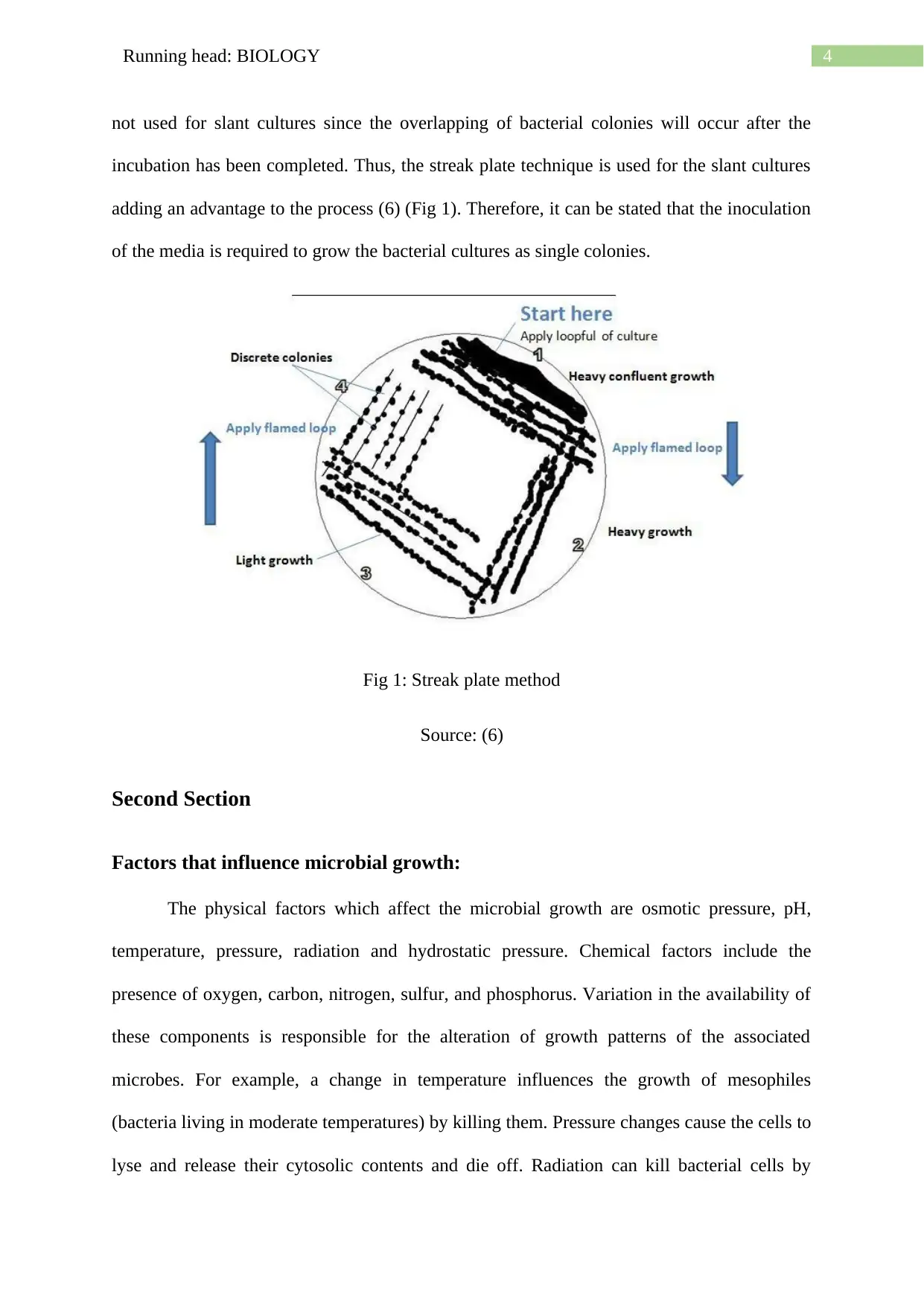
not used for slant cultures since the overlapping of bacterial colonies will occur after the
incubation has been completed. Thus, the streak plate technique is used for the slant cultures
adding an advantage to the process (6) (Fig 1). Therefore, it can be stated that the inoculation
of the media is required to grow the bacterial cultures as single colonies.
Fig 1: Streak plate method
Source: (6)
Second Section
Factors that influence microbial growth:
The physical factors which affect the microbial growth are osmotic pressure, pH,
temperature, pressure, radiation and hydrostatic pressure. Chemical factors include the
presence of oxygen, carbon, nitrogen, sulfur, and phosphorus. Variation in the availability of
these components is responsible for the alteration of growth patterns of the associated
microbes. For example, a change in temperature influences the growth of mesophiles
(bacteria living in moderate temperatures) by killing them. Pressure changes cause the cells to
lyse and release their cytosolic contents and die off. Radiation can kill bacterial cells by
5Running head: BIOLOGY
causing mutation in their genetic material (7). The osmotic pressure of the surroundings also
maintains the growth of bacterial cells by maintaining their osmotic balance. Changes in the
surrounding water concentration kill the cells by either lysis of shrinkage. Chemical factors
mainly include the nutrient concentration associated with the growth media (8).
Carbohydrates, proteins, and lipids are required for the development of bacterial cell walls
and the rest of the internal components. Imbalances in the nutrient concentrations can cause
the cells to die or show reduced growth.
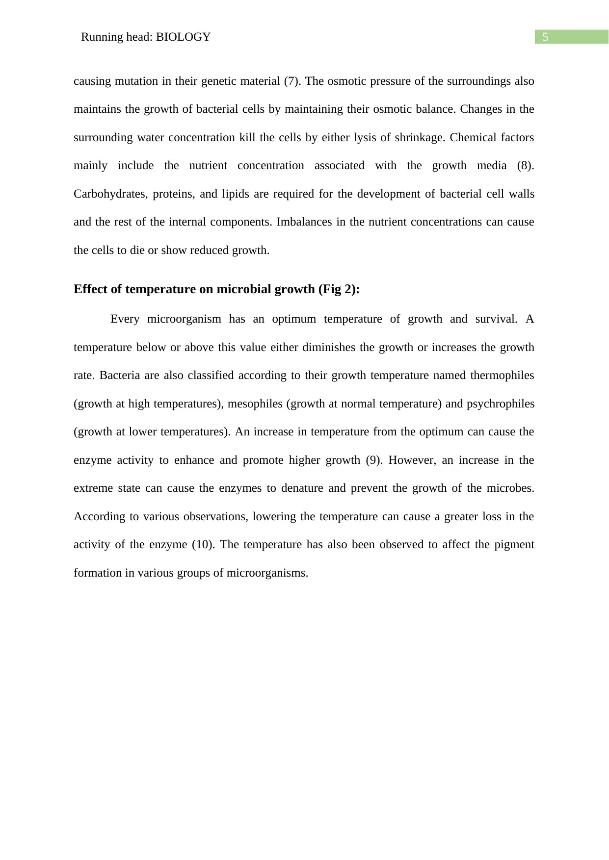
Effect of temperature on microbial growth (Fig 2):
Every microorganism has an optimum temperature of growth and survival. A
temperature below or above this value either diminishes the growth or increases the growth
rate. Bacteria are also classified according to their growth temperature named thermophiles
(growth at high temperatures), mesophiles (growth at normal temperature) and psychrophiles
(growth at lower temperatures). An increase in temperature from the optimum can cause the
enzyme activity to enhance and promote higher growth (9). However, an increase in the
extreme state can cause the enzymes to denature and prevent the growth of the microbes.
According to various observations, lowering the temperature can cause a greater loss in the
activity of the enzyme (10). The temperature has also been observed to affect the pigment
formation in various groups of microorganisms.
causing mutation in their genetic material (7). The osmotic pressure of the surroundings also
maintains the growth of bacterial cells by maintaining their osmotic balance. Changes in the
surrounding water concentration kill the cells by either lysis of shrinkage. Chemical factors
mainly include the nutrient concentration associated with the growth media (8).
Carbohydrates, proteins, and lipids are required for the development of bacterial cell walls
and the rest of the internal components. Imbalances in the nutrient concentrations can cause
the cells to die or show reduced growth.
Effect of temperature on microbial growth (Fig 2):
Every microorganism has an optimum temperature of growth and survival. A
temperature below or above this value either diminishes the growth or increases the growth
rate. Bacteria are also classified according to their growth temperature named thermophiles
(growth at high temperatures), mesophiles (growth at normal temperature) and psychrophiles
(growth at lower temperatures). An increase in temperature from the optimum can cause the
enzyme activity to enhance and promote higher growth (9). However, an increase in the
extreme state can cause the enzymes to denature and prevent the growth of the microbes.
According to various observations, lowering the temperature can cause a greater loss in the
activity of the enzyme (10). The temperature has also been observed to affect the pigment
formation in various groups of microorganisms.
⊘ This is a preview!⊘
Do you want full access?
Subscribe today to unlock all pages.
Trusted by 1+ million students worldwide
6Running head: BIOLOGY
Fig 2: Effect of temperature on microbial growth
Source: (9)
Measuring microbial growth (Haemocytometer and Colorimeter):
Microorganisms are not visible to the naked eye. However, they exist and are visible
under the microscope after staining. Thus, there is also a way to count them and estimate the
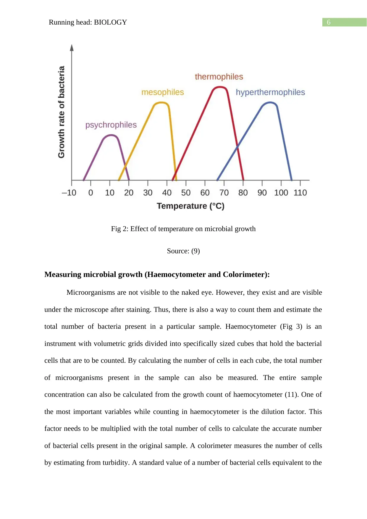
total number of bacteria present in a particular sample. Haemocytometer (Fig 3) is an
instrument with volumetric grids divided into specifically sized cubes that hold the bacterial
cells that are to be counted. By calculating the number of cells in each cube, the total number
of microorganisms present in the sample can also be measured. The entire sample
concentration can also be calculated from the growth count of haemocytometer (11). One of
the most important variables while counting in haemocytometer is the dilution factor. This
factor needs to be multiplied with the total number of cells to calculate the accurate number
of bacterial cells present in the original sample. A colorimeter measures the number of cells
by estimating from turbidity. A standard value of a number of bacterial cells equivalent to the
Fig 2: Effect of temperature on microbial growth
Source: (9)
Measuring microbial growth (Haemocytometer and Colorimeter):
Microorganisms are not visible to the naked eye. However, they exist and are visible
under the microscope after staining. Thus, there is also a way to count them and estimate the
total number of bacteria present in a particular sample. Haemocytometer (Fig 3) is an
instrument with volumetric grids divided into specifically sized cubes that hold the bacterial
cells that are to be counted. By calculating the number of cells in each cube, the total number
of microorganisms present in the sample can also be measured. The entire sample
concentration can also be calculated from the growth count of haemocytometer (11). One of
the most important variables while counting in haemocytometer is the dilution factor. This
factor needs to be multiplied with the total number of cells to calculate the accurate number
of bacterial cells present in the original sample. A colorimeter measures the number of cells
by estimating from turbidity. A standard value of a number of bacterial cells equivalent to the
Paraphrase This Document
Need a fresh take? Get an instant paraphrase of this document with our AI Paraphraser
7Running head: BIOLOGY
observed absorbance of turbidity is recorded (12). This value is then used to estimate the
number of cells present in the chosen culture.
Fig 3: Grid of a haemocytometer with cells
Source: (11)
Nutritional requirements of bacteria
Every organism requires nutrients for their growth and survival. As they grow, they
start metabolizing new nutrients that are present in their surrounding media. The major
component of a growth media is the nutrient composition which is measured in milligrams.
This composition is specific for different growth media required for bacteria growth. Bacteria
require both macronutrients and micronutrients for their growth and survival. Bacteria require
8Running head: BIOLOGY
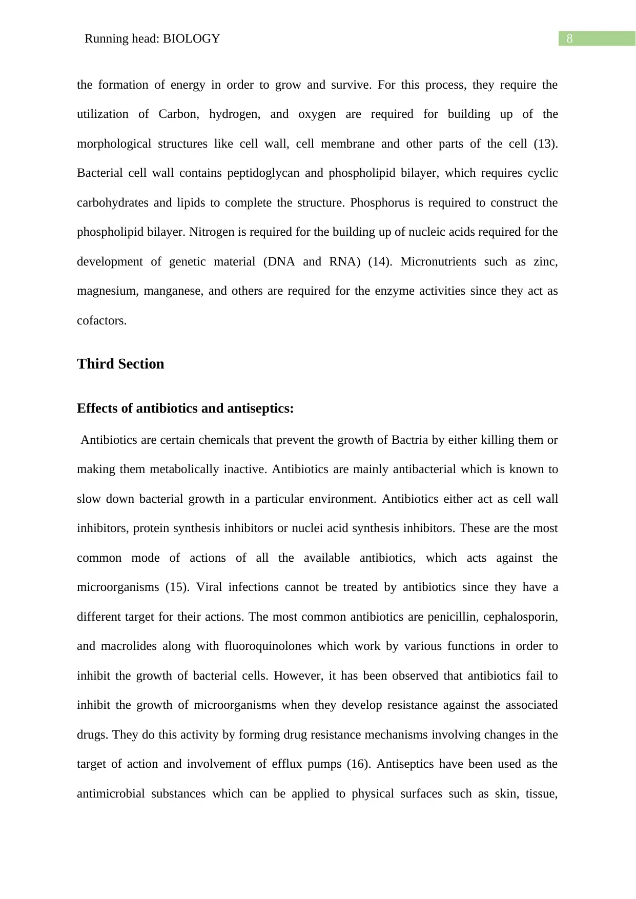
the formation of energy in order to grow and survive. For this process, they require the
utilization of Carbon, hydrogen, and oxygen are required for building up of the
morphological structures like cell wall, cell membrane and other parts of the cell (13).
Bacterial cell wall contains peptidoglycan and phospholipid bilayer, which requires cyclic
carbohydrates and lipids to complete the structure. Phosphorus is required to construct the
phospholipid bilayer. Nitrogen is required for the building up of nucleic acids required for the
development of genetic material (DNA and RNA) (14). Micronutrients such as zinc,
magnesium, manganese, and others are required for the enzyme activities since they act as
cofactors.
Third Section
Effects of antibiotics and antiseptics:
Antibiotics are certain chemicals that prevent the growth of Bactria by either killing them or
making them metabolically inactive. Antibiotics are mainly antibacterial which is known to
slow down bacterial growth in a particular environment. Antibiotics either act as cell wall
inhibitors, protein synthesis inhibitors or nuclei acid synthesis inhibitors. These are the most
common mode of actions of all the available antibiotics, which acts against the
microorganisms (15). Viral infections cannot be treated by antibiotics since they have a
different target for their actions. The most common antibiotics are penicillin, cephalosporin,
and macrolides along with fluoroquinolones which work by various functions in order to
inhibit the growth of bacterial cells. However, it has been observed that antibiotics fail to
inhibit the growth of microorganisms when they develop resistance against the associated
drugs. They do this activity by forming drug resistance mechanisms involving changes in the
target of action and involvement of efflux pumps (16). Antiseptics have been used as the
antimicrobial substances which can be applied to physical surfaces such as skin, tissue,
⊘ This is a preview!⊘
Do you want full access?
Subscribe today to unlock all pages.
Trusted by 1+ million students worldwide
9Running head: BIOLOGY
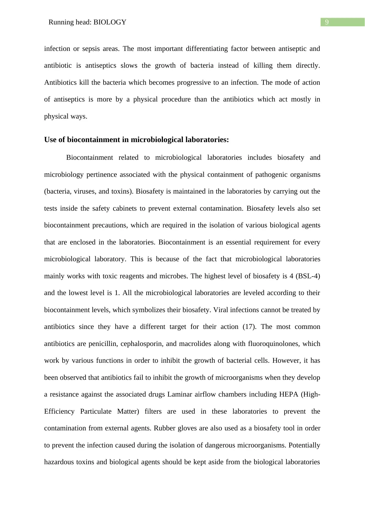
infection or sepsis areas. The most important differentiating factor between antiseptic and
antibiotic is antiseptics slows the growth of bacteria instead of killing them directly.
Antibiotics kill the bacteria which becomes progressive to an infection. The mode of action
of antiseptics is more by a physical procedure than the antibiotics which act mostly in
physical ways.
Use of biocontainment in microbiological laboratories:
Biocontainment related to microbiological laboratories includes biosafety and
microbiology pertinence associated with the physical containment of pathogenic organisms
(bacteria, viruses, and toxins). Biosafety is maintained in the laboratories by carrying out the
tests inside the safety cabinets to prevent external contamination. Biosafety levels also set
biocontainment precautions, which are required in the isolation of various biological agents
that are enclosed in the laboratories. Biocontainment is an essential requirement for every
microbiological laboratory. This is because of the fact that microbiological laboratories
mainly works with toxic reagents and microbes. The highest level of biosafety is 4 (BSL-4)
and the lowest level is 1. All the microbiological laboratories are leveled according to their
biocontainment levels, which symbolizes their biosafety. Viral infections cannot be treated by
antibiotics since they have a different target for their action (17). The most common
antibiotics are penicillin, cephalosporin, and macrolides along with fluoroquinolones, which
work by various functions in order to inhibit the growth of bacterial cells. However, it has
been observed that antibiotics fail to inhibit the growth of microorganisms when they develop
a resistance against the associated drugs Laminar airflow chambers including HEPA (High-
Efficiency Particulate Matter) filters are used in these laboratories to prevent the
contamination from external agents. Rubber gloves are also used as a biosafety tool in order
to prevent the infection caused during the isolation of dangerous microorganisms. Potentially
hazardous toxins and biological agents should be kept aside from the biological laboratories
Paraphrase This Document
Need a fresh take? Get an instant paraphrase of this document with our AI Paraphraser
10Running head: BIOLOGY
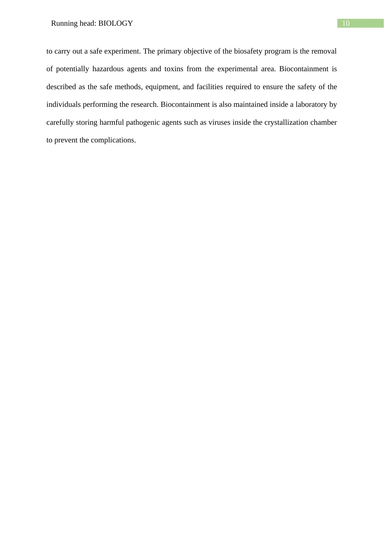
to carry out a safe experiment. The primary objective of the biosafety program is the removal
of potentially hazardous agents and toxins from the experimental area. Biocontainment is
described as the safe methods, equipment, and facilities required to ensure the safety of the
individuals performing the research. Biocontainment is also maintained inside a laboratory by
carefully storing harmful pathogenic agents such as viruses inside the crystallization chamber
to prevent the complications.
11Running head: BIOLOGY
References
1. Basu, S.; Bose, C.; Ojha, N.; Das, N.; Das, J.; Pal, M.; and Khurana, S;. 2015. Evolution of
bacterial and fungal growth media. Bioinformation; 11(4), 182.
2. Alazhari, M.; Sharma, T.; Heath, A.; Cooper, R. and Paine, K.; 2018. Application of
expanded perlite encapsulated bacteria and growth media for self-healing
concrete. Construction and Building Materials; 160, pp.610-619.
3. Harris, T.M.; Rumaseb, A.; Beissbarth, J.; Barzi, F.; Leach, A.J. and Smith-Vaughan,
H.C.; 2017. Culture of non-typeable Haemophilus influenzae from the nasopharynx: Not all
media are equal. Journal of microbiological methods; 137, pp.3-5.
4. Talaro, K.P. and Chess, B.; 2018. Foundations in microbiology.; McGraw-Hill.
5. Brock, D.A.; Canas, A.; Jones, K.; Queller, D.C. and Strassmann, J.E.; 2017. Exposure to
dense bacteria lawns does not cause the social amoeba Dictyostelium discoideum to carry
bacteria through the social stage (No. e2698v1).; PeerJ Preprints.
6. Baird, B.; 2018. Testing Bacterial Antibiotic Production under Carbohydrate and Protein
Starvation.
7. Marshall, J.J.; Strategic Partnerships Alliance LLC; 2017. Sterilizing radiation system for
use with door handle.; U.S. Patent Application 15/597,996.
8. Page, R. and Peti, W.; 2016. Toxin-antitoxin systems in bacterial growth arrest and
persistence. Nature chemical biology; 12(4), p.208.
9. Jung, J.Y.; Lee, H.J.; Chun, B.H. and Jeon, C.O.; 2016. Effects of temperature on bacterial
communities and metabolites during fermentation of myeolchi-Aekjeot, a traditional korean
fermented anchovy sauce. PloS one; 11(3), p.e0151351.
⊘ This is a preview!⊘
Do you want full access?
Subscribe today to unlock all pages.
Trusted by 1+ million students worldwide
1 out of 13


Your All-in-One AI-Powered Toolkit for Academic Success.
+13062052269
info@desklib.com
Available 24*7 on WhatsApp / Email
![[object Object]](/_next/static/media/star-bottom.7253800d.svg)
Unlock your academic potential
Copyright © 2020–2026 A2Z Services. All Rights Reserved. Developed and managed by ZUCOL.