Research Paper: Thesis and Outline on MRI Risks in Pregnancy
VerifiedAdded on 2022/10/13
|5
|905
|7
Homework Assignment
AI Summary
This assignment presents a thesis statement and outline for a research paper on the use of Magnetic Resonance Imaging (MRI) during pregnancy. The thesis argues that while there is limited evidence of harm, the research will investigate the benefits and risks of the procedure. The introduction explains the use of MRI in pregnancy to detect illnesses in the mother and fetus, highlighting its advantages over ultrasound. The body of the outline discusses factors influencing MRI risks, including radiofrequency energy, magnetic field strength, and scan time. It also addresses the risks associated with gadolinium contrast and the potential for fetal harm, along with the safety considerations. The conclusion recommends MRI without contrast during the first trimester and suggests further research into the safety of MRI in later stages of pregnancy, considering varying conditions and the impact of the acoustic environment on fetal development.
Running head: THESIS AND OUTLINE
Thesis and Outline
Student Name
University Name
Author Note
Thesis and Outline
Student Name
University Name
Author Note
Paraphrase This Document
Need a fresh take? Get an instant paraphrase of this document with our AI Paraphraser
1
Thesis and Outline
Table of Contents
Thesis statement.........................................................................................................................2
Introduction................................................................................................................................2
Why is it used?.......................................................................................................................2
How does it help physicians?.................................................................................................2
MRI during pregrancy................................................................................................................2
MRI risks....................................................................................................................................3
Conclusion..................................................................................................................................3
References..................................................................................................................................4
2
Thesis and Outline
Thesis statement
There is little evidence present on whether MRI is harmful during the diagnosis of
pregnant women; this research study will investigate the pros and cons of the procedure.
Introduction
Why is it used?
Magnetic Resonance Imaging is used during pregnancy to detect the presence of any
illnesses in the mother and the fetus (Ray et al., 2016).
How does it help physicians?
It helps the physician to observe the growth of the baby inside the womb. Despite this,
there are affirmations that the rays may turn to be harmful for the fetus. Unlike ultrasound,
the fetal lie position, the oligohydramnios, the overlying bone, or the obesity of the mother
does not restrict the MRI. The MRI can comprehensively image the fetus from every required
angle and plane, thereby providing a larger field of view of fetus along with the placenta in
addition to excellent resolution of the soft tissue of brain, lungs, heart, and abdomen (Bulas,
Dorothy & Alexia Egloff, 2013).
MRI during pregrancy
In a recent study evaluated about 1.4 million births along with periodic exposure to
the magnetic resonance imaging in first trimester of the pregnancy, compared the data with
the non-exposed controls, and observed no increased risk to the fetus or the mother (Ray et
al., 2016). However, more research is required to understand the prevalence of harms while
diagnosing pregnancy and the safety procedures that need to be undertaken. There are several
factors influencing the risk associated while conducting an MRI like the radiofrequency
⊘ This is a preview!⊘
Do you want full access?
Subscribe today to unlock all pages.

Trusted by 1+ million students worldwide
3
Thesis and Outline
energy variability, magnetic field strength, scan time and gradient magnetic field along with
observing the effects of MRI during second and third trimester (Nissan et al., 2019).
MRI risks
Recent data has shown that MRI scans with gadolinium contrast successfully
enhanced the quality of images during pregnancy however; an increased risk including
adverse rheumatologic effects, inflammation and harmful effects on skin has been reported
resulting in stillbirth along with neonatal death (Kanda et al., 2016). Deposition of
Gadolinium contrast has also been reported in the brain of the fetus, which is extremely
harmful (Choi & Moon, 2019). Concerns has also been expressed regarding the safety of the
MRI exposure because of the heating caused by the augmented magnetic fields which might
affect the sensitive tissues of a fetus along with prolonged exposure to MRI’s loud acoustic
environment (Gulani et al., 2017).
Conclusion
In conclusion, MRI during the first trimester in general is not harmful however; the
administration of contrast to enhance the resolution and quality of imaging is probably
dangerous for the growing fetus. Therefore, to negate the chances of any adverse effects,
MRI without the use of the contrast dye is recommended for the comprehensive view of the
fetus within the mother during the first trimester. More research is required to ensure the
safety of MRI scans during the second and third trimesters of pregnancy along with studies
with varying MRI conditions, intensities of magnetic field, duration of the MRI scan timing
and effect of the loud acoustic environment on the fetal growth before recommending the
procedure for women in advanced stages of pregnancy.
Paraphrase This Document
Need a fresh take? Get an instant paraphrase of this document with our AI Paraphraser
4
Thesis and Outline
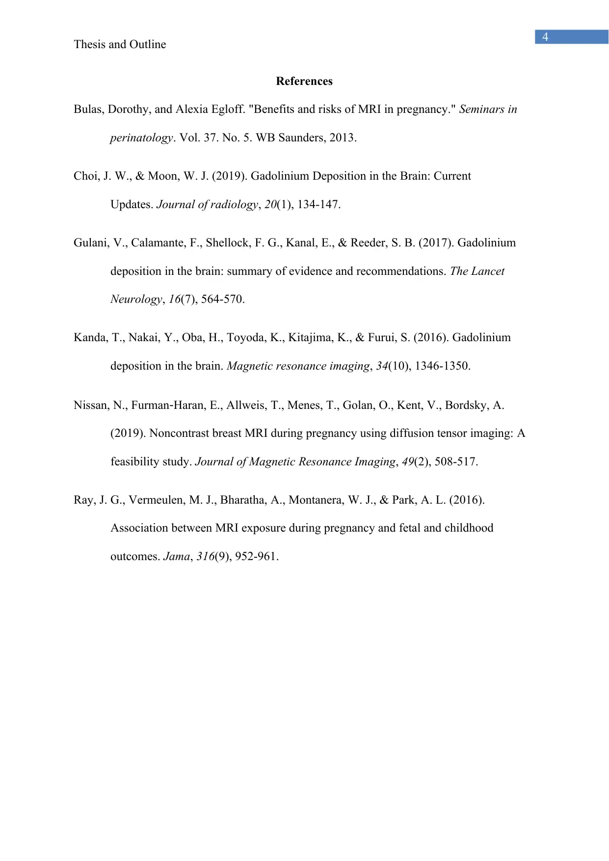
References
Bulas, Dorothy, and Alexia Egloff. "Benefits and risks of MRI in pregnancy." Seminars in
perinatology. Vol. 37. No. 5. WB Saunders, 2013.
Choi, J. W., & Moon, W. J. (2019). Gadolinium Deposition in the Brain: Current
Updates. Journal of radiology, 20(1), 134-147.
Gulani, V., Calamante, F., Shellock, F. G., Kanal, E., & Reeder, S. B. (2017). Gadolinium
deposition in the brain: summary of evidence and recommendations. The Lancet
Neurology, 16(7), 564-570.
Kanda, T., Nakai, Y., Oba, H., Toyoda, K., Kitajima, K., & Furui, S. (2016). Gadolinium
deposition in the brain. Magnetic resonance imaging, 34(10), 1346-1350.
Nissan, N., Furman‐Haran, E., Allweis, T., Menes, T., Golan, O., Kent, V., Bordsky, A.
(2019). Noncontrast breast MRI during pregnancy using diffusion tensor imaging: A
feasibility study. Journal of Magnetic Resonance Imaging, 49(2), 508-517.
Ray, J. G., Vermeulen, M. J., Bharatha, A., Montanera, W. J., & Park, A. L. (2016).
Association between MRI exposure during pregnancy and fetal and childhood
outcomes. Jama, 316(9), 952-961.
1 out of 5
Related Documents
Your All-in-One AI-Powered Toolkit for Academic Success.
+13062052269
info@desklib.com
Available 24*7 on WhatsApp / Email
![[object Object]](/_next/static/media/star-bottom.7253800d.svg)
Unlock your academic potential
Copyright © 2020–2026 A2Z Services. All Rights Reserved. Developed and managed by ZUCOL.


