Report on MRI Scanning Techniques: Development, Uses, Limitations

Verified

Added on  2023/06/15

|1
|809
|143
Report
AI Summary
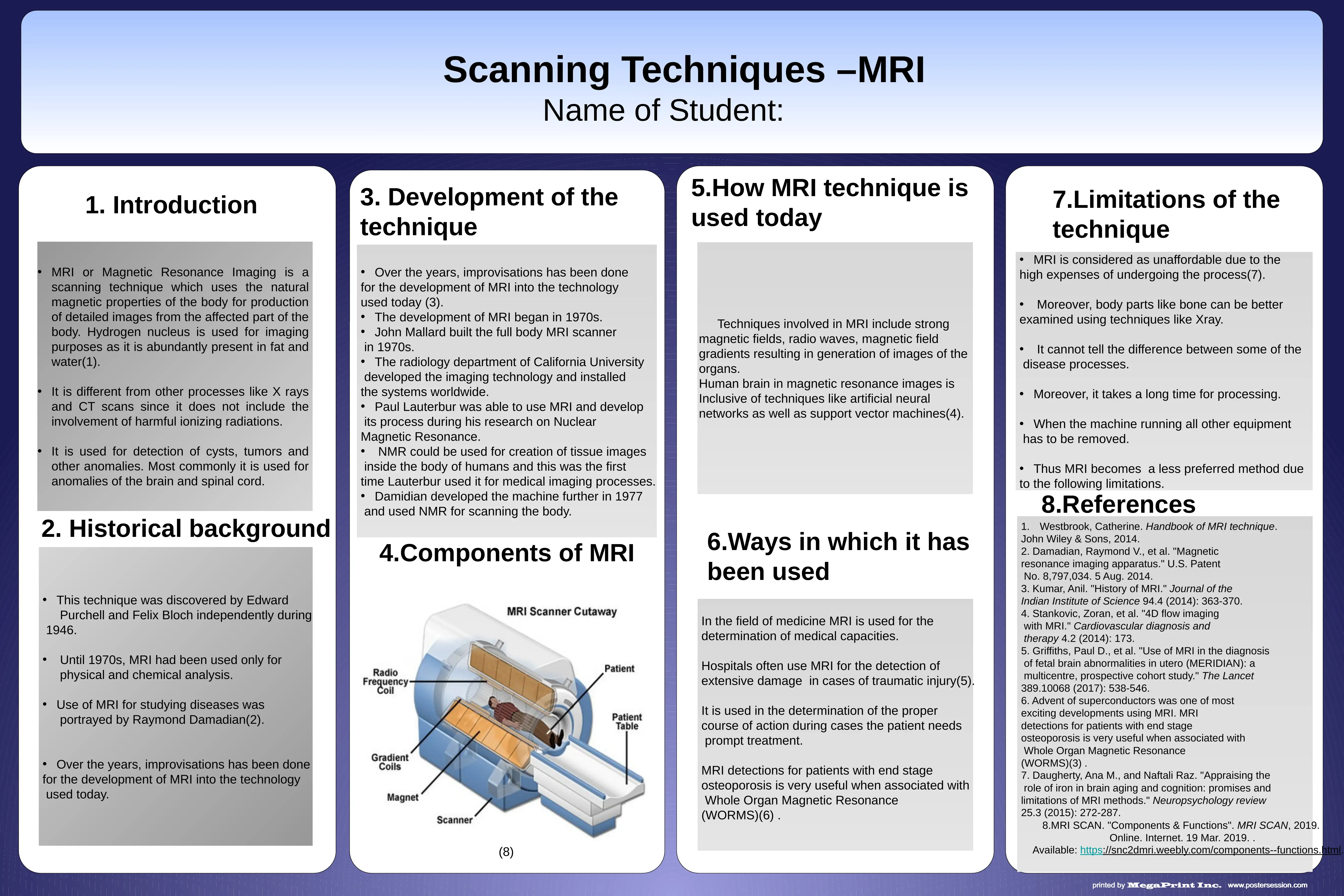
This report provides a detailed overview of Magnetic Resonance Imaging (MRI) scanning techniques, which utilize the body's natural magnetic properties to create detailed images. Unlike X-rays and CT scans, MRI does not involve harmful ionizing radiation, making it a preferred method for detecting cysts, tumors, and other anomalies, particularly in the brain and spinal cord. The report traces the historical development of MRI from the 1970s, highlighting key contributions by researchers like John Mallard, Paul Lauterbur, and Raymond Damadian. It outlines the components of an MRI machine and discusses how the technique is used in modern medicine, including its application in diagnosing traumatic injuries and studying diseases. The report also addresses the limitations of MRI, such as its high cost, long processing time, and inability to differentiate between certain disease processes, noting that alternative methods like X-rays may be more suitable for examining bones. Despite these limitations, MRI remains a crucial tool in determining appropriate medical actions and assessing medical capacities.
Document Page
4.Components of MRI
2. Historical background 6.Ways in which it has
been used
Scanning Techniques –MRI
Name of Student:
7.Limitations of the
technique
1. Introduction 3. Development of the
technique
5.How MRI technique is
used today
References
This technique was discovered by Edward
Purchell and Felix Bloch independently during
1946.
Until 1970s, MRI had been used only for
physical and chemical analysis.
Use of MRI for studying diseases was
portrayed by Raymond Damadian(2).
Over the years, improvisations has been done
for the development of MRI into the technology
used today.
Over the years, improvisations has been done
for the development of MRI into the technology
used today (3).
The development of MRI began in 1970s.
John Mallard built the full body MRI scanner
in 1970s.
The radiology department of California University
developed the imaging technology and installed
the systems worldwide.
Paul Lauterbur was able to use MRI and develop
its process during his research on Nuclear
Magnetic Resonance.
NMR could be used for creation of tissue images
inside the body of humans and this was the first
time Lauterbur used it for medical imaging processes.
Damidian developed the machine further in 1977
and used NMR for scanning the body.
Techniques involved in MRI include strong
magnetic fields, radio waves, magnetic field
gradients resulting in generation of images of the
organs.
Human brain in magnetic resonance images is
Inclusive of techniques like artificial neural
networks as well as support vector machines(4).
In the field of medicine MRI is used for the
determination of medical capacities.
Hospitals often use MRI for the detection of
extensive damage in cases of traumatic injury(5).
It is used in the determination of the proper
course of action during cases the patient needs
prompt treatment.
MRI detections for patients with end stage
osteoporosis is very useful when associated with
Whole Organ Magnetic Resonance
(WORMS)(6) .
MRI is considered as unaffordable due to the
high expenses of undergoing the process(7).
Moreover, body parts like bone can be better
examined using techniques like Xray.
It cannot tell the difference between some of the
disease processes.
Moreover, it takes a long time for processing.
When the machine running all other equipment
has to be removed.
Thus MRI becomes a less preferred method due
to the following limitations.
1. Westbrook, Catherine. Handbook of MRI technique.
John Wiley & Sons, 2014.
2. Damadian, Raymond V., et al. "Magnetic
resonance imaging apparatus." U.S. Patent
No. 8,797,034. 5 Aug. 2014.
3. Kumar, Anil. "History of MRI." Journal of the
Indian Institute of Science 94.4 (2014): 363-370.
4. Stankovic, Zoran, et al. "4D flow imaging
with MRI." Cardiovascular diagnosis and
therapy 4.2 (2014): 173.
5. Griffiths, Paul D., et al. "Use of MRI in the diagnosis
of fetal brain abnormalities in utero (MERIDIAN): a
multicentre, prospective cohort study." The Lancet
389.10068 (2017): 538-546.
6. Advent of superconductors was one of most
exciting developments using MRI. MRI
detections for patients with end stage
osteoporosis is very useful when associated with
Whole Organ Magnetic Resonance
(WORMS)(3) .
7. Daugherty, Ana M., and Naftali Raz. "Appraising the
role of iron in brain aging and cognition: promises and
limitations of MRI methods." Neuropsychology review
25.3 (2015): 272-287.
8.MRI SCAN. "Components & Functions". MRI SCAN, 2019.
Online. Internet. 19 Mar. 2019. .
Available: https://snc2dmri.weebly.com/components--functions.html.
MRI or Magnetic Resonance Imaging is a
scanning technique which uses the natural
magnetic properties of the body for production
of detailed images from the affected part of the
body. Hydrogen nucleus is used for imaging
purposes as it is abundantly present in fat and
water(1).
It is different from other processes like X rays
and CT scans since it does not include the
involvement of harmful ionizing radiations.
It is used for detection of cysts, tumors and
other anomalies. Most commonly it is used for
anomalies of the brain and spinal cord.
(8)
8.References
tabler-icon-diamond-filled.svg

Paraphrase This Document

Need a fresh take? Get an instant paraphrase of this document with our AI Paraphraser
[object Object]