University Case Study: Diagnosis, Treatment of Neuroblastoma
VerifiedAdded on 2022/09/01
|10
|1607
|21
Case Study
AI Summary
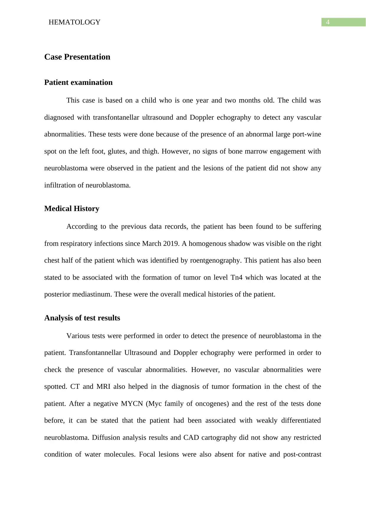
This case study focuses on a one-year-old child presenting with a port-wine spot and respiratory infections, ultimately diagnosed with weakly differentiated neuroblastoma. The patient underwent various diagnostic tests, including transfontanellar ultrasound, Doppler echography, CT scans, and MRI, to assess for vascular abnormalities and tumor presence. While initial findings revealed a tumor in the posterior mediastinum, no neuroblastoma involvement was observed in the bone marrow. The child's medical history included respiratory infections since March 2019, with a shadow identified on the right chest via roentgenography. The diagnosis was confirmed by negative MYCN results and other tests. Treatment primarily involved chemotherapy based on intermediate and low-risk neuroblastoma protocols, with mild leukopenia and diarrhea managed effectively. The case study discusses the etiology, epidemiology, prevalence, and complications of neuroblastoma, including the ethical dilemma and prognosis. The child was discharged from the hospital after recovery.

Running head: HEMATOLOGY
NEUROBLASTOMA, A CASE STUDY
Name of the Student
Name of the University
Author Note
NEUROBLASTOMA, A CASE STUDY
Name of the Student
Name of the University
Author Note
Secure Best Marks with AI Grader
Need help grading? Try our AI Grader for instant feedback on your assignments.

1HEMATOLOGY
Abstract
This case study talks about a child who is suffering from respiratory diseases for a
long time and also has been found to show a port wine spot on some organs of the body. This
child is diagnosed with the presence of neuroblastoma by various medical tests. However,
neuroblastoma is not found to be associated with the patient. The child was having mild
leukopenia which was treated with specific therapy. Finally, the child was discharged from
the hospital.

2HEMATOLOGY
Table of Contents
Case Presentation.......................................................................................................................4
Patient examination................................................................................................................4
Medical History......................................................................................................................4
Analysis of test results...........................................................................................................4
Planning of diagnosis.............................................................................................................5
Differential Diagnosis................................................................................................................5
Support for considered conditions.........................................................................................5
Support for additional investigations.....................................................................................5
Pathophysiology.........................................................................................................................5
Treatment...................................................................................................................................6
Discussion..................................................................................................................................6
Etiology..................................................................................................................................6
Epidemiology.........................................................................................................................6
Prevalence..............................................................................................................................7
Complications........................................................................................................................7
Prognosis................................................................................................................................7
Ethical dilemma.....................................................................................................................7
Conclusion..................................................................................................................................7
References..................................................................................................................................9

3HEMATOLOGY
Secure Best Marks with AI Grader
Need help grading? Try our AI Grader for instant feedback on your assignments.

4HEMATOLOGY
Case Presentation
Patient examination
This case is based on a child who is one year and two months old. The child was
diagnosed with transfontanellar ultrasound and Doppler echography to detect any vascular
abnormalities. These tests were done because of the presence of an abnormal large port-wine
spot on the left foot, glutes, and thigh. However, no signs of bone marrow engagement with
neuroblastoma were observed in the patient and the lesions of the patient did not show any
infiltration of neuroblastoma.
Medical History
According to the previous data records, the patient has been found to be suffering
from respiratory infections since March 2019. A homogenous shadow was visible on the right
chest half of the patient which was identified by roentgenography. This patient has also been
stated to be associated with the formation of tumor on level Tn4 which was located at the
posterior mediastinum. These were the overall medical histories of the patient.
Analysis of test results
Various tests were performed in order to detect the presence of neuroblastoma in the
patient. Transfontannellar Ultrasound and Doppler echography were performed in order to
check the presence of vascular abnormalities. However, no vascular abnormalities were
spotted. CT and MRI also helped in the diagnosis of tumor formation in the chest of the
patient. After a negative MYCN (Myc family of oncogenes) and the rest of the tests done
before, it can be stated that the patient had been associated with weakly differentiated
neuroblastoma. Diffusion analysis results and CAD cartography did not show any restricted
condition of water molecules. Focal lesions were also absent for native and post-contrast

5HEMATOLOGY
tests. No organic pathological variations were found to be associated with intracranial
anatomic components.
Planning of diagnosis
After looking at the symptoms of the patient, the diagnosis plan was established as
discussed below.
Differential Diagnosis
Support for considered conditions
The diagnostic tests involved MRI, CT scan, MYCN genetic test, Trepanobiopsy and
Claude Bernard Horner test for the identification of neuroblastoma in the patient. According
to scientific studies, it can be stated that the test for the presence of the MYCN gene in an
amplified form is a diagnostic feature of neuroblastoma (1). MRI and CT scans are the two
imaging techniques used to visualize the presence of any type of tumor in a particular region
(2). Claude Bernard Horner's test was done in order to test the patient for any types of
disorders associated with the central nervous system (3). This patient was found to be
associated with having a possibility of Klipper-Trenaunay syndrome according to the
previously stated test. Condition of muscle tones, lymph nodes, breathing and heart rates and
the sizes of liver and spleen were considered as the diagnostic tests for neuroblastoma for this
child.
Support for additional investigations
Differential diagnosis of neuroblastoma was done by additional diagnostic tests,
which involved chemotherapy, and microscopic tests for the identification of ipsilateral
metastasis in the lymph nodes. These tests were also used by various physiologists in order to
identify the association of neuroblastoma in a patient (4).

6HEMATOLOGY
Pathophysiology
Pediatric neuroblastoma has been found as the most common extracranial solid
tumor-associated with infants. This is a malignant condition of the embryo which is
associated with the sympathetic nervous system which arises from neuroblasts (5). These
cells have been observed to invaginate in the embryo, migrate along the neural axis and
finally accumulate in the adrenal medulla, sympathetic medulla, and other sites. The
distribution pattern of these cells has been found to correlate with the primary neuroblastoma
presentation sites. However, in this case, which involves the diagnosis of a child having the
possibility of neuroblastoma, was found to be absent at a significant level. All the tests which
were performed to diagnose the presence of neuroblastoma found the presence of weakly
differentiated neuroblastoma.
Treatment
In this case study, chemotherapy has been used as the treatment process for
neuroblastoma according to the protocol for intermediate and low-risk Neuroblastoma. This
process has been supported by various case studies involving the treatment of neuroblastoma
(6). The child had mild leukopenia associated with diarrhea which was found to be controlled
well.
Discussion
Etiology
Much information about the etiology of neuroblastoma is unknown since it is linked
to the genetic tendency of inheritance. This disease has also been found to be associated with
the autosomal dominant pattern of inheritance with the amplification of N-MYC oncogene in
Paraphrase This Document
Need a fresh take? Get an instant paraphrase of this document with our AI Paraphraser

7HEMATOLOGY
20% of the patients approximately (7). The progression of this disease is not exactly the same
as other tumor-associated cancer.
Epidemiology
Seven to ten percent of childhood cancers have been found to be associated with
Neuroblastoma. Every year, it has been found that 800 new cases are being diagnosed
throughout the world for neuroblastoma (6). Neuroblastoma has also been observed to
account for 50% of all cancers in infants.
Prevalence
Neuroblastoma has been found to be present in 8% to 105 of all childhood tumors.
According to the statistical records, 500 new cases have been found to be reported annually
for neuroblastoma in infants (4).
Complications
The complications of neuroblastoma include the spreading of the disease in other
parts of the body (bone marrow, liver, bone, and skin). The compression of the spinal cord is
another associate complication of the disease (3).
Prognosis
A child's survival rate if neuroblastoma in 80% and has been found to be dependent on many
factors associated with risks of a grouping of the tumor.
Ethical dilemma
An ethical dilemma is a paradox that can arise due to the decision-making problem
between the moral imperatives of the child and the parents.

8HEMATOLOGY
Conclusion
On a concluding note, it can be stated that; neuroblastoma is one of the most prevalent
cancers occurring in the infant population of a particular region. However, for this case study,
the child has been found to be diagnosed for neuroblastoma. MRI, CT scanning and other
medical tests were performed for the identification of neuroblastoma. Weak associations of
neuroblastoma were observed in the child and the child was discharged from the hospital
after getting recovered.

9HEMATOLOGY
References
1. Olsen RR, Otero JH, Garcia-Lopez J, Wallace K, Finkelstein D, Rehg JE, Yin Z, Wang
YD, Freeman KW. MYCN induces neuroblastoma in primary neural crest cells. Oncogene.
2017 Aug;36(35):5075.
2. Brisse HJ, Blanc T, Schleiermacher G, Mosseri V, Philippe-Chomette P, Janoueix-Lerosey
I, Pierron G, Lapouble E, Peuchmaur M, Fréneaux P, Galmiche L. Radiogenomics of
neuroblastomas: Relationships between imaging phenotypes, tumor genomic profile and
survival. PloS one. 2017 Sep 25;12(9):e0185190.
3. da Mota Gomes M. ON THE SHOULDERS OF GIANTS: THE CASE OF THE CLAUDE
BERNARD HORNER SYNDROME. Revista Brasileira de Neurologia. 2019 Jan;55(1).
4. Grasso S, Cangelosi D, Chapelle J, Alzona M, Centonze G, Lamolinara A, Salemme V,
Angelini C, Morellato A, Saglietto A, Bianchi FT. The SRCIN1/p140Cap adaptor protein
negatively regulates the aggressiveness of neuroblastoma. Cell Death & Differentiation. 2019
Jul 8:1-8.
5. Caputi FF, Candeletti S, Romualdi P. Epigenetic approaches in neuroblastoma disease
pathogenesis. InNeuroblastoma-Current State and Recent Updates 2017 Oct 25. IntechOpen.
6. Ladenstein R, Pötschger U, Pearson AD, Brock P, Luksch R, Castel V, Yaniv I, Papadakis
V, Laureys G, Malis J, Balwierz W. Busulfan and melphalan versus carboplatin, etoposide,
and melphalan as high-dose chemotherapy for high-risk neuroblastoma
(HR-NBL1/SIOPEN): an international, randomised, multi-arm, open-label, phase 3 trial. The
lancet oncology. 2017 Apr 1;18(4):500-14.
7. Schleiermacher G. Biology of Neuroblastoma. InNeuroblastoma 2020 (pp. 17-28).
Springer, Cham.
1 out of 10
Related Documents

Your All-in-One AI-Powered Toolkit for Academic Success.
+13062052269
info@desklib.com
Available 24*7 on WhatsApp / Email
Unlock your academic potential
© 2024 | Zucol Services PVT LTD | All rights reserved.