University Nursing Case Study: STEMI, Risk Factors, and Nursing Care
VerifiedAdded on 2021/04/17
|13
|3945
|118
Case Study
AI Summary
This nursing case study focuses on a patient experiencing ST-elevation myocardial infarction (STEMI). It begins by exploring the patient's risk factors, differentiating between modifiable (smoking, lack of exercise) and non-modifiable (age, gender) elements. The study then delves into the pathophysiology of STEMI and non-STEMI, explaining the mechanisms of myocardial damage and the role of blood clots. It further examines the homeostatic mechanisms that lead to specific symptoms like radiating pain, pallor, and clamminess. A detailed nursing care plan is presented, including subjective and objective data assessment, with a focus on pain relief, myocardial damage prevention, and the MONA treatment protocol (Morphine, Oxygen, Nitro-glycerine, Aspirin). The care plan emphasizes the importance of monitoring vital signs, providing patient education, and addressing potential complications, offering a comprehensive approach to managing STEMI patients.
Running head: Nursing Case Study
Nursing Assignment- Case study of Mr. Sipois
Name of the Student
Name of the University
Author Note
Nursing Assignment- Case study of Mr. Sipois
Name of the Student
Name of the University
Author Note
Paraphrase This Document
Need a fresh take? Get an instant paraphrase of this document with our AI Paraphraser
1Nursing Case Study
Risk Factors and Health promotion (for Myocardial infaction)
STEMI or ST elevation myocardial infarction is a type of heart attack that is caused
due to blockage of blood for a prolonged time to a significant portion of the heart (Heart.org
2018). Acute coronary syndromes are usually differentiated based on the absence or presence
of ST elevation on the ECG, and is called Non STEMI and STEMI respectively. Patients
suffered from Acute Myocardial Infarction require immediate therapy without delay with
percutaneous coronary intervention PPCI done within 90 minutes of first medical contact
(Camaro & de Boer 2015, p. 243).
The term ‘risk factor’ was coined by the researchers of Framingham Heart Study,
USA for cardiovascular diseases (CVD). The studies showed factors like high blood
cholesterol, smoking, hypertension, ECG abnormalities can increase the risk for CVD. The
risk factors can be divided into modifiable and non modifiable risks (metalyse.com 2018).
Several modifiable risk factors are associated with Myocardial Infarction, like:
smoking, abnormal blood lipids, diabetes, hypertension, abdominal obesity, inadequate
physical activity, psychosocial factors, inadequate consumption of fibre, alcohol
consumption.
Smoking is considered as a significant risk for myocardial infarction. Studies show
that the risk of cardiac arrest can increase directly proportionally to the number of cigarettes
smoked without any upper limit. Smoking or inhaling products other than cigarettes can also
increase risks for myocardial infarction, although not as much as cigarettes (Aygül et al.
2008, p. 4). Studies by Aygül et al. (2009, p 5) supports this data, showing that smoking was
found to be more prevalent among STEMI patients. This suggests that the smoking habit of
the patient might be related to the condition.
2Nursing Case Study
Studies show that regular exercise in moderate to intense levels can reduce the risk of
myocardial infaction. Though regular exercise, the normal functioning of the cardiovascular
system can be maintained. It also improves respiration and, cardiac output as well as
maintains an efficient heart rate and stroke volume and therefore prevents the onset of
cardiovascular diseases (Agarwal 2018, p. 541). The patient admitted that he does very little
exercise, and works as a computer programmer (which possibly keeps him restricted to his
work desk for prolonged periods of time). This can also explain the onset of STEMI in the
patient.
Other modifiable risk factors like hypertension, obesity, diabetes, abnormal blood
lipids have not been reported for the patient, however it is important to check his dietary
intake as well as any signs of psychosocial factors like depression, stress and adverse life
effects. Studies have linked cardiovascular disease with psychosocial stressors and poor diet
(Lagraauw et al. 2015, p. 19; Reedy et al. 2014, p. 882)
Non modifiable risks includes: age, gender, genetic factors, racial and ethnic
background. Studies by Dhingra and Vasan (2012, p. 88) shows that age is an independent
risk factor for CVD and can increase by the presence of other risk factors. The risk of CVD
increases significantly after the age of 65. Also, the propensity of CVD earlier in life is more
probable for males than in females. A family history of CVD in a first degree relative can
also increase risks for cardiovascular problems. The patient is 55 years of age, which places
him closer to the non modifiable risk for CVD, as well as his gender. Any family history of
CVD should be investigated.
Intervention strategies for the patient should include: immediate cessation of smoking,
including daily- regular to vigorous exercise, including fibre in diet and restricting the intake
of fats. The patient can be referred to a diet consultant as well as an occupational therapist
⊘ This is a preview!⊘
Do you want full access?
Subscribe today to unlock all pages.

Trusted by 1+ million students worldwide
3Nursing Case Study
and lifestyle modification support. He also might need regular follow-up with general
physician to monitor his cardiac activity.
Pathophysiology (for STEMI and non-STEMI)
STEMI is caused due to a complete and persistence blockage of the coronary artery
(the vessel that supplies blood to the muscles of the heart) by the formation of a blood clot
(thrombus). This blockage leads to damage to the cardiac muscles (and is called a myocardial
damage), and the damage increases with the length of time of the blockage, due to increasing
damaged muscles of the heart. In animal models, the myocardial injury due to a blockage of
the coronary artery spreads in the form of waves, starting from the inner layers of the cardiac
muscles to the outer layers, and during infarction, the full thickness of the cardiac muscles are
affected. Among patients who have survived STEMI, the infracted or damaged muscle is
gradually replaced with a scar tissue (by a process called fibrosis), and the damage is
determined by the stroke volume and efficiency of the heart (Ncbi.nlm.nih.gov 2018).
Non-STEMI or NSTEMI is caused by an imbalance between the myocardial demand
and supply of oxygen. This is caused due to a lowered blood flow through the coronary
vessels. This can be caused by a partial or non-occlusive blood clot (or thrombus) that forms
in a disrupted plaque. The clot causes a near complete blockage of coronary artery. Different
sequence of events can lead to NSTEMI like: rupture of a plaque with superimposed blood
clot which is non-occlusive causing vascular obstruction; dynamic obstruction of cardiac
vessels; narrowing of the arteries (reducing the flow of blood); inflammatory reaction;
conditions (like hypoxemia, hypotension and hypovolemia) that can cause lowering of blood
flow through the coronary vessels. The most common cause of NSTEMI is the rupture of a
plaque, causing a blood clot, which prevents or reduces the flow of blood. The rupture
usually occurs near the shoulder region and contains a large number of inflammatory cells.
Paraphrase This Document
Need a fresh take? Get an instant paraphrase of this document with our AI Paraphraser
4Nursing Case Study
The lack of ST elevation in NSTEMI is because the entire thickness of the cardiac walls is
not involved, unlike in the case of STEMI (Montalescot et al. 2007, p. 1410). Abnormalities
in ECG therefore are not observed in case of NSTEMI, or might only have subtle changes
(Ncbi.nlm.nih.gov 2018). Studies by Montalescot et al. (2007, p 1416) shows that both
STEMI and NSTEMI has similar prognosis.
Homeostatic Mechanisms (mechanisms that leads to specific symptoms in STEMI)
a. Radiating pain to shoulder and left arm
During an episode of heart attack, a sharp radiating pain shooting to the shoulder and
left arm is caused due to a confusion of the nervous system. The palpation on the left side of
the chest is because the anatomical position of the heart slightly tilted to the left. During an
episode of heart attack, the nerves that supply the heart and left arm are confused (as signals
from the cardiac neurons are felt to originate also from the left arm), since they arise from the
same spinal segment. This confusion leads to the sensation of pain to be felt in the left arm,
and is also known as referred pain (Banharak et al. 2018, p. 19; Mata et al. 2014, p. 377).
The pain is caused when nervous signals from the cardiac muscles enter the spinal nerve
which it shares from the sensory nerve from the left hand. Therefore the sensation of pain
travelling from the cardiac nerve is felt in the arm, even though the signal did not originate
there.
b. Pallor
Pallor is referred to as the pale coloration of the skin that can be caused due to an
illness, stress, shock, and anaemia. This can be caused due to a reduction in the amount of
oxyhemoglobin (oxygenated haemoglobin) and increase in the amount of carbamino
haemoglobin (haemoglobin bound to carbon dioxide) in the skin and mucous membrane.
5Nursing Case Study
Oxyhemoglobin is oxygen bound haemoglobin in blood, which has a bright red colour, while
carbaminohemoglobin is dull in colour. A rise in the concentration of carbamino
haemoglobin in the blood causes the discoloration of the skin. Pallor generally prominent in
the face and palms. This is also caused to the reduced blood flow to the peripheral parts of the
body. The sites where pallor can be observed are: lower conjunctiva, tip and dorsum of
tongue, soft palate, nail beds, palmar or plantar creases, and skin. Pallor can also be caused
due to anaemia, or reduction in the haemoglobin concentration in the blood.
c. Clamminess
Clamminess refers to a cold, most and sticky condition of the skin. This occurs when
the temperature of the kin goes below normal and is associated with moisture. The skin also
looks pale, and can occur due to cardiovascular problems or a decrease in the flow of blood
through the peripheral organs like skin or appendages in order to conserve blood supply to
the internal organs. Blockage of cardiac vessels can also lead to clamminess, as the body tries
to restrict the blood to the skin and peripheral organs. During STEMI, the flow of blood to
the coronary artery decreases, this leads to a condition of shock. The reduction of the flow of
blood to the skin results in the drop in the skin temperature, as well as the blood pressure,
which causes shock. Increase in the vascular resistance of peripheral organs also can cause
reduction in the blood supply thereby leading to clamminess (Williams and Wilkins 2018).
Nursing Care
Nursing Care plan for STEMI requires two types of data: subjective data and
objective data. The subjective data consists of information from the patient, and include
factors like chest pain, feeling a sense of pressure on the chest, PQRST pain assessment
(provoke/precipitate/palliate, quality, radiate, severity of symptoms, time). Objective data can
⊘ This is a preview!⊘
Do you want full access?
Subscribe today to unlock all pages.

Trusted by 1+ million students worldwide
6Nursing Case Study
include clinical findings such as: ST elevation in ECG report, decreased oxygenation of
blood, left ventricular failure and tachycardia.
The care plan for STEMI should focus on the following factors: providing relief from
pain and ischemic signs or symptoms; preventing myocardial damage; eliminating respiratory
dysfunction; establish and maintain adequate blood flow to the tissues, alleviating anxiety;
detecting complications early or preventing them; controlling or removing chest pain,
restoring heart rate and blood circulation to adequate levels; adequate activity level to enable
basic self care is achieved; proper understanding of the disease, treatment plan and prognosis
is achieved. The priorities of care should be highest for ensuring relief from pain and anxiety,
followed by reduction in the myocardial stress and workload. The next priorities should
include the prevention or detection of fatal dysrhythmias, assisting and treating complications
caused due to it and promotion of cardiac health and self care (Nice.org.uk 2018).
As a part of intervention, the nurses can administer oxygen and medications to reduce
symptoms; encourage bed rest, providing back rest for the patient to reduce discomfort of the
chest. The patient should also be encouraged to frequently change positions to prevent
accumulation of fluids in the base of lungs. Skin temperature and peripheral pulse should be
regularly checked to identify signs of palliation or clamminess and to monitor flow of blood.
Cardiac rhythm, rate, heart sounds, blood pressure, chest pain, respiratory systems, urinary
output, skin colour change and clinical reports should be monitored regularly (Nice.org.uk
2018).
Different types of treatment/management can be applicable for the treatment of STEMI,
which are discussed below:
1. Morphine Oxygen Nitro-glycerine Aspirin (MONA). Aspirin is used to dilute or
thinning the blood. A total of 324mg of Aspirin can be administered for this process.
Paraphrase This Document
Need a fresh take? Get an instant paraphrase of this document with our AI Paraphraser
7Nursing Case Study
Nitro-glycerine can be administered with Aspirin to dialate the blood vessels and
remove obstruction and blockage of blood. 0.4mg tablet of nitro-glycerine can be
administered sublingually to alleviate chest pain. A second dosage can be
administered after 5 minutes if the pain does not subside. The blood pressure of the
patient should also be monitored and the systolic pressure should be below 90 mm of
Hg. Oxygen is given if the oxygenation of blood is below 94% or if the patient feels
out of breath. 2 L of NC can be administered initially. However, caution must be
applied while administering oxygen as it can cause vasoconstriction, reducing the
flow of blood even further. Morphine is administered if the chest pain is not alleviated
by aspirin and nitro-glycerine.
2. 3 or 5 Lead Monitoring: The patient needs to be placed on cardiac monitoring
immediately to identify worsening of condition.
3. Cardiac Catheterization with Percutaneous Coronary Intervention (PCI): In the
catheterization lab, the blood clot can be located, and a stent can be placed to regain
the blood flow to the heart.
4. Heparin: This is an anticoagulant, and dissolves blood clot that blocks the vessels. For
STEMI, heparin should be administered at 60 units per kg (up to a maximum of 4,000
units) for bolus and 12 units per kg for continuous infusion.
5. Monitoring Cardiac Enzymes (Troponin I, Creatinin Kinase MB): Elevated levels of
these enzymes are generally the indicators of stress or injury to the cardiac muscles.
Troponin enzyme plays an important role in the interaction between the myosin and
actin filaments of the cardiac muscle. Destruction of the cells (due to stress or injury)
leads to the release of the enzymes in the surrounding area. Creatinine Kinase in the
cardiac muscle cells converts ATP to ADP, thereby releasing energy for the
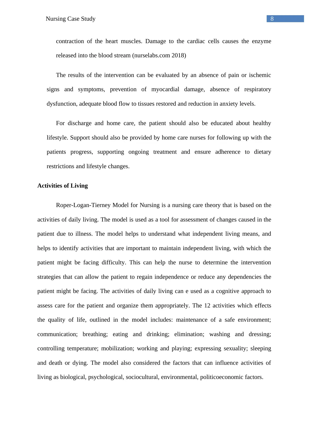
8Nursing Case Study
contraction of the heart muscles. Damage to the cardiac cells causes the enzyme
released into the blood stream (nurselabs.com 2018)
The results of the intervention can be evaluated by an absence of pain or ischemic
signs and symptoms, prevention of myocardial damage, absence of respiratory
dysfunction, adequate blood flow to tissues restored and reduction in anxiety levels.
For discharge and home care, the patient should also be educated about healthy
lifestyle. Support should also be provided by home care nurses for following up with the
patients progress, supporting ongoing treatment and ensure adherence to dietary
restrictions and lifestyle changes.
Activities of Living
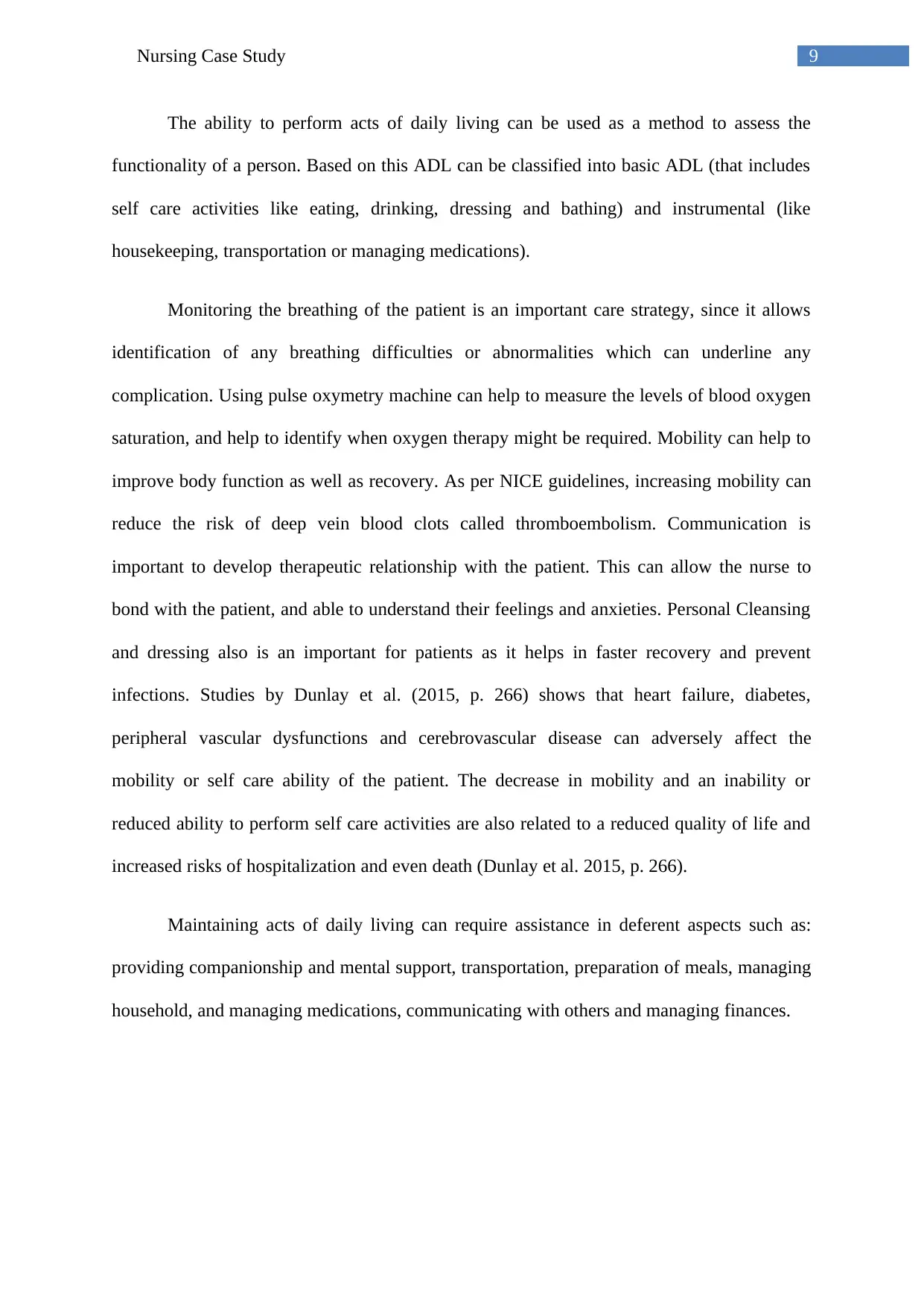
Roper-Logan-Tierney Model for Nursing is a nursing care theory that is based on the
activities of daily living. The model is used as a tool for assessment of changes caused in the
patient due to illness. The model helps to understand what independent living means, and
helps to identify activities that are important to maintain independent living, with which the
patient might be facing difficulty. This can help the nurse to determine the intervention
strategies that can allow the patient to regain independence or reduce any dependencies the
patient might be facing. The activities of daily living can e used as a cognitive approach to
assess care for the patient and organize them appropriately. The 12 activities which effects
the quality of life, outlined in the model includes: maintenance of a safe environment;
communication; breathing; eating and drinking; elimination; washing and dressing;
controlling temperature; mobilization; working and playing; expressing sexuality; sleeping
and death or dying. The model also considered the factors that can influence activities of
living as biological, psychological, sociocultural, environmental, politicoeconomic factors.
⊘ This is a preview!⊘
Do you want full access?
Subscribe today to unlock all pages.

Trusted by 1+ million students worldwide
9Nursing Case Study
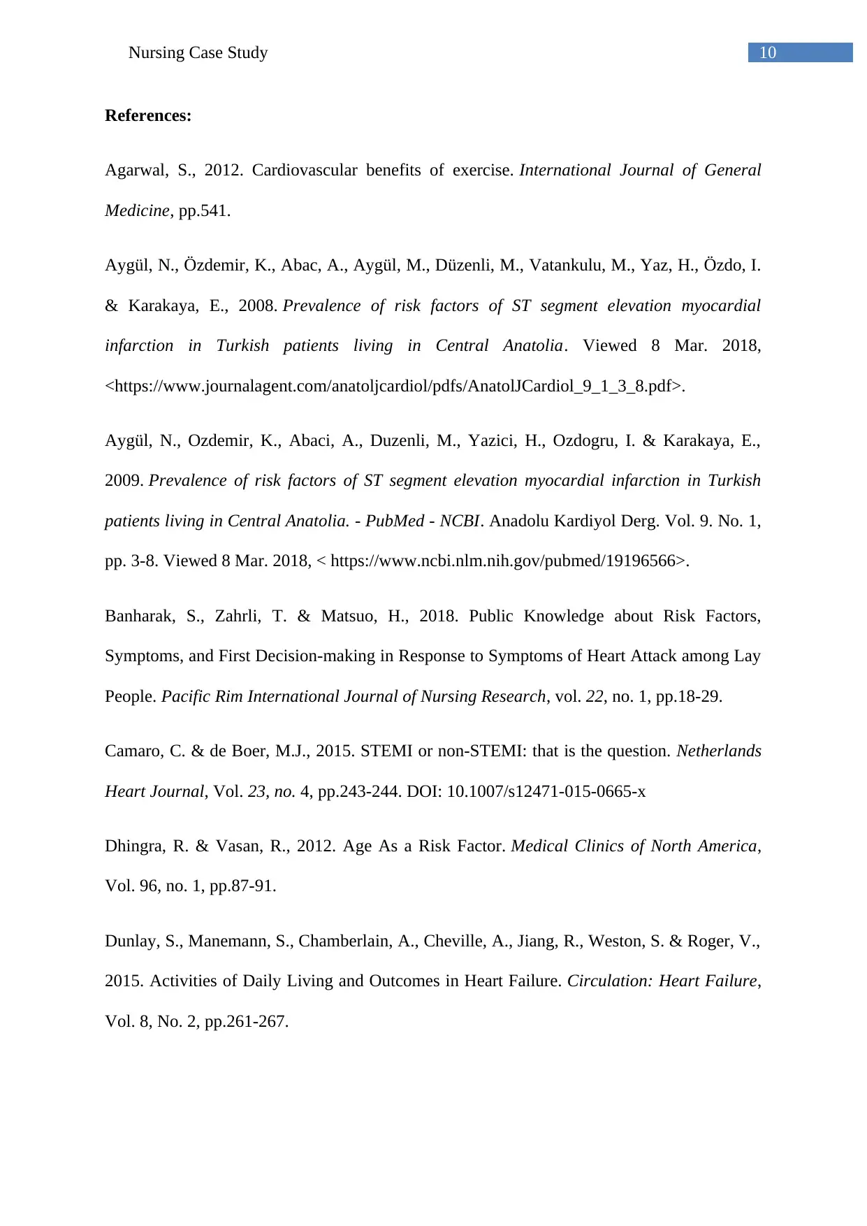
The ability to perform acts of daily living can be used as a method to assess the
functionality of a person. Based on this ADL can be classified into basic ADL (that includes
self care activities like eating, drinking, dressing and bathing) and instrumental (like
housekeeping, transportation or managing medications).
Monitoring the breathing of the patient is an important care strategy, since it allows
identification of any breathing difficulties or abnormalities which can underline any
complication. Using pulse oxymetry machine can help to measure the levels of blood oxygen
saturation, and help to identify when oxygen therapy might be required. Mobility can help to
improve body function as well as recovery. As per NICE guidelines, increasing mobility can
reduce the risk of deep vein blood clots called thromboembolism. Communication is
important to develop therapeutic relationship with the patient. This can allow the nurse to
bond with the patient, and able to understand their feelings and anxieties. Personal Cleansing
and dressing also is an important for patients as it helps in faster recovery and prevent
infections. Studies by Dunlay et al. (2015, p. 266) shows that heart failure, diabetes,
peripheral vascular dysfunctions and cerebrovascular disease can adversely affect the
mobility or self care ability of the patient. The decrease in mobility and an inability or
reduced ability to perform self care activities are also related to a reduced quality of life and
increased risks of hospitalization and even death (Dunlay et al. 2015, p. 266).
Maintaining acts of daily living can require assistance in deferent aspects such as:
providing companionship and mental support, transportation, preparation of meals, managing
household, and managing medications, communicating with others and managing finances.
Paraphrase This Document
Need a fresh take? Get an instant paraphrase of this document with our AI Paraphraser
10Nursing Case Study
References:
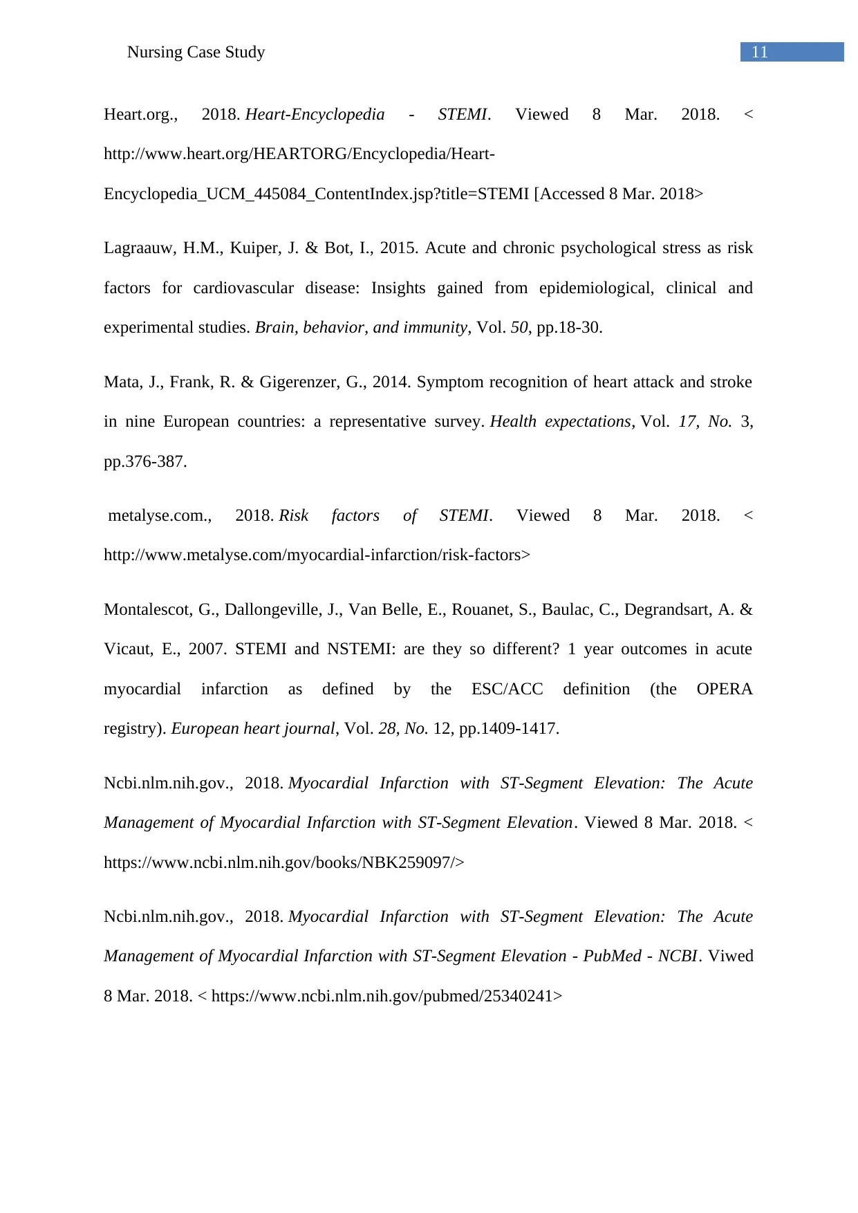
Agarwal, S., 2012. Cardiovascular benefits of exercise. International Journal of General
Medicine, pp.541.
Aygül, N., Özdemir, K., Abac, A., Aygül, M., Düzenli, M., Vatankulu, M., Yaz, H., Özdo, I.
& Karakaya, E., 2008. Prevalence of risk factors of ST segment elevation myocardial
infarction in Turkish patients living in Central Anatolia. Viewed 8 Mar. 2018,
<https://www.journalagent.com/anatoljcardiol/pdfs/AnatolJCardiol_9_1_3_8.pdf>.
Aygül, N., Ozdemir, K., Abaci, A., Duzenli, M., Yazici, H., Ozdogru, I. & Karakaya, E.,
2009. Prevalence of risk factors of ST segment elevation myocardial infarction in Turkish
patients living in Central Anatolia. - PubMed - NCBI. Anadolu Kardiyol Derg. Vol. 9. No. 1,
pp. 3-8. Viewed 8 Mar. 2018, < https://www.ncbi.nlm.nih.gov/pubmed/19196566>.
Banharak, S., Zahrli, T. & Matsuo, H., 2018. Public Knowledge about Risk Factors,
Symptoms, and First Decision-making in Response to Symptoms of Heart Attack among Lay
People. Pacific Rim International Journal of Nursing Research, vol. 22, no. 1, pp.18-29.
Camaro, C. & de Boer, M.J., 2015. STEMI or non-STEMI: that is the question. Netherlands
Heart Journal, Vol. 23, no. 4, pp.243-244. DOI: 10.1007/s12471-015-0665-x
Dhingra, R. & Vasan, R., 2012. Age As a Risk Factor. Medical Clinics of North America,
Vol. 96, no. 1, pp.87-91.
Dunlay, S., Manemann, S., Chamberlain, A., Cheville, A., Jiang, R., Weston, S. & Roger, V.,
2015. Activities of Daily Living and Outcomes in Heart Failure. Circulation: Heart Failure,
Vol. 8, No. 2, pp.261-267.
11Nursing Case Study
Heart.org., 2018. Heart-Encyclopedia - STEMI. Viewed 8 Mar. 2018. <
http://www.heart.org/HEARTORG/Encyclopedia/Heart-
Encyclopedia_UCM_445084_ContentIndex.jsp?title=STEMI [Accessed 8 Mar. 2018>
Lagraauw, H.M., Kuiper, J. & Bot, I., 2015. Acute and chronic psychological stress as risk
factors for cardiovascular disease: Insights gained from epidemiological, clinical and
experimental studies. Brain, behavior, and immunity, Vol. 50, pp.18-30.
Mata, J., Frank, R. & Gigerenzer, G., 2014. Symptom recognition of heart attack and stroke
in nine European countries: a representative survey. Health expectations, Vol. 17, No. 3,
pp.376-387.
metalyse.com., 2018. Risk factors of STEMI. Viewed 8 Mar. 2018. <
http://www.metalyse.com/myocardial-infarction/risk-factors>
Montalescot, G., Dallongeville, J., Van Belle, E., Rouanet, S., Baulac, C., Degrandsart, A. &
Vicaut, E., 2007. STEMI and NSTEMI: are they so different? 1 year outcomes in acute
myocardial infarction as defined by the ESC/ACC definition (the OPERA
registry). European heart journal, Vol. 28, No. 12, pp.1409-1417.
Ncbi.nlm.nih.gov., 2018. Myocardial Infarction with ST-Segment Elevation: The Acute
Management of Myocardial Infarction with ST-Segment Elevation. Viewed 8 Mar. 2018. <
https://www.ncbi.nlm.nih.gov/books/NBK259097/>
Ncbi.nlm.nih.gov., 2018. Myocardial Infarction with ST-Segment Elevation: The Acute
Management of Myocardial Infarction with ST-Segment Elevation - PubMed - NCBI. Viwed
8 Mar. 2018. < https://www.ncbi.nlm.nih.gov/pubmed/25340241>
⊘ This is a preview!⊘
Do you want full access?
Subscribe today to unlock all pages.

Trusted by 1+ million students worldwide
1 out of 13
Related Documents
Your All-in-One AI-Powered Toolkit for Academic Success.
+13062052269
info@desklib.com
Available 24*7 on WhatsApp / Email
![[object Object]](/_next/static/media/star-bottom.7253800d.svg)
Unlock your academic potential
Copyright © 2020–2026 A2Z Services. All Rights Reserved. Developed and managed by ZUCOL.


