Muscles Involved in Reaching for a Glass in an Overhead Cabinet

Verified

Added on  2021/04/24

|8
|1008
|32
Report
AI Summary
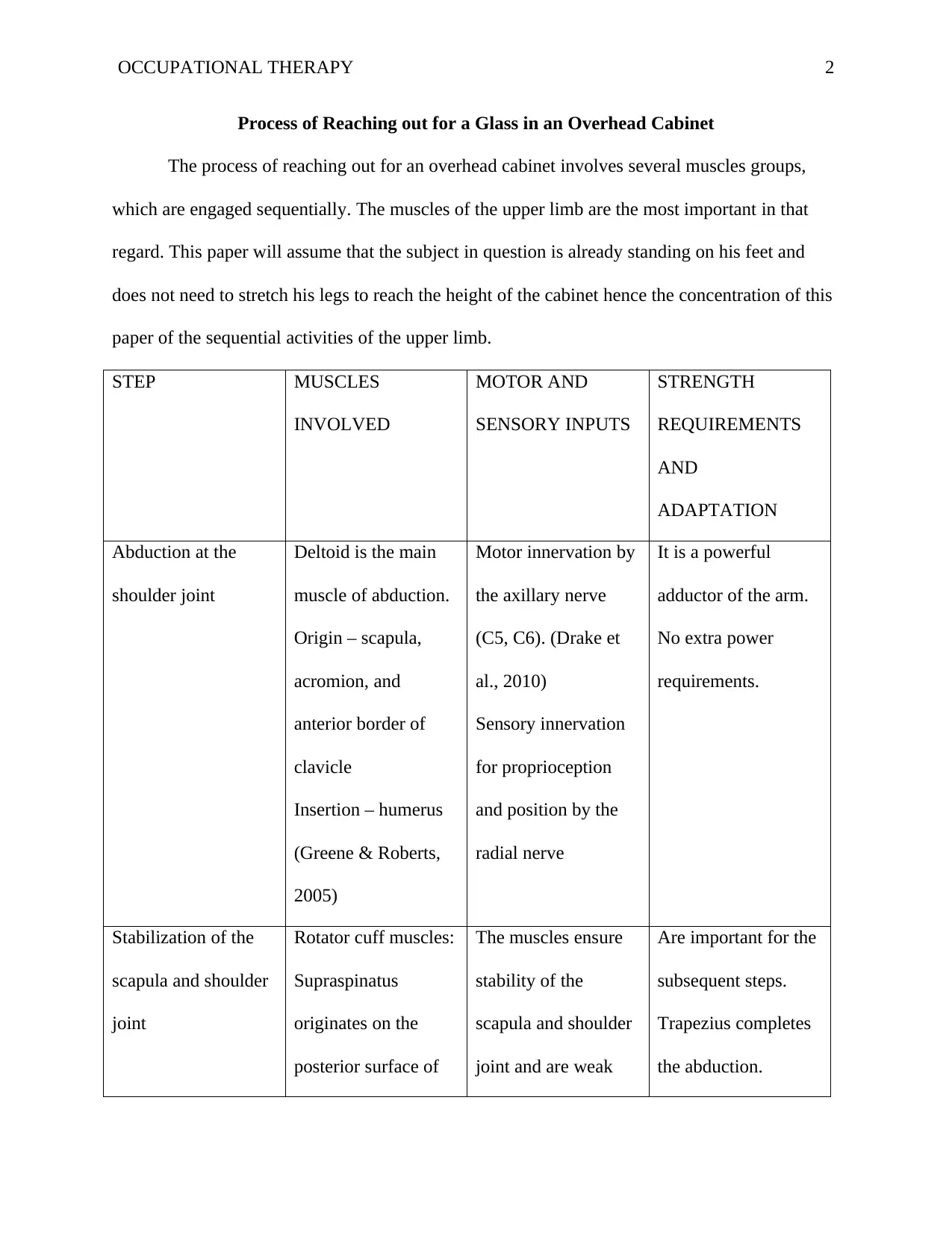
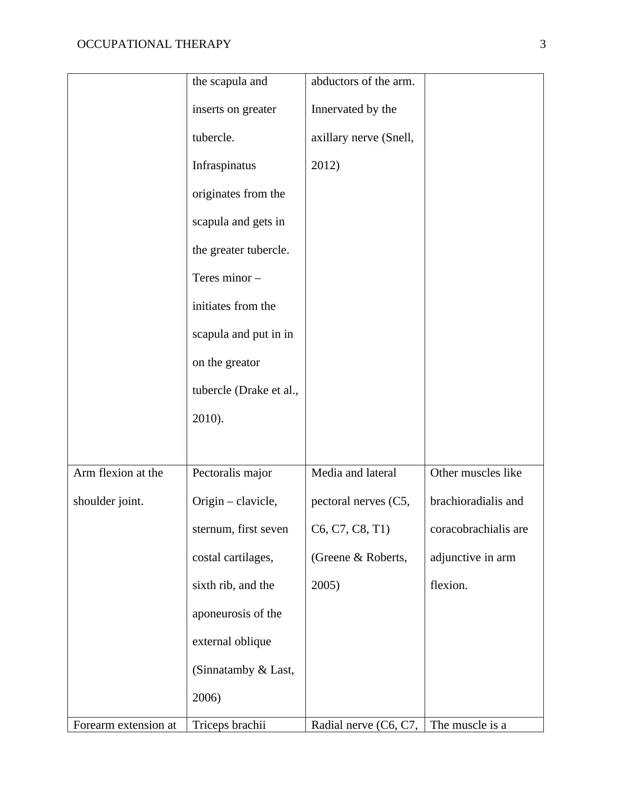
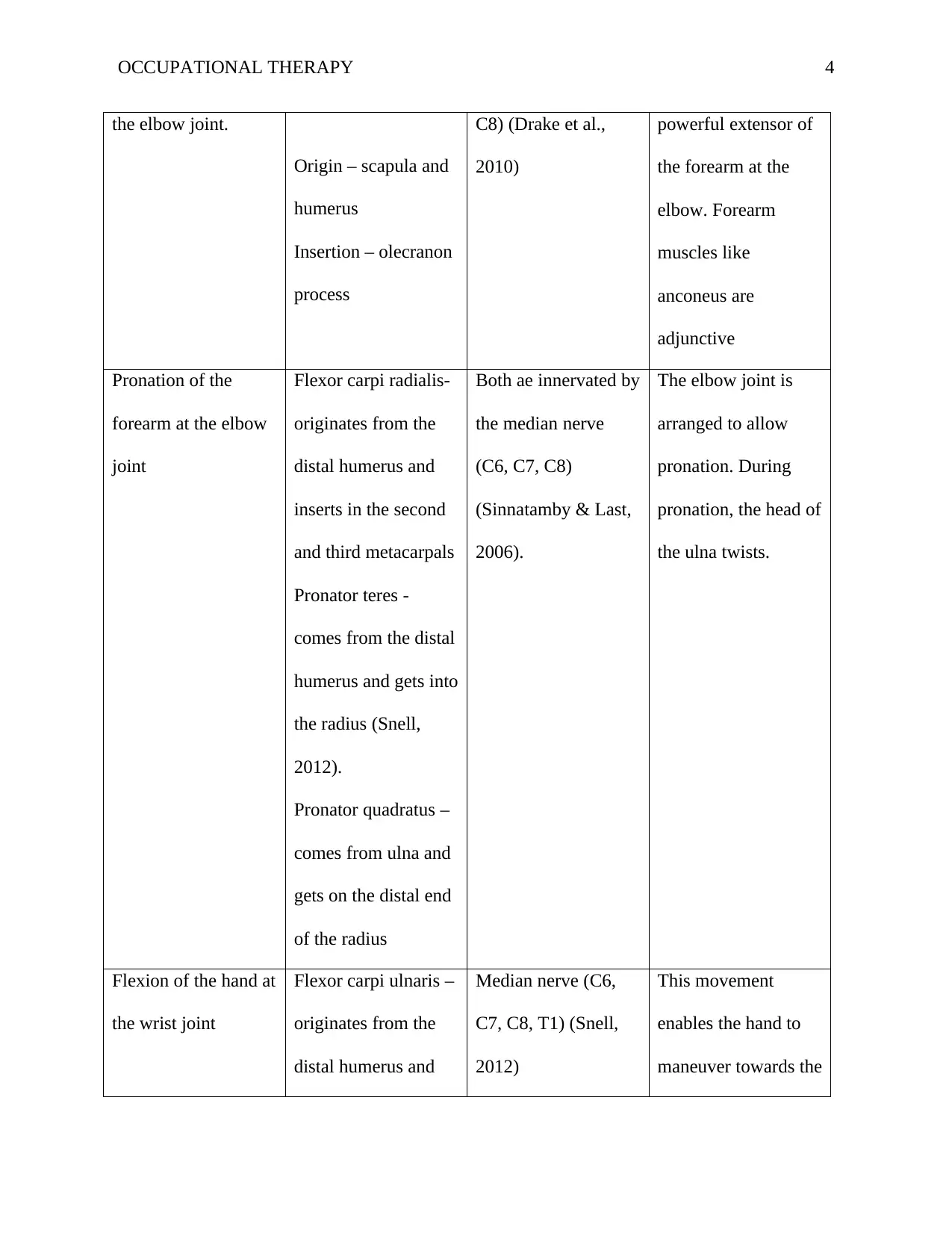
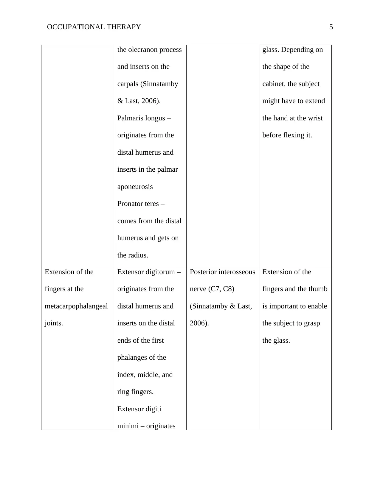
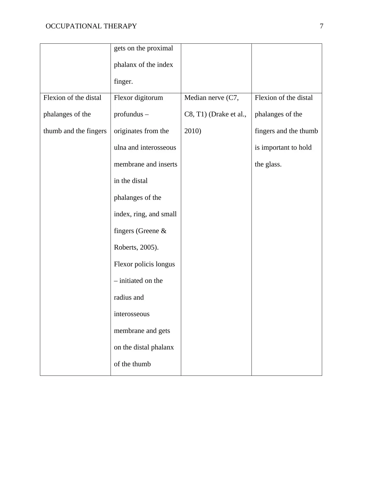
This report details the sequential muscle activations required to reach for a glass in an overhead cabinet, focusing on the upper limb. It begins with the assumption that the subject is standing and examines each step, from shoulder abduction (deltoid) and stabilization (rotator cuff, trapezius) to arm flexion (pectoralis major), forearm extension (triceps brachii), pronation (flexor carpi radialis, pronator teres, pronator quadratus), hand flexion (flexor carpi ulnaris, palmaris longus), finger extension (extensor digitorum, extensor digiti minimi, extensor policis brevis, extensor policis longus, extensor indicis), and finally, finger flexion (flexor digitorum profundus, flexor policis longus) to grasp the glass. The report outlines the muscles involved, their origins, insertions, motor and sensory inputs, and strength requirements, referencing key anatomical texts. The elbow joint's pronation capabilities and the metacarpophalangeal joints' extension role are also highlighted. The analysis provides a comprehensive understanding of the biomechanics of this common daily task.
Document Page
Running Head: OCCUPATIONAL THERAPY 1
Occupational Therapy
Name
Institutional Affiliation
tabler-icon-diamond-filled.svg

Paraphrase This Document

Need a fresh take? Get an instant paraphrase of this document with our AI Paraphraser
Document Page
OCCUPATIONAL THERAPY 2
Process of Reaching out for a Glass in an Overhead Cabinet
The process of reaching out for an overhead cabinet involves several muscles groups,
which are engaged sequentially. The muscles of the upper limb are the most important in that
regard. This paper will assume that the subject in question is already standing on his feet and
does not need to stretch his legs to reach the height of the cabinet hence the concentration of this
paper of the sequential activities of the upper limb.
STEP MUSCLES
INVOLVED
MOTOR AND
SENSORY INPUTS
STRENGTH
REQUIREMENTS
AND
ADAPTATION
Abduction at the
shoulder joint
Deltoid is the main
muscle of abduction.
Origin – scapula,
acromion, and
anterior border of
clavicle
Insertion – humerus
(Greene & Roberts,
2005)
Motor innervation by
the axillary nerve
(C5, C6). (Drake et
al., 2010)
Sensory innervation
for proprioception
and position by the
radial nerve
It is a powerful
adductor of the arm.
No extra power
requirements.
Stabilization of the
scapula and shoulder
joint
Rotator cuff muscles:
Supraspinatus
originates on the
posterior surface of
The muscles ensure
stability of the
scapula and shoulder
joint and are weak
Are important for the
subsequent steps.
Trapezius completes
the abduction.
Document Page
OCCUPATIONAL THERAPY 3
the scapula and
inserts on greater
tubercle.
Infraspinatus
originates from the
scapula and gets in
the greater tubercle.
Teres minor –
initiates from the
scapula and put in in
on the greator
tubercle (Drake et al.,
2010).
abductors of the arm.
Innervated by the
axillary nerve (Snell,
2012)
Arm flexion at the
shoulder joint.
Pectoralis major
Origin – clavicle,
sternum, first seven
costal cartilages,
sixth rib, and the
aponeurosis of the
external oblique
(Sinnatamby & Last,
2006)
Media and lateral
pectoral nerves (C5,
C6, C7, C8, T1)
(Greene & Roberts,
2005)
Other muscles like
brachioradialis and
coracobrachialis are
adjunctive in arm
flexion.
Forearm extension at Triceps brachii Radial nerve (C6, C7, The muscle is a
Document Page
OCCUPATIONAL THERAPY 4
the elbow joint.
Origin – scapula and
humerus
Insertion – olecranon
process
C8) (Drake et al.,
2010)
powerful extensor of
the forearm at the
elbow. Forearm
muscles like
anconeus are
adjunctive
Pronation of the
forearm at the elbow
joint
Flexor carpi radialis-
originates from the
distal humerus and
inserts in the second
and third metacarpals
Pronator teres -
comes from the distal
humerus and gets into
the radius (Snell,
2012).
Pronator quadratus –
comes from ulna and
gets on the distal end
of the radius
Both ae innervated by
the median nerve
(C6, C7, C8)
(Sinnatamby & Last,
2006).
The elbow joint is
arranged to allow
pronation. During
pronation, the head of
the ulna twists.
Flexion of the hand at
the wrist joint
Flexor carpi ulnaris –
originates from the
distal humerus and
Median nerve (C6,
C7, C8, T1) (Snell,
2012)
This movement
enables the hand to
maneuver towards the
tabler-icon-diamond-filled.svg

Paraphrase This Document

Need a fresh take? Get an instant paraphrase of this document with our AI Paraphraser
Document Page
OCCUPATIONAL THERAPY 5
the olecranon process
and inserts on the
carpals (Sinnatamby
& Last, 2006).
Palmaris longus –
originates from the
distal humerus and
inserts in the palmar
aponeurosis
Pronator teres –
comes from the distal
humerus and gets on
the radius.
glass. Depending on
the shape of the
cabinet, the subject
might have to extend
the hand at the wrist
before flexing it.
Extension of the
fingers at the
metacarpophalangeal
joints.
Extensor digitorum –
originates from the
distal humerus and
inserts on the distal
ends of the first
phalanges of the
index, middle, and
ring fingers.
Extensor digiti
minimi – originates
Posterior interosseous
nerve (C7, C8)
(Sinnatamby & Last,
2006).
Extension of the
fingers and the thumb
is important to enable
the subject to grasp
the glass.
Document Page
OCCUPATIONAL THERAPY 6
from the distal
humerus and gets on
the distal end of the
first phalanx of the
little finger.
Extensor policis
brevis – comes from
the posterior radius
and gets on the
proximal phalanx of
the thumb (Snell,
2012).
Extensor policis
longus – initiated
from the posterior
surface of the ulna
and gets on the distal
phalanx of the thumb
(Sinnatamby & Last,
2006).
Extensor indicis –
comes from the
posterior ulna and
Document Page
OCCUPATIONAL THERAPY 7
gets on the proximal
phalanx of the index
finger.
Flexion of the distal
phalanges of the
thumb and the fingers
Flexor digitorum
profundus –
originates from the
ulna and interosseous
membrane and inserts
in the distal
phalanges of the
index, ring, and small
fingers (Greene &
Roberts, 2005).
Flexor policis longus
– initiated on the
radius and
interosseous
membrane and gets
on the distal phalanx
of the thumb
Median nerve (C7,
C8, T1) (Drake et al.,
2010)
Flexion of the distal
phalanges of the
fingers and the thumb
is important to hold
the glass.
tabler-icon-diamond-filled.svg

Paraphrase This Document

Need a fresh take? Get an instant paraphrase of this document with our AI Paraphraser
Document Page
OCCUPATIONAL THERAPY 8
References
Drake, R. L., Vogl, W., Mitchell, A. W. M., & Gray, H. (2010). Gray's anatomy for students.
Philadelphia: Churchill Livingstone/Elsevier.
Greene, D. P., & Roberts, S. L. (2005). Kinesiology: Movement in the context of activity. St.
Louis: Elsevier Mosby.
Sinnatamby, C. S., & Last, R. J. (2006). Last's Anatomy: Regional and applied. Edinburgh:
Elsevier/Churchill Livingstone.
Snell, R. S. (2012). Clinical anatomy by regions. Baltimore, MD: Lippincott Williams &
Wilkins.
chevron_up_icon
1 out of 8
circle_padding
hide_on_mobile
zoom_out_icon
[object Object]