Indian Radiographers' Perceptions of Patient Care and Safety in MRI
VerifiedAdded on 2022/08/12
|57
|13384
|19
Report
AI Summary
This report presents a study on the perceptions of Indian radiographers regarding patient care and safety in MRI procedures. Conducted using descriptive and survey research designs, the study gathered data from 29 radiographers in India, revealing that patient safety is a primary concern. The findings indicate that demographic factors, excluding education, do not significantly influence radiographers' views on patient health and safety, while education plays a crucial role in skill and knowledge levels related to MRI safety procedures. The work environment also emerged as a key determinant of adherence to safety guidelines. The radiographers demonstrate a strong understanding of the importance of patient safety and care before, during, and after imaging procedures. The report includes an introduction to MRI safety, literature review of MRI risks and prevention, research methodology, results and findings from questionnaires and interviews, and discussion and conclusion. The study highlights the importance of continuous training and adherence to safety protocols to ensure patient well-being.
Perceptions of Indian Radiographers about Patient Care and Safety In MRI
Name
Institution
Tutor
Date
Name
Institution
Tutor
Date
Paraphrase This Document
Need a fresh take? Get an instant paraphrase of this document with our AI Paraphraser
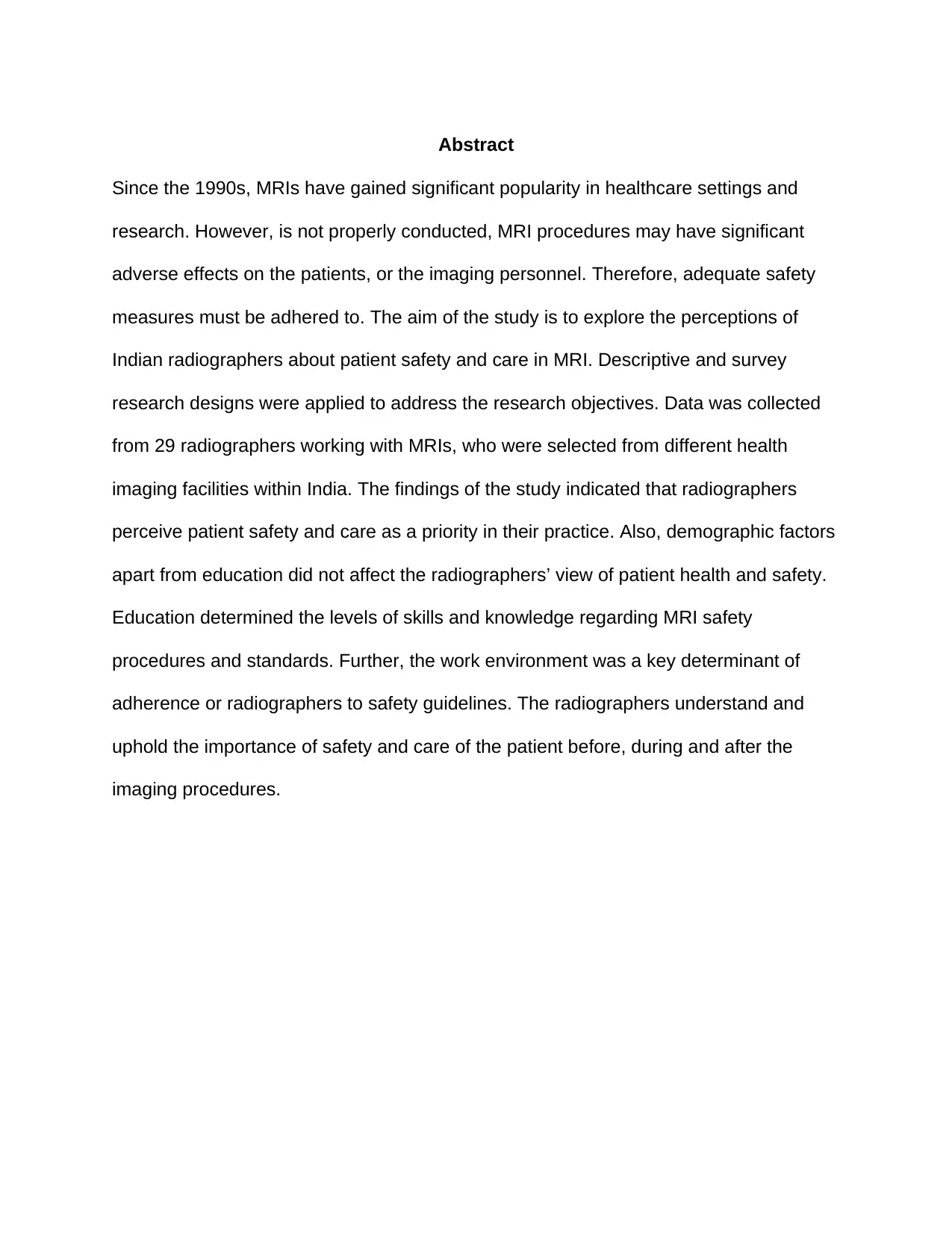
Abstract
Since the 1990s, MRIs have gained significant popularity in healthcare settings and
research. However, is not properly conducted, MRI procedures may have significant
adverse effects on the patients, or the imaging personnel. Therefore, adequate safety
measures must be adhered to. The aim of the study is to explore the perceptions of
Indian radiographers about patient safety and care in MRI. Descriptive and survey
research designs were applied to address the research objectives. Data was collected
from 29 radiographers working with MRIs, who were selected from different health
imaging facilities within India. The findings of the study indicated that radiographers
perceive patient safety and care as a priority in their practice. Also, demographic factors
apart from education did not affect the radiographers’ view of patient health and safety.
Education determined the levels of skills and knowledge regarding MRI safety
procedures and standards. Further, the work environment was a key determinant of
adherence or radiographers to safety guidelines. The radiographers understand and
uphold the importance of safety and care of the patient before, during and after the
imaging procedures.
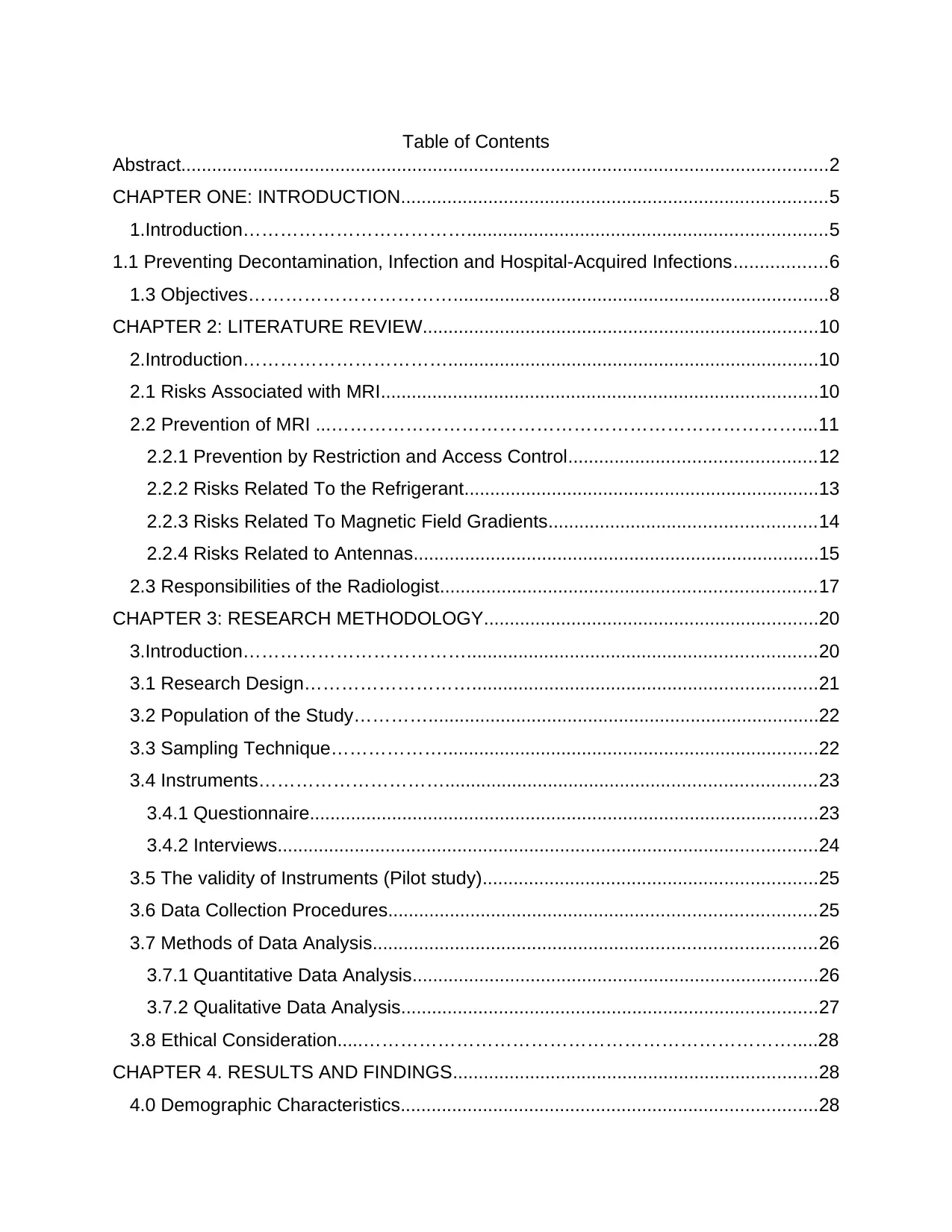
Table of Contents
Abstract..............................................................................................................................2
CHAPTER ONE: INTRODUCTION...................................................................................5
1.Introduction………………………………......................................................................5
1.1 Preventing Decontamination, Infection and Hospital-Acquired Infections..................6
1.3 Objectives…………………………….........................................................................8
CHAPTER 2: LITERATURE REVIEW.............................................................................10
2.Introduction……………………………........................................................................10
2.1 Risks Associated with MRI.....................................................................................10
2.2 Prevention of MRI ...…………………………………………………………………....11
2.2.1 Prevention by Restriction and Access Control................................................12
2.2.2 Risks Related To the Refrigerant.....................................................................13
2.2.3 Risks Related To Magnetic Field Gradients....................................................14
2.2.4 Risks Related to Antennas...............................................................................15
2.3 Responsibilities of the Radiologist.........................................................................17
CHAPTER 3: RESEARCH METHODOLOGY.................................................................20
3.Introduction………………………………....................................................................20
3.1 Research Design………………………...................................................................21
3.2 Population of the Study…………............................................................................22
3.3 Sampling Technique……………….........................................................................22
3.4 Instruments…………………………........................................................................23
3.4.1 Questionnaire...................................................................................................23
3.4.2 Interviews.........................................................................................................24
3.5 The validity of Instruments (Pilot study).................................................................25
3.6 Data Collection Procedures...................................................................................25
3.7 Methods of Data Analysis......................................................................................26
3.7.1 Quantitative Data Analysis...............................................................................26
3.7.2 Qualitative Data Analysis.................................................................................27
3.8 Ethical Consideration.....…………………………………………………………….....28
CHAPTER 4. RESULTS AND FINDINGS.......................................................................28
4.0 Demographic Characteristics.................................................................................28
⊘ This is a preview!⊘
Do you want full access?
Subscribe today to unlock all pages.

Trusted by 1+ million students worldwide
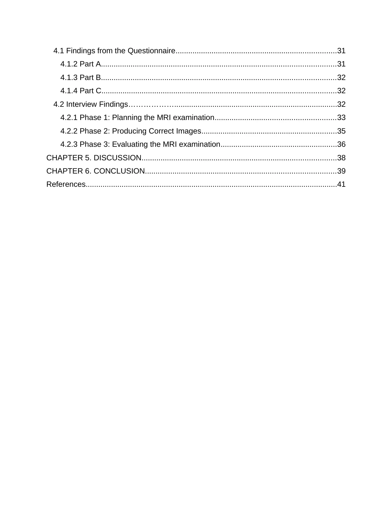
4.1 Findings from the Questionnaire............................................................................31
4.1.2 Part A...............................................................................................................31
4.1.3 Part B...............................................................................................................32
4.1.4 Part C...............................................................................................................32
4.2 Interview Findings……………….............................................................................32
4.2.1 Phase 1: Planning the MRI examination.........................................................33
4.2.2 Phase 2: Producing Correct Images................................................................35
4.2.3 Phase 3: Evaluating the MRI examination.......................................................36
CHAPTER 5. DISCUSSION............................................................................................38
CHAPTER 6. CONCLUSION..........................................................................................39
References.......................................................................................................................41
Paraphrase This Document
Need a fresh take? Get an instant paraphrase of this document with our AI Paraphraser
CHAPTER ONE: INTRODUCTION
1.Introduction
In the recent two decades, the safety and care of patients have managed to garner a lot
of attention. The Institute of Medicine report published in 1999, "To Err is Human",
indicated that between 44,000 and 98000 patients die in American Hospitals as a result
of preventable errors (Lark, Kirkpatrick, and Chung, 2018). Notably, many broad
epidemiological types of research have claimed that a made or the increased level of
risk is mainly because of mistakes that occur within the current medical environment
(Hartwig et al. 2009; Ko and Yu, 2015; Mello and Hemenway, 2004 and; Hiivala et al.,
2016). Improving the safety level of patients has become paramount within all
healthcare services and procedures. A recent case study revelation of medical
misconduct involving MRI, in a Taiwan hospital, clearly states that the risk sources tend
to lack adequate clinical training and inaccurate and insufficient communication
amongst the MRI staff and clinical team (Lee, Lin and Chan, 2015).
There is a rising consideration of MRI by practitioners due to the exquisite soft-tissue
delineation provided by the imaging modality. The MRI process involves exposure to
powerful static magnetic fields and gradient fields and radiofrequency energy. Without
going into the details and technical aspects of its operation, an MRI machine generates
magnetic fields to provide high definition images of all parts of the human body (Kanal
et al. 2007; Sammet 2016). The advantage compared to other imaging devices, it emits
no radiation that could harm patients or professional users. Its major drawback is that it
is a very powerful electromagnet which necessarily requires perfect control of the
environment near the installation. It is essential for staff, visitors and patients to
understand the primary hazards associated with the environment and select the safest
and rigorously protection procedures to follow (Durbridge 2011). Within the Indian
context, key safety considerations that are related to the patients are associated with
magnetic resonance imaging (MRI) should always consider the following aspects
(Durbridge 2011).
The associated behaviour of ferromagnetic objects when they are exposed to a
robust magnetic field as the forces can react over the ferromagnetic implant
thereby, resulting in it to transit in a different direction causing injury or potential
death. Additionally, many external ferromagnetic objects might also get
influenced by the strong magnetic fields and tend to become airborne and will
transmit rapidly towards the isocenter of the magnetic. This situation might also
cause injury or death (Durbridge, 2011).
Gradient or static magnetic fields might also have an impact over other medical
devices, whether external or implanted and can cause them to malfunction
(Durbridge 2011).
Acoustic sounds that are associated with frequently switching gradient coils can
also be a severe risk for the patients. This can be avoidable through
appropriately using noise reduction and hearing protection technologies
(Durbridge 2011).
The risks associated with radiofrequency (RF) include heating up of tissues
because of RF energy deposition, which can be measured as the specific
⊘ This is a preview!⊘
Do you want full access?
Subscribe today to unlock all pages.

Trusted by 1+ million students worldwide
absorption rate (SAR). Resultantly, this is more evident because of the
increasing field strengths mainly because of the increased frequency of the RF
pulses. Additionally, the energy of RF can also be deposited within wires, cables,
tattoos and skin patches causing these elements to get heated and potentially
result in burns to the patient (Callaghan et al. 2019; Durbridge 2011; Takahashi,
Fujimoto, Hamada, Tezuka, and Tanaka, 2016).
1.1 Preventing Decontamination, Infection and Hospital-Acquired Infections
As per a study undertaken by the Centres for Disease Control and Prevention (CDC),
basic control over infections within hospitals must include all the general principles to
avoid transmission within all patient care, as well as specific ways for preventing that,
are suspected or known to be infected within a transmittable microorganism (Centers for
Disease Control and Prevention, 2017). General principles to be applied by healthcare
professionals include:
Using personal and protective equipment at the time of exposure to that of
infectious material (Centers for Disease Control and Prevention, 2017);
Performing hand hygiene and using all five moments (World Health Organization,
2018) before and after touching the patient and appropriately using the solutions
for washing hands or disinfection (Centers for Disease Control and Prevention,
2017);
Assure staff awareness about issues that are related to patients in isolation
particularly, the contact and respiratory types of isolation (Centers for Disease
Control and Prevention, 2017);
Paraphrase This Document
Need a fresh take? Get an instant paraphrase of this document with our AI Paraphraser
Assuring safer procedures for injections and follow the rule 'one syringe, one
needle and one use' (Centers for Disease Control and Prevention, 2017).
Handling laundry garments and textiles very carefully and dispose of them off per
the hospital policy (Centers for Disease Control and Prevention, 2017);
Ensuring cleanliness of the materials related to sterilization and disinfection as
well as the environment of the patient (Centers for Disease Control and
Prevention, 2017).
Transmission prevention processes should also be used within patients who have been
infected or occupied with particular infectious agents and which include (Centers for
Disease Control and Prevention, 2017):
Precautions undertaken during contact in case of contact transmission occur in a
patient. This is achieved through isolating the patient, making use of personal
protective equipment, making use of disposable equipment and assuring
surfaces are appropriately cleaned.
Airborne precautionary measures in case infectious microorganisms transmit
through airborne routes like measles, tuberculosis or chickenpox. Droplet
precaution can be used along with restricting susceptible healthcare
professionals to access the room by immunizing all susceptible people.
Droplet precautions can be used for agents that are transmitted by respiratory
droplets such as talking, coughing or sneezing. This can be attained through the
continuous usage of facemasks, using personal protective equipment as well as
limiting patient transport and movement.
1.2 Aim
Patient care and safety is a priority in healthcare settings (Aspden Corrigan and
Wolcott, 2004; Levine et al. 2007). Radiologists with healthcare units all over India that
are involved in MRI needs to be aware of these considerations and should, as such,
undertake appropriate safety procedures that must be in place. With millions of MRI
scans being performed Indian hospitals coupled with growth within the MRI activity
between 2011 and 2015 (Arora, 2014), the need for ensuring best practice for the care
and safety of patients across the country has become empirical. The research study
aims to determine the perception of Indian Radiographers for MRI patient safety and
care.
1.3 Objectives
The survey aims to meet the following objectives:
1. To assess the knowledge of Indian radiographers regarding patient care and
safety in MRI procedures.
2. To investigate the perceptions of Indian Radiographers for regarding patient care
and safety in MRI procedures.
⊘ This is a preview!⊘
Do you want full access?
Subscribe today to unlock all pages.

Trusted by 1+ million students worldwide
CHAPTER 2: LITERATURE REVIEW
2.Introduction
There have been continuous and rapid developments in MRI techniques that have
widely been applied in the past two decades. The technical developments are
demonstrated by the optimization and improvements in conventional MRI techniques
such as MR fingerprinting and compressed sensing MRI and the new pulse sequences
such as DWIBS (Diffusion-weighted Whole-body Imaging with Background body signal
Suppression) and CEST-MRI (Chemical Exchange Saturation Transfer MRI). The rapid
development in MRI techniques has resulted in an increased application of MRI
techniques and the emergence of new knowledge in biomedical research and clinical
sciences (Dong, Andrews, Xie, and Yokoo, 2015). With the wide adoption and utilization
of MRI, patient safety and risk associated with the modality has been a significant issue
of concern for practitioners and researchers (Andersen, 2007; Lee, Lin and Chan,
2015).
2.1 Risks Associated with MRI
Any human endeavour creates risks. This is particularly the case for health-related
activities and medical imagery. The proper functioning of an imaging firm or service is
closely linked to controlling the risks that its activity generates. As patient and
professional safety is a fundamental issue in imaging procedures, it is, therefore,
essential to continually comply with safety rules and to prevent risks (European
Medicine Agency, 2017)
Paraphrase This Document
Need a fresh take? Get an instant paraphrase of this document with our AI Paraphraser
The projectile effect is certainly one of the most important risks in the MRI. The list of
objects responsible for accidents associated with MRI is long. It involves retaining
oxygen shells, helium cans, equipment for monitoring and assistance in resuscitation,
scissors, reflex hammers, stethoscopes, mobile phones, barrettes, keys, wheelchairs.
When they enter the magnetic field, these objects undergo a violent attraction and reach
speeds of several meters per second (Cross, Hoff, and Kanal, 2018).). The main
accidents reported in the literature concern projectiles with a large mass, such as
oxygen shells. In 2001, a 6-year-old boy died in the United States from head trauma
following a shock from a ferromagnetic oxygen shell that penetrated the magnet at high
speed (Bellin et al., 2002). Other cases of oxygen shells projected in an MRI, with
material damage, but without victims have been reported in the literature. If the danger
increases with the mass of the projectile, even small objects such as coins can be
thrown into the magnet and injure the patient.
Another risk concerns metallic implants as well as implantable medical devices or even
intracorporeal metallic foreign bodies. In such a case, the associated threat comes from
displacements or rotational movements of these foreign bodies or implants.
Haemorrhages caused by the displacement of intracranial ferromagnetic vascular clips,
intraocular metallic foreign bodies, bullets or fragments of shells resulting from to
exposure electromagnetic field causes significant harm. Malfunctions of certain
implanted devices (pacemakers, neurostimulators, cochlear implants, insulin pumps can
cause considerable injury (International Organization for Standardization, 2012).
2.2 Prevention of MRI Risks'
The time-varying gradient fields, main magnetic fields involved, and radiofrequency
energy involved in MRIs require a variety of unique safety considerations. The
practitioner who works in the MRI environment needs to understand the
translation/torque, projectile effect, human health effects and personal items required for
the MRI environment (Durbridge, 2011). There are various strategies for achieving
safety in the MRI environment, such as restricting access to the area, taking precaution
when handling equipment and providing all safety information before entering into the
area.
2.2.1 Prevention by Restriction and Access Control
Accessing the room where the examination will take place should be restricted to
people who are not aware of the relative constraints associated with intense magnetic
fields. The danger it represents is always materialized by signs placed over the
examination room door, or even of the surrounding premises. The magnetic field limit of
0.5 Millitesla (mT) is considered to be an "exclusion zone" prohibited for people with
particular active medical devices (pacemaker for example), ferromagnetic objects and
certain electronic equipment. Such a limitation is generally a part of the magnet room
because of the progress of the shielding: "active" shielding at the level of the magnet
(additional magnetic field coils) and "passive" shielding fixed on the walls, ceilings or
floor if necessary (metal plates). If this might not be the case with higher fields, this limit
might be marked over the ground (Crook and Robinson, 2009; Expert Panel on MR
Safety et al. 2013).
⊘ This is a preview!⊘
Do you want full access?
Subscribe today to unlock all pages.

Trusted by 1+ million students worldwide
1 out of 57
Related Documents
Your All-in-One AI-Powered Toolkit for Academic Success.
+13062052269
info@desklib.com
Available 24*7 on WhatsApp / Email
![[object Object]](/_next/static/media/star-bottom.7253800d.svg)
Unlock your academic potential
Copyright © 2020–2026 A2Z Services. All Rights Reserved. Developed and managed by ZUCOL.


