Analysis of PCR, Cloning, and Protein Expression in Biology

Verified

Added on  2023/01/17

|5
|755
|29
Practical Assignment
AI Summary
This assignment focuses on analyzing a molecular biology experiment involving PCR amplification, cloning, and protein expression using the pGEX6P2 vector. The first task requires students to interpret and draw a gel electrophoresis image based on the results of restriction enzyme digestions and PCR reactions, labeling bands with their sizes. The second task involves analyzing a target DNA sequence, identifying primer binding sites, and determining the direction of primer extension. Furthermore, students must address discrepancies in the expected protein size, determine the last five amino acids of the protein, and design improved primers to correct errors in the original primer design. The assignment assesses the understanding of plasmid maps, restriction digestion, PCR principles, and protein translation, along with the ability to troubleshoot experimental outcomes and design solutions.
Document Page
Task 1:
Make a copy of the image of a gel given below. You can then draw appropriate bands in each lane based
on the description of what is loaded in each lane (given below). You can draw these bands by hand on a
printed version of the image or add the bands to the image using Powerpoint or similar software. If you
do create a hand-drawn version remember to take a photo and then insert that photo into the
document you submit.
We understand that your diagram will not be able to portray the bands to their exact base-pair size, and
that in some cases you may not be able to calculate an exact size for your bands. Just do your best in
terms of positioning the bands in the lanes with respect to the standard markers, but please do not
panic if the bands are a couple of mm away from “perfect” in your diagram. LABEL each band with its
size (in bp) – we will tolerate a ±10 bp difference between your value and the true value.
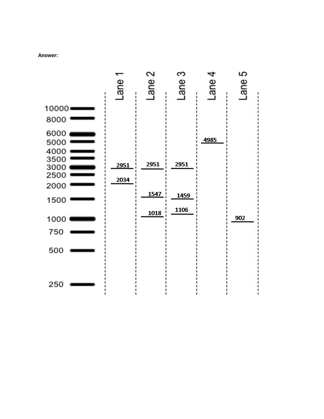
Lane 1 – pGEX6P2 with NO insert, digested with Btg I and Pst I
Lane 2 – pGEX6P2 with the PCR product inserted in the correct orientation (the coding sequence
is in the same direction as the GST encoded by pGEX), digested with Btg I and Pst I
Lane 3 – pGEX6P2 with the PCR product inserted in the incorrectorientation (the coding
sequence is in the opposite direction compared to the GST encoded by pGEX), digested with
Btg I and Pst I
Lane 4: In this lane draw the fragments that would result from a PCR that uses all of the
necessary PCR components (including pGEX6P2 with no insert as template, dNTPs, Taq
polymerase, buffer, water) and ONLY the SCREEN-R primer (no SCREEN-F primer).
Lane 5: In this lane draw the fragments that would result from a PCR that uses all of the
necessary PCR components (including pGEX6P2 with the PCR product inserted as template,
dNTPs, Taq polymerase, buffer, water) and BOTH the SCREEN-F and SCREEN-R primers.
* Note: all the sites are marked on the plasmid map.
tabler-icon-diamond-filled.svg

Paraphrase This Document

Need a fresh take? Get an instant paraphrase of this document with our AI Paraphraser
Document Page
Answer:
Document Page
Task 2:
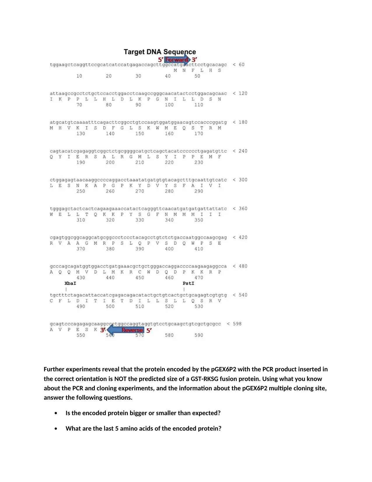
Copy the target DNA sequence image given below. Highlight where the RKSG-Fwd and RKSG-Rev
primers bind. Indicate whether the primers are the SAME sequence as shown on the diagram or the
COMPLEMENTARY sequence to the one shown in the diagram. Put an arrow on the end of each
annotated region to show the direction the primer points (the end where the polymerase will bind
and begin extension). Label each end of the primer 5’ or 3’.
Document Page
Further experiments reveal that the protein encoded by the pGEX6P2 with the PCR product inserted in
the correct orientation is NOT the predicted size of a GST-RKSG fusion protein. Using what you know
about the PCR and cloning experiments, and the information about the pGEX6P2 multiple cloning site,
answer the following questions.
Is the encoded protein bigger or smaller than expected?
What are the last 5 amino acids of the encoded protein?
tabler-icon-diamond-filled.svg

Paraphrase This Document

Need a fresh take? Get an instant paraphrase of this document with our AI Paraphraser
Document Page
Answer:
The expected protein size is larger due to lack of insertion of stop codon at the product insertion site.
The last 5 amino acids of the encoded protein are RAAAS
Provide the sequences of a pair of primers that would give a PCR product that, when cloned into
pGEX6P2, would result in the plasmid encoding a protein of the desired size. Indicate which is the 5’ end
and which is the 3’ end for both primers. Describe the differences between these primers and the
original RKSG-Fwd and RKSG-Rev primers and how these differences correct the error(s) in the original
primer design.
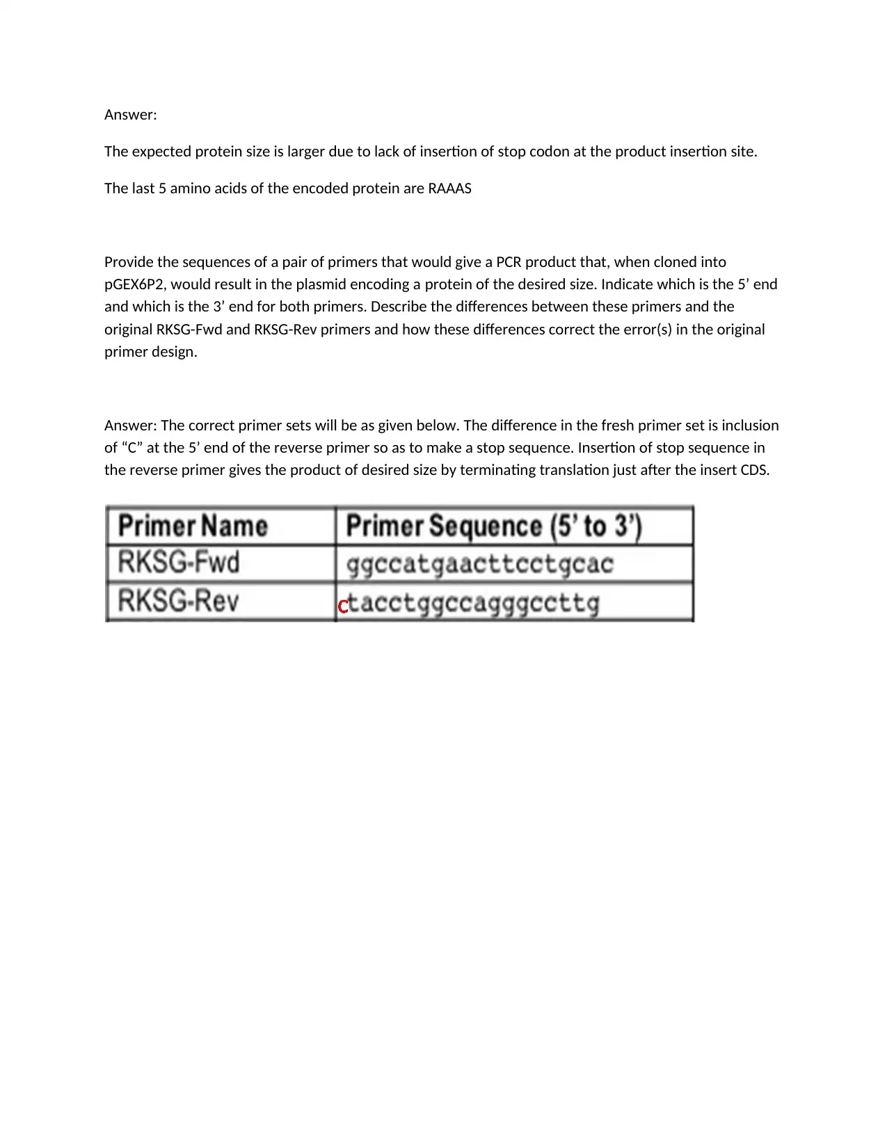
Answer: The correct primer sets will be as given below. The difference in the fresh primer set is inclusion
of “C” at the 5’ end of the reverse primer so as to make a stop sequence. Insertion of stop sequence in
the reverse primer gives the product of desired size by terminating translation just after the insert CDS.
chevron_up_icon
1 out of 5
circle_padding
hide_on_mobile
zoom_out_icon
[object Object]