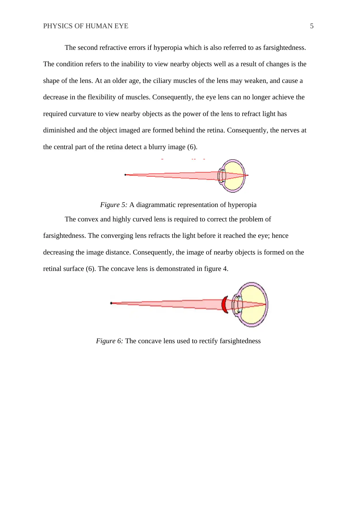
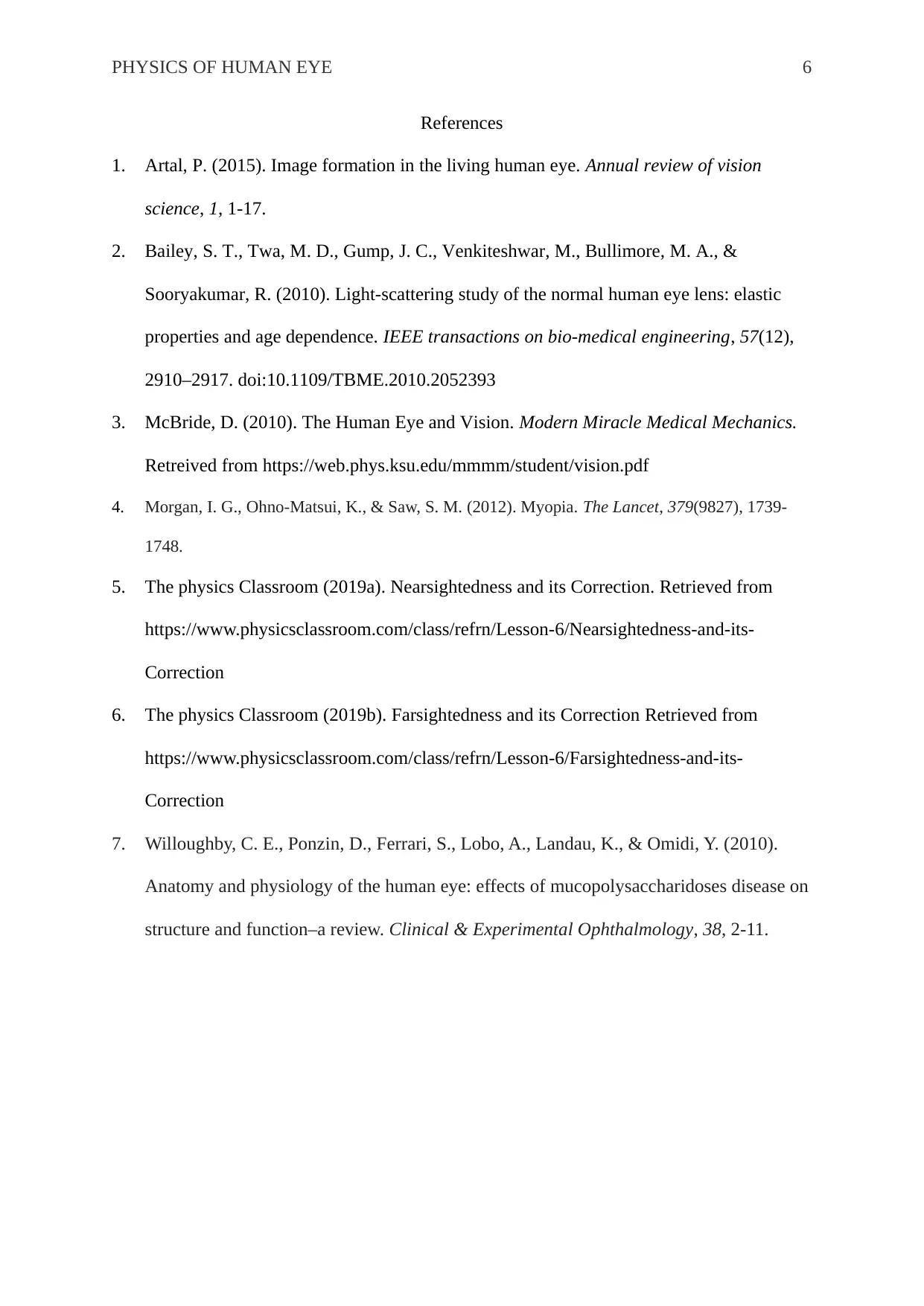
Physics of the Human Eye: Vision, Refractive Errors, and Correction
VerifiedAdded on 2022/09/06
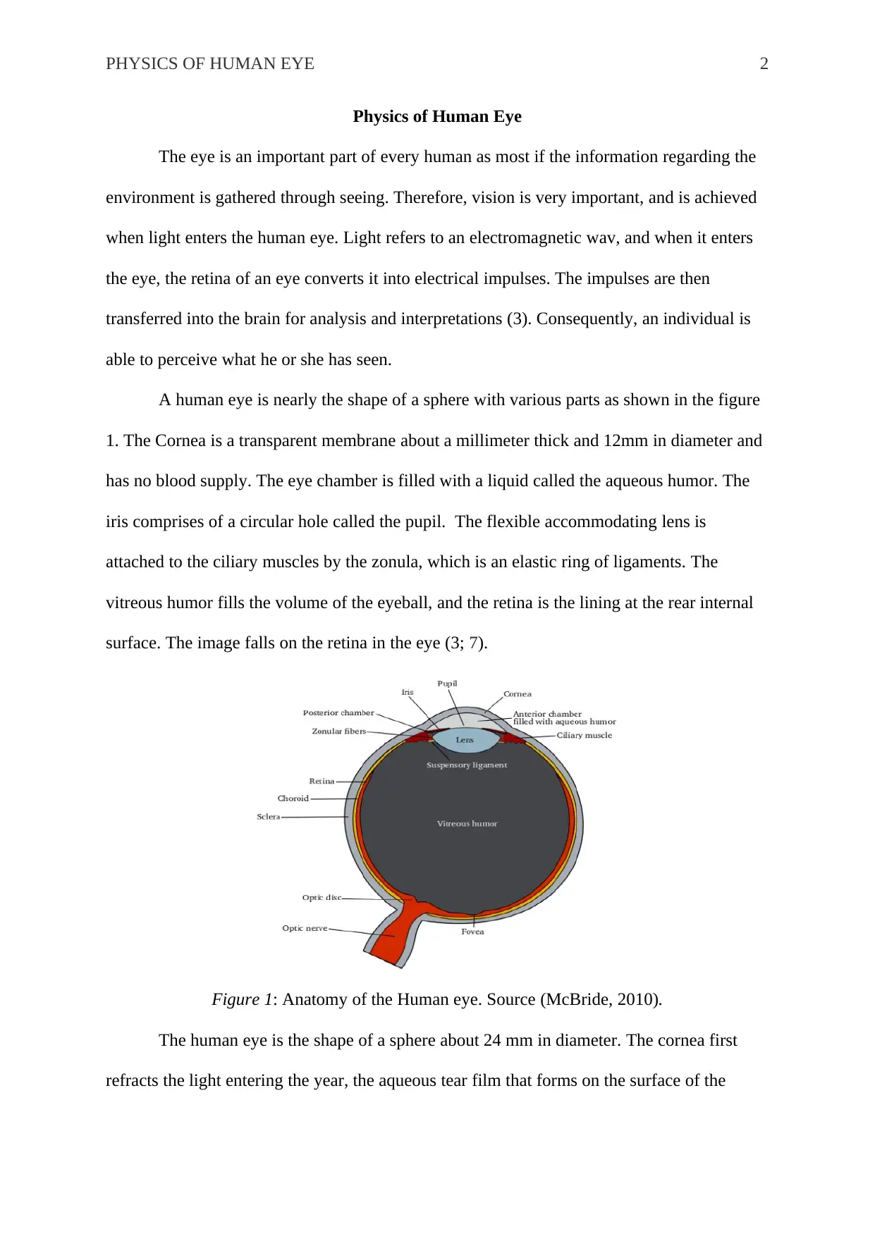
|6
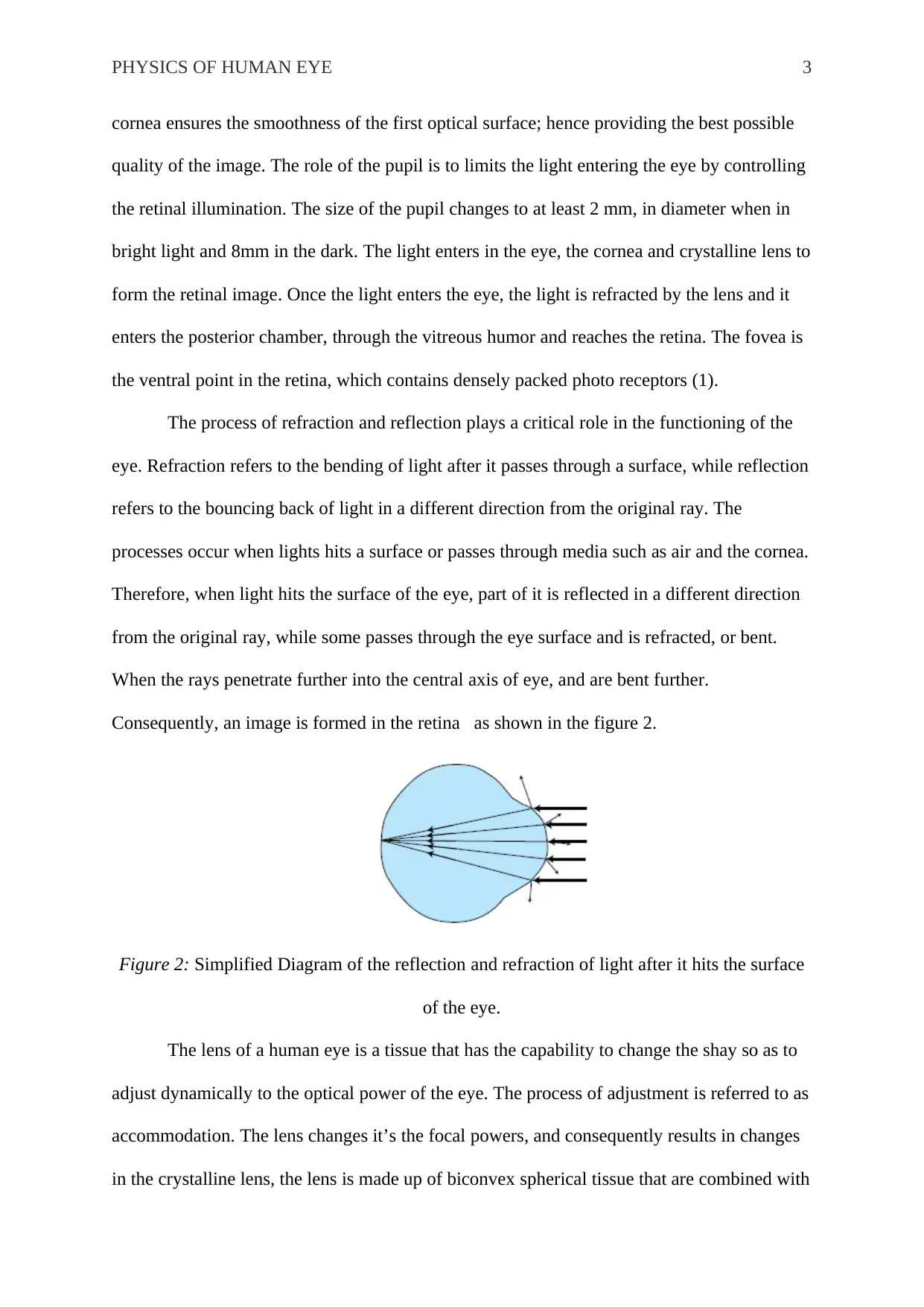
|1366
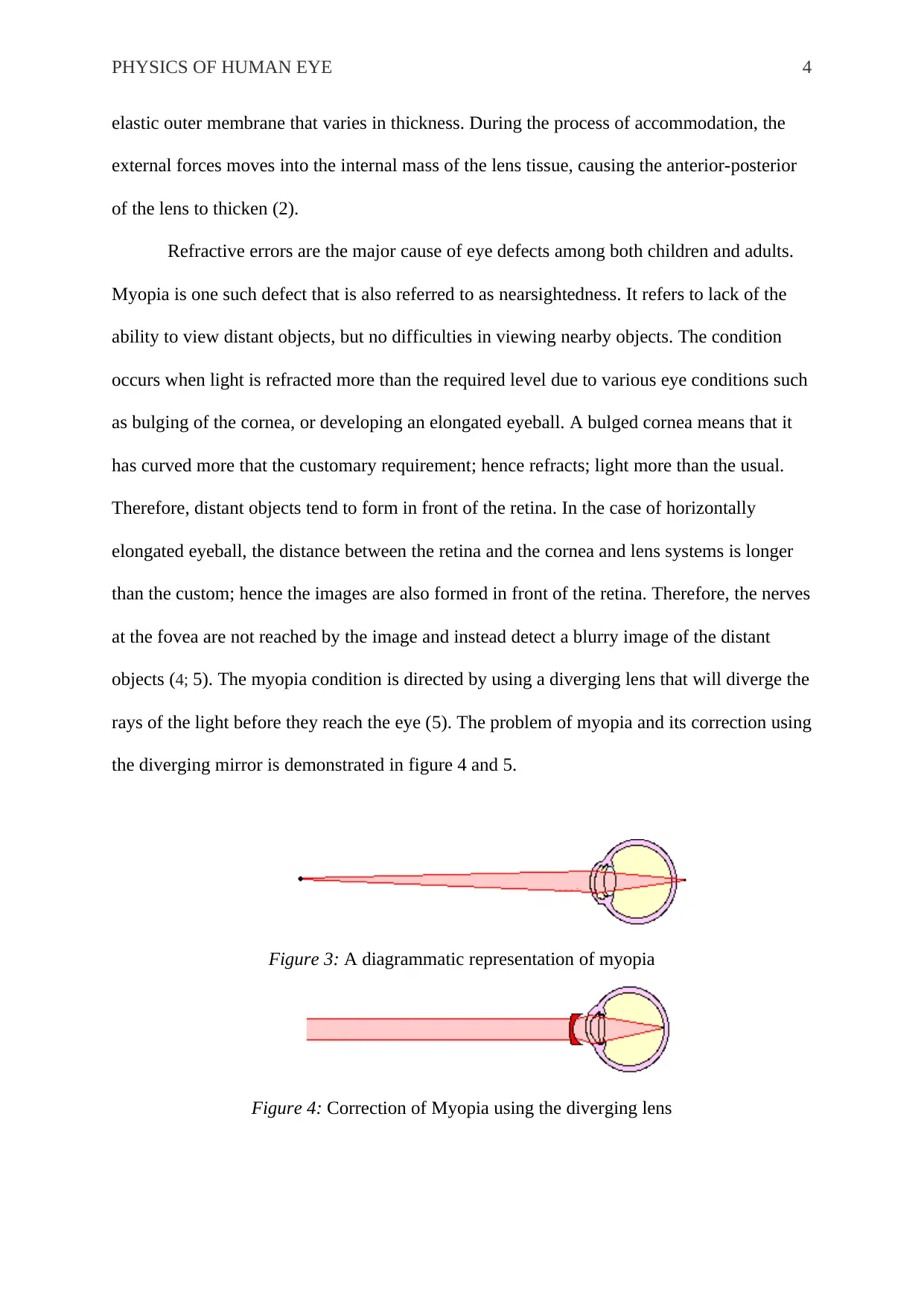
|24
Report
AI Summary
This report provides a comprehensive overview of the physics of the human eye. It begins with an introduction to the importance of vision and how light is converted into electrical impulses for brain analysis. The report then details the anatomy of the human eye, including the cornea, pupil, lens, and retina, and explains how light enters and is processed within the eye. The processes of refraction and reflection are explained, highlighting their role in image formation on the retina. The report further discusses refractive errors, such as myopia (nearsightedness) and hyperopia (farsightedness), explaining their causes and how they are corrected using diverging and converging lenses, respectively. Diagrams are included to illustrate the concepts. The report concludes with a list of relevant references.
1 out of 6


Your All-in-One AI-Powered Toolkit for Academic Success.
+13062052269
info@desklib.com
Available 24*7 on WhatsApp / Email
![[object Object]](/_next/static/media/star-bottom.7253800d.svg)
Copyright © 2020–2026 A2Z Services. All Rights Reserved. Developed and managed by ZUCOL.