Medical Case Study: Pleural Effusion, Diagnosis and Treatment
VerifiedAdded on 2022/08/18
|8
|1655
|19
Case Study
AI Summary
This case study presents a 68-year-old male, A.B., admitted with pleural effusion, experiencing shortness of breath, chest pain, weakness, and a dry cough. His vital signs indicate hypertension, high respiratory rate, fever, and low SpO2. The case explores the pathophysiology, signs, and symptoms of pleural effusion, including the role of pneumonia. The physician performs a thoracentesis, and the solution details the procedure, calculation of IV flow rate for cefuroxime, and nursing interventions to promote pulmonary secretion clearing. The study also addresses reporting Klebsiella non-sensitivity to cefuroxime, chest tube management, and interventions for maintaining the chest tube system. The document includes answers to questions, references, and a discussion on potential complications and nursing assessments. The patient's case involves pneumonia and the need for antibiotics, along with a focus on monitoring and maintaining the patient's respiratory health.
Running head: PLEURAL EFFUSION
PLEURAL EFFUSION
Name of the Student
Name of the University
Author note
PLEURAL EFFUSION
Name of the Student
Name of the University
Author note
Paraphrase This Document
Need a fresh take? Get an instant paraphrase of this document with our AI Paraphraser
1PLEURAL EFFUSION
Answer 1
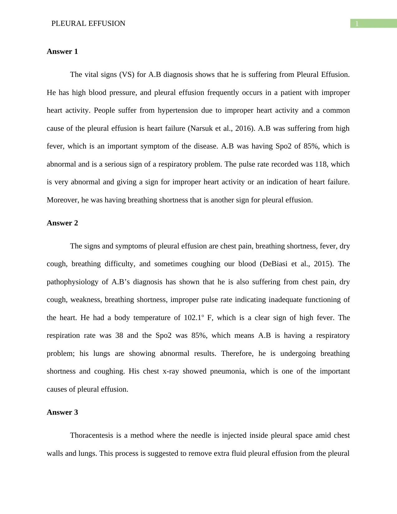
The vital signs (VS) for A.B diagnosis shows that he is suffering from Pleural Effusion.
He has high blood pressure, and pleural effusion frequently occurs in a patient with improper
heart activity. People suffer from hypertension due to improper heart activity and a common
cause of the pleural effusion is heart failure (Narsuk et al., 2016). A.B was suffering from high
fever, which is an important symptom of the disease. A.B was having Spo2 of 85%, which is
abnormal and is a serious sign of a respiratory problem. The pulse rate recorded was 118, which
is very abnormal and giving a sign for improper heart activity or an indication of heart failure.
Moreover, he was having breathing shortness that is another sign for pleural effusion.
Answer 2
The signs and symptoms of pleural effusion are chest pain, breathing shortness, fever, dry
cough, breathing difficulty, and sometimes coughing our blood (DeBiasi et al., 2015). The
pathophysiology of A.B’s diagnosis has shown that he is also suffering from chest pain, dry
cough, weakness, breathing shortness, improper pulse rate indicating inadequate functioning of
the heart. He had a body temperature of 102.1o F, which is a clear sign of high fever. The
respiration rate was 38 and the Spo2 was 85%, which means A.B is having a respiratory
problem; his lungs are showing abnormal results. Therefore, he is undergoing breathing
shortness and coughing. His chest x-ray showed pneumonia, which is one of the important
causes of pleural effusion.
Answer 3
Thoracentesis is a method where the needle is injected inside pleural space amid chest
walls and lungs. This process is suggested to remove extra fluid pleural effusion from the pleural
2PLEURAL EFFUSION
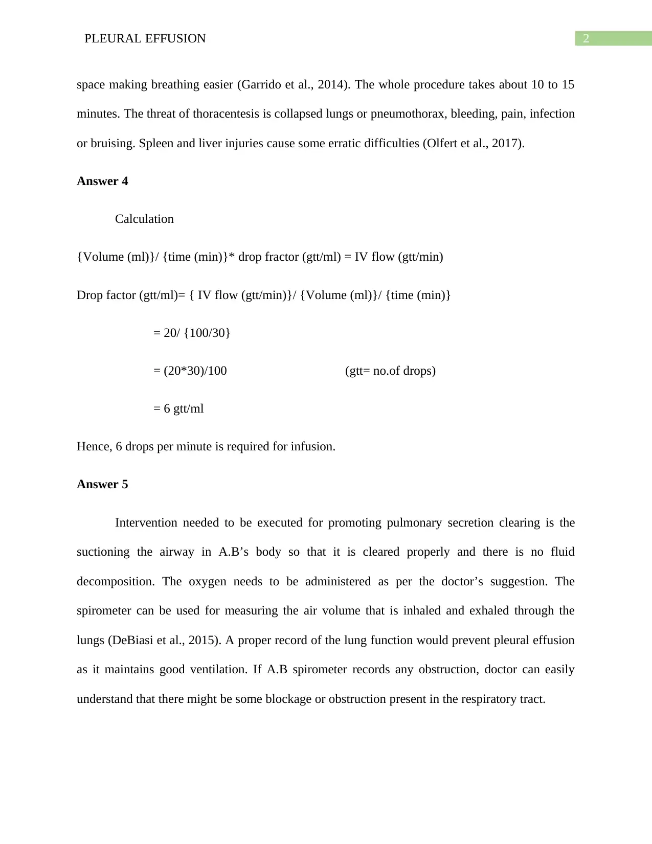
space making breathing easier (Garrido et al., 2014). The whole procedure takes about 10 to 15
minutes. The threat of thoracentesis is collapsed lungs or pneumothorax, bleeding, pain, infection
or bruising. Spleen and liver injuries cause some erratic difficulties (Olfert et al., 2017).
Answer 4
Calculation
{Volume (ml)}/ {time (min)}* drop fractor (gtt/ml) = IV flow (gtt/min)
Drop factor (gtt/ml)= { IV flow (gtt/min)}/ {Volume (ml)}/ {time (min)}
= 20/ {100/30}
= (20*30)/100 (gtt= no.of drops)
= 6 gtt/ml
Hence, 6 drops per minute is required for infusion.
Answer 5
Intervention needed to be executed for promoting pulmonary secretion clearing is the
suctioning the airway in A.B’s body so that it is cleared properly and there is no fluid
decomposition. The oxygen needs to be administered as per the doctor’s suggestion. The
spirometer can be used for measuring the air volume that is inhaled and exhaled through the
lungs (DeBiasi et al., 2015). A proper record of the lung function would prevent pleural effusion
as it maintains good ventilation. If A.B spirometer records any obstruction, doctor can easily
understand that there might be some blockage or obstruction present in the respiratory tract.
⊘ This is a preview!⊘
Do you want full access?
Subscribe today to unlock all pages.

Trusted by 1+ million students worldwide
3PLEURAL EFFUSION
Answer 6
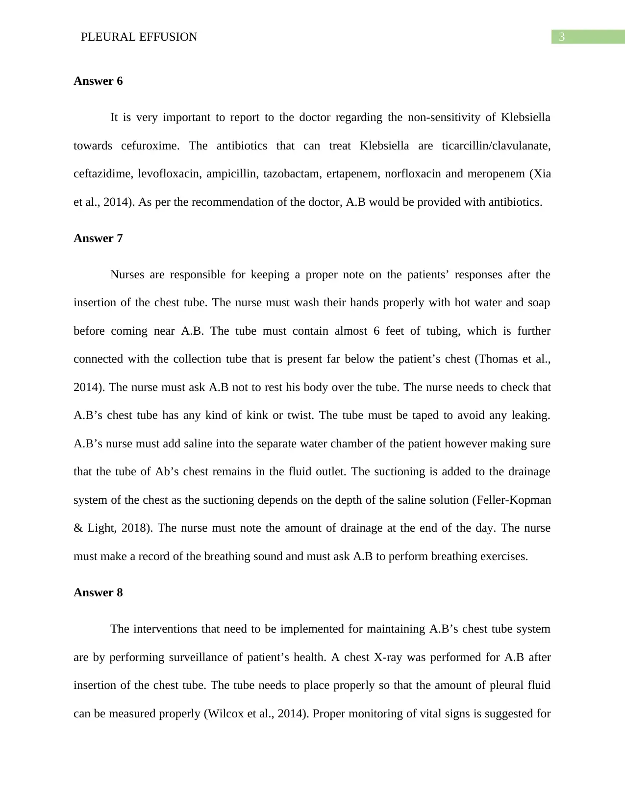
It is very important to report to the doctor regarding the non-sensitivity of Klebsiella
towards cefuroxime. The antibiotics that can treat Klebsiella are ticarcillin/clavulanate,
ceftazidime, levofloxacin, ampicillin, tazobactam, ertapenem, norfloxacin and meropenem (Xia
et al., 2014). As per the recommendation of the doctor, A.B would be provided with antibiotics.
Answer 7
Nurses are responsible for keeping a proper note on the patients’ responses after the
insertion of the chest tube. The nurse must wash their hands properly with hot water and soap
before coming near A.B. The tube must contain almost 6 feet of tubing, which is further
connected with the collection tube that is present far below the patient’s chest (Thomas et al.,
2014). The nurse must ask A.B not to rest his body over the tube. The nurse needs to check that
A.B’s chest tube has any kind of kink or twist. The tube must be taped to avoid any leaking.
A.B’s nurse must add saline into the separate water chamber of the patient however making sure
that the tube of Ab’s chest remains in the fluid outlet. The suctioning is added to the drainage
system of the chest as the suctioning depends on the depth of the saline solution (Feller-Kopman
& Light, 2018). The nurse must note the amount of drainage at the end of the day. The nurse
must make a record of the breathing sound and must ask A.B to perform breathing exercises.
Answer 8
The interventions that need to be implemented for maintaining A.B’s chest tube system
are by performing surveillance of patient’s health. A chest X-ray was performed for A.B after
insertion of the chest tube. The tube needs to place properly so that the amount of pleural fluid
can be measured properly (Wilcox et al., 2014). Proper monitoring of vital signs is suggested for
Paraphrase This Document
Need a fresh take? Get an instant paraphrase of this document with our AI Paraphraser
4PLEURAL EFFUSION
assessing improvement or deterioration. Pain management is recommended. After inserting the
chest tube, A.B would go through pain. Inappropriate analgesic management would lead to
inhibition movement of lung expansion. The suction could be attached to the sealed drain to
facilitate the drainage of pleural effusions. Chest tube clamping is also recommended. The
drainage bottle must be kept under the chest level to drain out a huge volume of pleural effusion
for avoiding the risk of pulmonary edema re-expansion (Xia et al., 2014)
Answer 9
1. True
2. False
3. True
4. True
5. False
6. False
7. True
8. True
Answer 10
The assessment performed by nurses is to do a further checkup. There can be a possibility
that A.B’s chest tube has opened up or it was contaminated. The pleural fluid was accumulated
in the chest. As A.B was sitting, it can be assessed that there might be something wrong with the
chest tube. The doctor needs to be informed about the situation.
5PLEURAL EFFUSION
Answer 11
The chest tube found contaminated and disconnected needs immediate attention. The tube
needs to be clamed quickly, and the end of the chest tube must be placed in the sterile water. As
the tube in contaminated washing with sterile water is strongly suggested before, the hospital
arranges for a fresh tube (Feller-Kopman & Light, 2018). It is also strongly recommended that
both the ends of the tube must be washed with alcohol and then again reconnected in the same
way.
⊘ This is a preview!⊘
Do you want full access?
Subscribe today to unlock all pages.

Trusted by 1+ million students worldwide
6PLEURAL EFFUSION
References
Barsuk, J. H., Cohen, E. R., Williams, M. V., Scher, J., Feinglass, J., McGaghie, W. C., ... &
Wayne, D. B. (2016). The effect of simulation‐based mastery learning on thoracentesis
referral patterns. Journal of hospital medicine, 11(11), 792-795.
DeBiasi, E. M., Pisani, M. A., Murphy, T. E., Araujo, K., Kookoolis, A., Argento, A. C., &
Puchalski, J. (2015). Mortality among patients with pleural effusion undergoing
thoracentesis. European Respiratory Journal, 46(2), 495-502.
Feller-Kopman, D., & Light, R. (2018). Pleural disease. New England Journal of
Medicine, 378(8), 740-751.
Garrido, V. V., Viedma, E. C., Villar, A. F., de Pablo Gafas, A., Rodríguez, E. P., Pérez, J. M.
P., ... & Cuadrado, L. V. (2014). Recommendations of diagnosis and treatment of pleural
effusion. Update. Archivos de Bronconeumología (English Edition), 50(6), 235-249.
Olfert, J. A., Penz, E. D., Manns, B. J., Mishra, E. K., Davies, H. E., Miller, R. F., ... & Rahman,
N. M. (2017). Cost‐effectiveness of indwelling pleural catheter compared with talc in
malignant pleural effusion. Respirology, 22(4), 764-770.
Thomas, R., Francis, R., Davies, H. E., & Lee, Y. G. (2014). Interventional therapies for
malignant pleural effusions: the present and the future. Respirology, 19(6), 809-822.
Wilcox, M. E., Chong, C. A., Stanbrook, M. B., Tricco, A. C., Wong, C., & Straus, S. E. (2014).
Does This Patient Have an Exudative Pleural Effusion?: The Rational Clinical
Examination Systematic Review. Jama, 311(23), 2422-2431.
Paraphrase This Document
Need a fresh take? Get an instant paraphrase of this document with our AI Paraphraser

7PLEURAL EFFUSION
Xia, H., Wang, X. J., Zhou, Q., Shi, H. Z., & Tong, Z. H. (2014). Efficacy and safety of talc
pleurodesis for malignant pleural effusion: a meta-analysis. PLoS One, 9(1).
Xia, H., Wang, X. J., Zhou, Q., Shi, H. Z., & Tong, Z. H. (2014). Efficacy and safety of talc
pleurodesis for malignant pleural effusion: a meta-analysis. PLoS One, 9(1).
1 out of 8
Related Documents
Your All-in-One AI-Powered Toolkit for Academic Success.
+13062052269
info@desklib.com
Available 24*7 on WhatsApp / Email
![[object Object]](/_next/static/media/star-bottom.7253800d.svg)
Unlock your academic potential
Copyright © 2020–2026 A2Z Services. All Rights Reserved. Developed and managed by ZUCOL.


