Case Study Analysis: Primary Biliary Cholangitis (PBC) - PBC 12
VerifiedAdded on 2022/11/26
|12
|3193
|88
Case Study
AI Summary
This case study focuses on a 42-year-old female patient, Pamela, suffering from liver cirrhosis due to Primary Biliary Cholangitis (PBC). The assignment explores the patient's background, including her lack of improvement with Ursodeoxycholic acid (UDCA) and associated symptoms like fatigue and itching. It delves into the pathophysiology of PBC, highlighting its autoimmune nature, the role of T cells, and genetic and environmental risk factors. The study examines the management of PBC, including UDCA, Obeticholic acid, and liver transplants, along with the importance of nursing interventions in patient care. The case study also discusses the limitations of current treatments and the need for further research, including the potential use of budesonide and methotrexate. The nurses' role in patient education, symptom monitoring, and medication administration is emphasized. The assignment critically appraises the evidence-based literature, providing recommendations for future nursing practice and a holistic perspective on patient care.

Running heads: PBC 1
Student name
Student No.
Unit
Title: Primary Biliary Cholangitis
Student name
Student No.
Unit
Title: Primary Biliary Cholangitis
Paraphrase This Document
Need a fresh take? Get an instant paraphrase of this document with our AI Paraphraser
PBC 2
Introduction
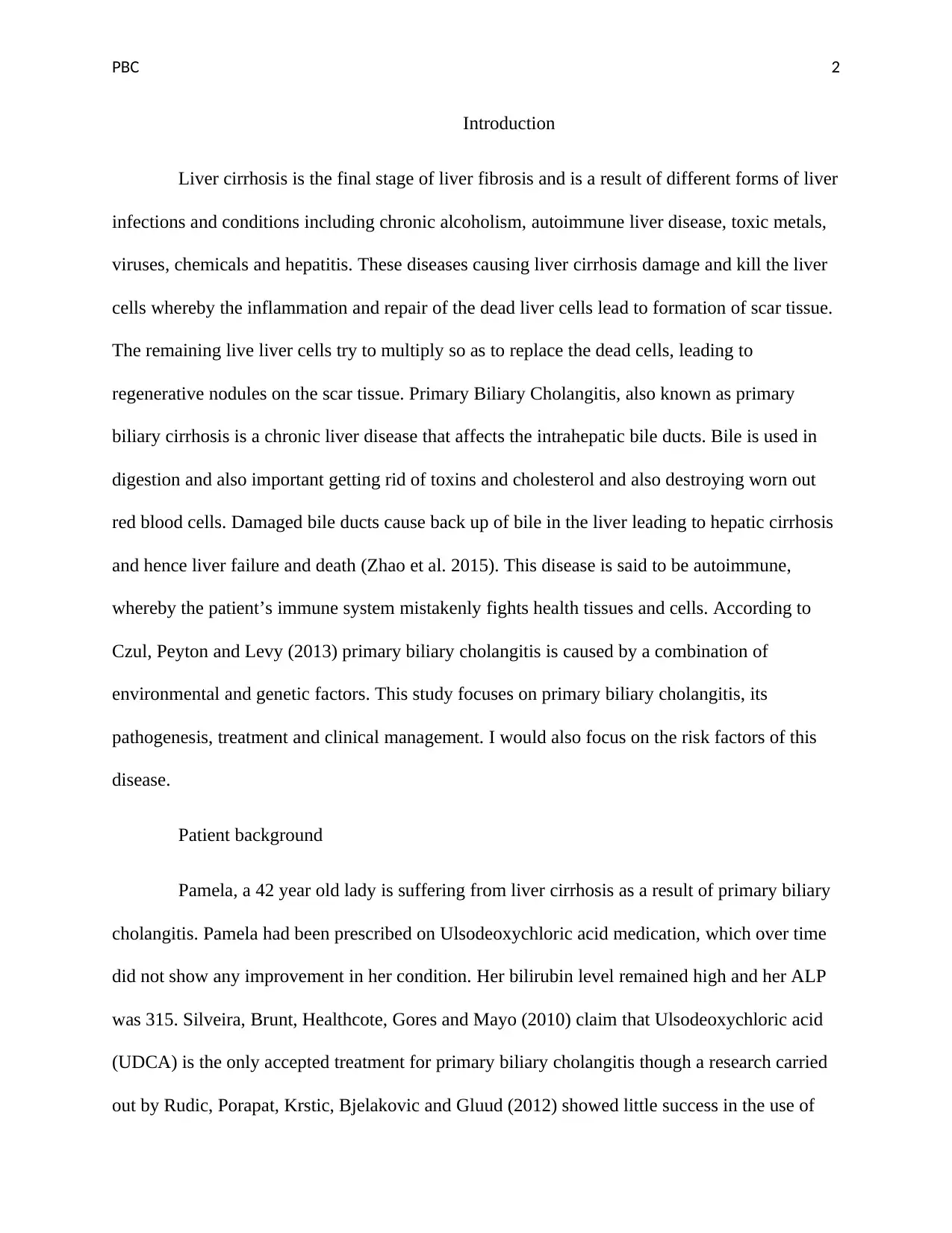
Liver cirrhosis is the final stage of liver fibrosis and is a result of different forms of liver
infections and conditions including chronic alcoholism, autoimmune liver disease, toxic metals,
viruses, chemicals and hepatitis. These diseases causing liver cirrhosis damage and kill the liver
cells whereby the inflammation and repair of the dead liver cells lead to formation of scar tissue.
The remaining live liver cells try to multiply so as to replace the dead cells, leading to
regenerative nodules on the scar tissue. Primary Biliary Cholangitis, also known as primary
biliary cirrhosis is a chronic liver disease that affects the intrahepatic bile ducts. Bile is used in
digestion and also important getting rid of toxins and cholesterol and also destroying worn out
red blood cells. Damaged bile ducts cause back up of bile in the liver leading to hepatic cirrhosis
and hence liver failure and death (Zhao et al. 2015). This disease is said to be autoimmune,
whereby the patient’s immune system mistakenly fights health tissues and cells. According to
Czul, Peyton and Levy (2013) primary biliary cholangitis is caused by a combination of
environmental and genetic factors. This study focuses on primary biliary cholangitis, its
pathogenesis, treatment and clinical management. I would also focus on the risk factors of this
disease.
Patient background
Pamela, a 42 year old lady is suffering from liver cirrhosis as a result of primary biliary
cholangitis. Pamela had been prescribed on Ulsodeoxychloric acid medication, which over time
did not show any improvement in her condition. Her bilirubin level remained high and her ALP
was 315. Silveira, Brunt, Healthcote, Gores and Mayo (2010) claim that Ulsodeoxychloric acid
(UDCA) is the only accepted treatment for primary biliary cholangitis though a research carried
out by Rudic, Porapat, Krstic, Bjelakovic and Gluud (2012) showed little success in the use of
PBC 3
UDCA in the treatment of primary biliary cholangitis. Pamela frequently complained of being
tired, itching which caused her sleepless nights and experienced dry mouth despite drinking
amounts of water.
Pathophysiology
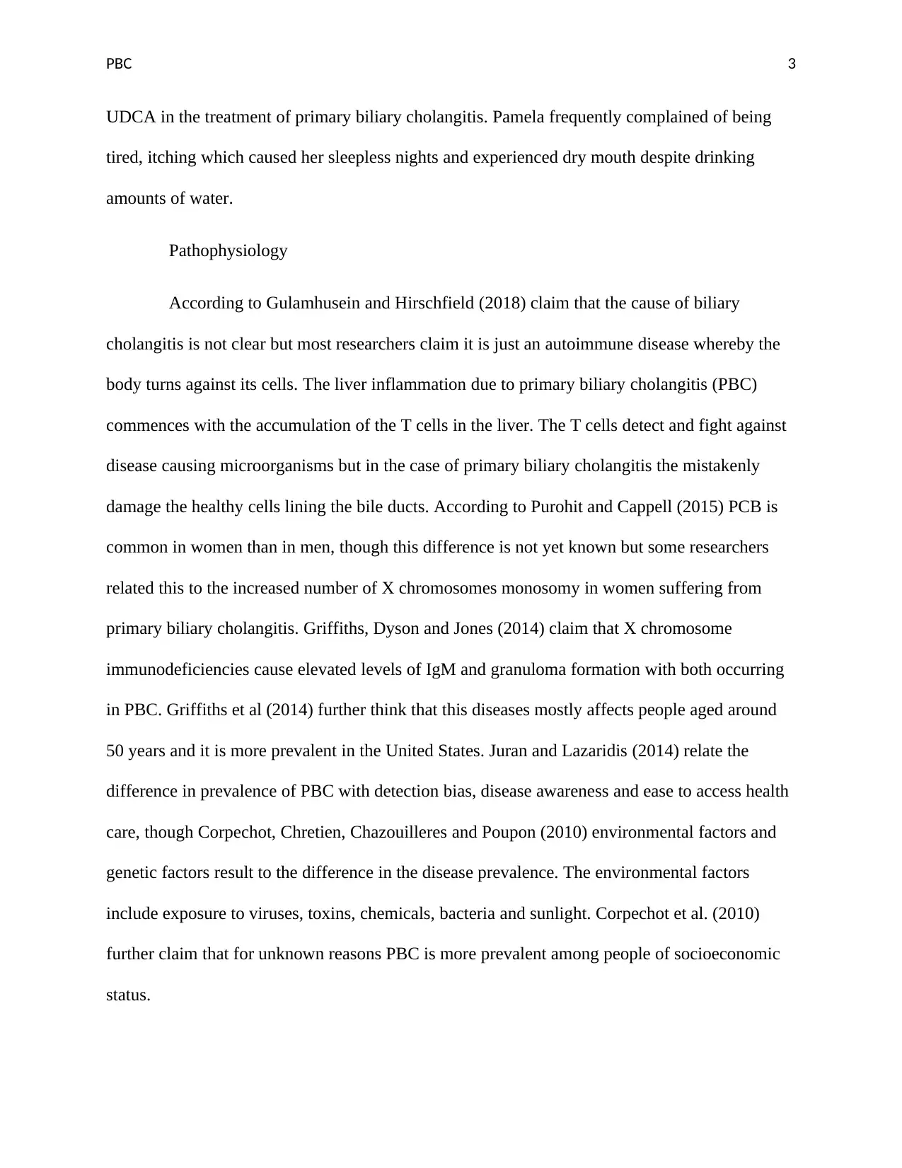
According to Gulamhusein and Hirschfield (2018) claim that the cause of biliary
cholangitis is not clear but most researchers claim it is just an autoimmune disease whereby the
body turns against its cells. The liver inflammation due to primary biliary cholangitis (PBC)
commences with the accumulation of the T cells in the liver. The T cells detect and fight against
disease causing microorganisms but in the case of primary biliary cholangitis the mistakenly
damage the healthy cells lining the bile ducts. According to Purohit and Cappell (2015) PCB is
common in women than in men, though this difference is not yet known but some researchers
related this to the increased number of X chromosomes monosomy in women suffering from
primary biliary cholangitis. Griffiths, Dyson and Jones (2014) claim that X chromosome
immunodeficiencies cause elevated levels of IgM and granuloma formation with both occurring
in PBC. Griffiths et al (2014) further think that this diseases mostly affects people aged around
50 years and it is more prevalent in the United States. Juran and Lazaridis (2014) relate the
difference in prevalence of PBC with detection bias, disease awareness and ease to access health
care, though Corpechot, Chretien, Chazouilleres and Poupon (2010) environmental factors and
genetic factors result to the difference in the disease prevalence. The environmental factors
include exposure to viruses, toxins, chemicals, bacteria and sunlight. Corpechot et al. (2010)
further claim that for unknown reasons PBC is more prevalent among people of socioeconomic
status.
⊘ This is a preview!⊘
Do you want full access?
Subscribe today to unlock all pages.

Trusted by 1+ million students worldwide
PBC 4
Smyk, Rigopoulou, Bogdanos (2013) srongly relate PBC with urinary tract infections.
In their study they claim that 48 percent of PBC patients had suffered from recurrent UTIs
before. They therefore studies the relationship between E. coli (leading cause of UTIs in women)
and PBC. They realized that E. coli triggers autoimmune response which in turn triggers cross
reactive response in T cells and B cells which occur in patients suffering from PBC. Studies by
Prince, Ducker and James (2010) showed a correlation between PBC and tobacco smoking. PBC
has a negative correlation with alcohol consumption. Prince et al. (2010) claim that people who
use hair dyes have a higher risk of contracting PBC though this contradicts with other
researchers.
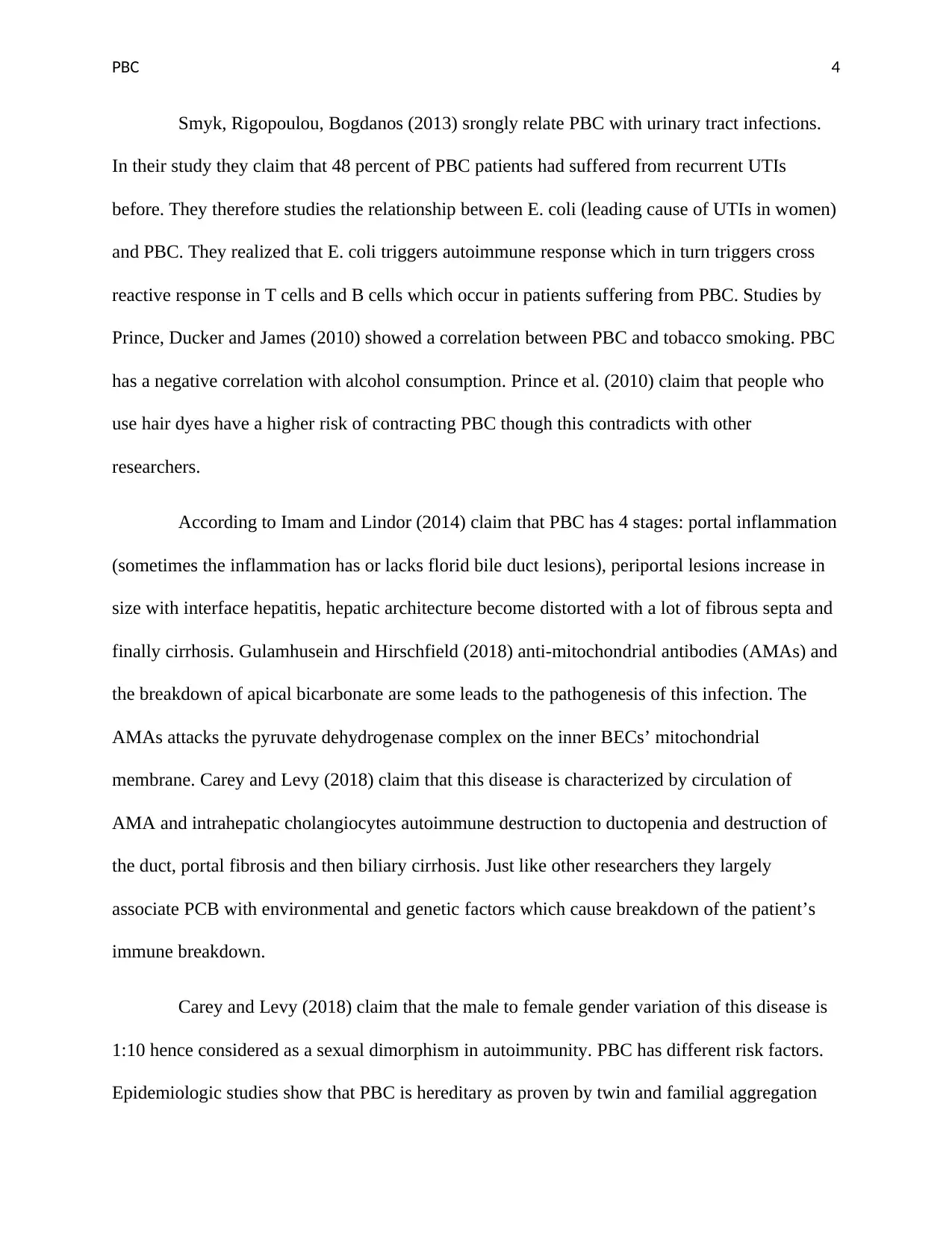
According to Imam and Lindor (2014) claim that PBC has 4 stages: portal inflammation
(sometimes the inflammation has or lacks florid bile duct lesions), periportal lesions increase in
size with interface hepatitis, hepatic architecture become distorted with a lot of fibrous septa and
finally cirrhosis. Gulamhusein and Hirschfield (2018) anti-mitochondrial antibodies (AMAs) and
the breakdown of apical bicarbonate are some leads to the pathogenesis of this infection. The
AMAs attacks the pyruvate dehydrogenase complex on the inner BECs’ mitochondrial
membrane. Carey and Levy (2018) claim that this disease is characterized by circulation of
AMA and intrahepatic cholangiocytes autoimmune destruction to ductopenia and destruction of
the duct, portal fibrosis and then biliary cirrhosis. Just like other researchers they largely
associate PCB with environmental and genetic factors which cause breakdown of the patient’s
immune breakdown.
Carey and Levy (2018) claim that the male to female gender variation of this disease is
1:10 hence considered as a sexual dimorphism in autoimmunity. PBC has different risk factors.
Epidemiologic studies show that PBC is hereditary as proven by twin and familial aggregation
Paraphrase This Document
Need a fresh take? Get an instant paraphrase of this document with our AI Paraphraser
PBC 5
researches. Among PBC cases a monozygotic twins accordance rate of 0.63 was the highest
record. Family history of this disease has been found to have a 1.33 percent and 9 percent
variation. Research from the genome wide association studies (GWAS) and iCHIP showed that
genes associated to PBC are from human leukocyte antigens. Carey and Levy (2018) also claim
that PBC has been associated with components regulating the body immunity. Studies indicate
that there are about 27 loci non-human leukocyte antigens linked with PBC. These genetic
variations are also shared with other autoimmune diseases, not PBC alone.
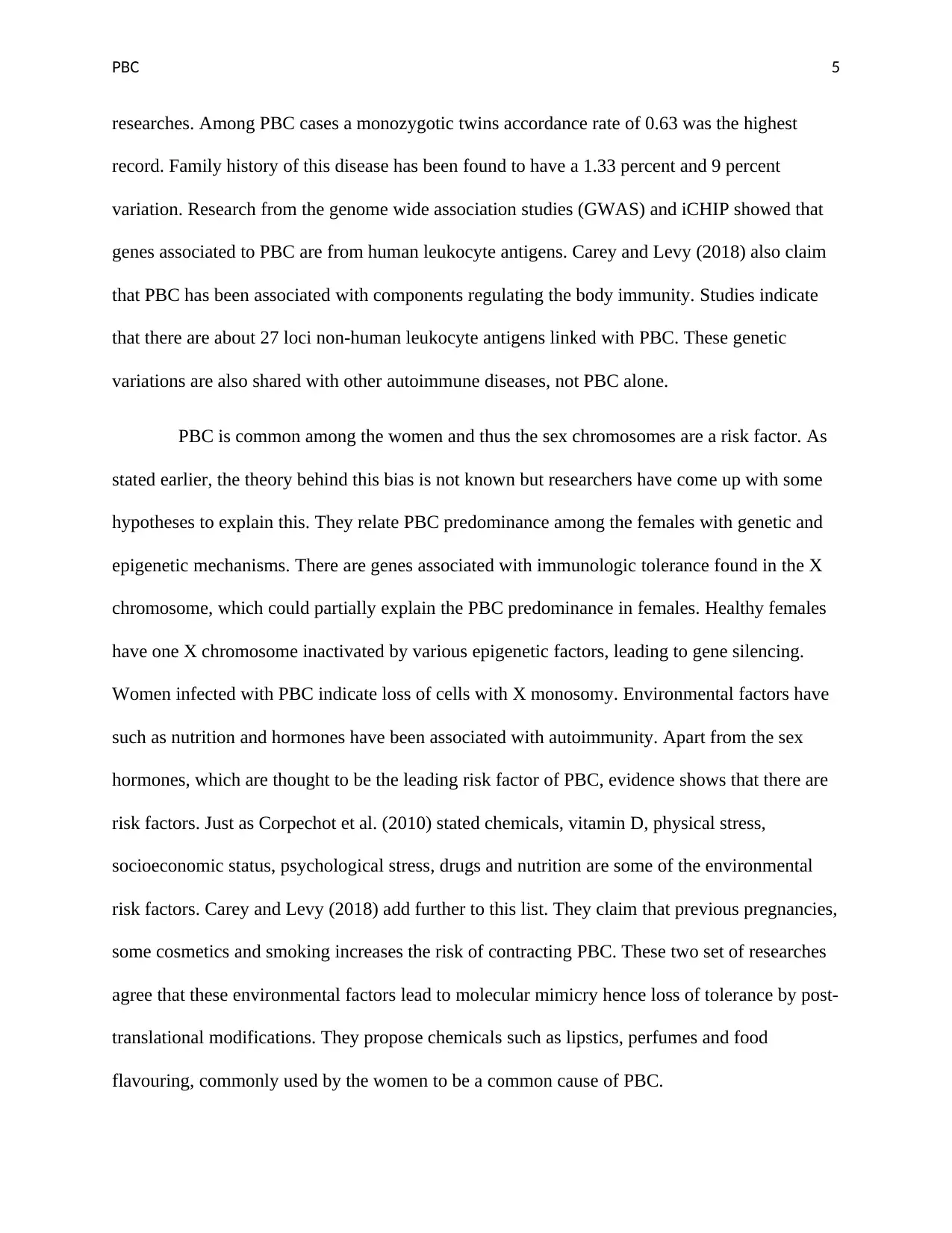
PBC is common among the women and thus the sex chromosomes are a risk factor. As
stated earlier, the theory behind this bias is not known but researchers have come up with some
hypotheses to explain this. They relate PBC predominance among the females with genetic and
epigenetic mechanisms. There are genes associated with immunologic tolerance found in the X
chromosome, which could partially explain the PBC predominance in females. Healthy females
have one X chromosome inactivated by various epigenetic factors, leading to gene silencing.
Women infected with PBC indicate loss of cells with X monosomy. Environmental factors have
such as nutrition and hormones have been associated with autoimmunity. Apart from the sex
hormones, which are thought to be the leading risk factor of PBC, evidence shows that there are
risk factors. Just as Corpechot et al. (2010) stated chemicals, vitamin D, physical stress,
socioeconomic status, psychological stress, drugs and nutrition are some of the environmental
risk factors. Carey and Levy (2018) add further to this list. They claim that previous pregnancies,
some cosmetics and smoking increases the risk of contracting PBC. These two set of researches
agree that these environmental factors lead to molecular mimicry hence loss of tolerance by post-
translational modifications. They propose chemicals such as lipstics, perfumes and food
flavouring, commonly used by the women to be a common cause of PBC.
PBC 6
Management of PBC
Cholesterol test, liver test and antibody tests for autoimmune disease signs are the
diagnostic method for PBC. Imaging tests such as ultrasound, MRCP, MRE and the ERCP could
be carried out to confirm the doctor’s diagnosis. According to Younossi et al. (2019), there is no
treatment for PBC but there are ways to manage it and prevent its complications. These ways
include use of UDCA. According to research, the presentation of PBC has changed over time. In
the pre-UDCA era people showed jaundice as the core symptom but this was in advanced stages
of the disease. Since diagnostic tools such as AMA detection were developed PBC patients are
now asymptomatic at diagnosis, showing symptoms like pruritus and fatigue even at the early
stages of the disease. UDCA therapy has been said to have brought a great impact in treatment of
PBC especially when administered at its early stages. For patients under the 1st and the 2nd stages
of PBC, studies indicate that the survival rates are the same. UDCA does not cure PBC but
improves the functioning of the liver. However, 40 percent of the patients show negative
response towards UDCA leading to liver transplant or even death.
Obeticholic acid is a new medication for PBC and can be given alone or combined with
ursodiol. Researchers claim that when administered for 12 months, it gives positive results.
Another medication is the tricor. Though Nevens, Andreone and Mazzella (2016) claim that
researcher do not know how this medicine works to relieve PBC symptoms, it helps relieve
itching and liver inflammation. When PBC is out of control and liver failure has been
experienced, liver transplant is carried out to prolong the patient’s life. When the patient
⊘ This is a preview!⊘
Do you want full access?
Subscribe today to unlock all pages.

Trusted by 1+ million students worldwide
PBC 7
experiences itching, the doctor administers treatments such as antihistamines, cholestyramine,
rifampin and opioid antagonists (Hirschfield et al. 2018). According to Selmi, Bowlus, Gershwin
and Coppel (2011) the nurse has big role to play in the management of this disease. The nurse
should advice the patient on how the medications are to be taken, for example Obeticholic acid
can be taken without regarding food. The nurses should be aware of any new signs or worsening
pruritus. Therefore they should be in touch with the patient at all time. In case of outpatient, the
health professionals should come up with as system whereby they make a follow up on the
patient’s progress. The health practitioners should also teach the patients how to recognize
changes in signs and symptoms and report them immediately.
According to Haung (2016) a lot of research is ongoing on the treatment and medication
of PBC. Drugs such as colchicine and methotrexate have been developed though they are still
under study. Rabahi et al. (2010) claim that budesonide is a medication that has been on research
to be used on PBC patients who do not show any response on UDCA. For the case of Pamela,
whose PBC was not responding to UDCA, budesonide should be administered to prevent further
complications of the disease. Rabahi et al. (2010) claim that research is ongoing to determine if
this drug could be administered together with UDCA. Leung, Bonis and Kaplan (2011) state that
though methotrexate has been used in treatment of PBC, little is known about its role and action.
They recommend this medication to be used for patients who do not respond to UDCA and show
characteristics of AIH. This medication can be used together with UDCA though Leung et al.
(2011) call for more research to understand better about the action of the medication. According
to Ali, Carey and Lindor (2015) FXR is a primary bile acids receptor that controls the
metabolism disposal and uptake of bile acid. There other novel therapeutic methods such as
Paraphrase This Document
Need a fresh take? Get an instant paraphrase of this document with our AI Paraphraser
PBC 8
PPAR-alpha that are under research and though to bring a turnaround in the management and
treatment of PBC (Trivedi, Hirschfield and Gershwin, 2016).
The approval of Obeticholic acid has been an important improvement in the treatment
of PCB. However, PBC being a disease without treatment there is a call for more research on this
condition. Advancements in technology have also led to development in accuracy in prognosis
and diagnosis of PBC. Though there has been recorded much improvements, increasing UDCA
response as far as PBC is concerned is a big challenge to researchers and health care
professionals.
PBC 9
References
Ali, A. H., Carey, E. J., & Lindor, K. D. (2015). Recent advances in the development of
farnesoid X receptor agonists. Annals of translational medicine, 3(1).
Carey, E. and Levy, C. (2018). Primary biliary cholangitis, an issue of clinics in liver disease.
Pennsylvania: Elsevier.
Corpechot, C., Chrétien, Y., Chazouillères, O., & Poupon, R. (2010). Demographic, lifestyle,
medical and familial factors associated with primary biliary cirrhosis. Journal of
hepatology, 53(1), 162-169.
Czul, F., Peyton, A., & Levy, C. (2013). Primary biliary cirrhosis: therapeutic advances. Clinics
in liver disease, 17(2), 229-242.
Griffiths, L., Dyson, J. K., & Jones, D. E. (2014, August). The new epidemiology of primary
biliary cirrhosis. In Seminars in liver disease (Vol. 34, No. 03, pp. 318-328). Thieme
Medical Publishers.
Gulamhusein, A. F. and Hirschfield, G.M. (2018). Pathophysiology of primary biliary
cholangitis. Best Pract Res Clin Gastroenterol;34-35:17-25. doi:
10.1016/j.bpg.2018.05.012.
Hirschfield, G. M., Dyson, J. K., Alexander, G., Chapman, M. H., Collier, J., Hübscher, S. and
Jones, D. (2018). The British Society of Gastroenterology/UK-PBC primary biliary
cholangitis treatment and management guidelines. Gut, 67(9), 1568–1594.
doi:10.1136/gutjnl-2017-315259
⊘ This is a preview!⊘
Do you want full access?
Subscribe today to unlock all pages.

Trusted by 1+ million students worldwide
PBC 10
Huang Y. Q. (2016). Recent advances in the diagnosis and treatment of primary biliary
cholangitis. World journal of hepatology, 8(33), 1419–1441.
doi:10.4254/wjh.v8.i33.1419
Imam, M. H., & Lindor, K. D. (2014, August). The natural history of primary biliary cirrhosis. In
Seminars in liver disease (Vol. 34, No. 03, pp. 329-333). Thieme Medical Publishers
Juran, B. D., & Lazaridis, K. N. (2014, August). Environmental factors in primary biliary
cirrhosis. In Seminars in liver disease (Vol. 34, No. 03, pp. 265-272). Thieme Medical
Publishers.
Leung, J., Bonis, P. A., & Kaplan, M. M. (2011). Colchicine or methotrexate, with ursodiol, are
effective after 20 years in a subset of patients with primary biliary cirrhosis. Clinical
Gastroenterology and Hepatology, 9(9), 776-780.
Nevens F, Andreone P, Mazzella G, et al. A Placebo-Controlled Trial of Obeticholic Acid in
Primary Biliary Cholangitis. N Engl J Med, 375:631.
Purohit, T., & Cappell, M. S. (2015). Primary biliary cirrhosis: Pathophysiology, clinical
presentation and therapy. World journal of hepatology, 7(7), 926–941.
doi:10.4254/wjh.v7.i7.926
Prince, M. I., Ducker, S. J., & James, O. F. W. (2010). Case–control studies of risk factors for
primary biliary cirrhosis in two United Kingdom populations. Gut, 59(4), 508-512.
Rabahi, N., Chretien, Y., Gaouar, F., Wendum, D., Serfaty, L., Chazouilleres, O., ... & Poupon,
R. (2010). Triple therapy with ursodeoxycholic acid, budesonide and mycophenolate
Paraphrase This Document
Need a fresh take? Get an instant paraphrase of this document with our AI Paraphraser
PBC 11
mofetil in patients with features of severe primary biliary cirrhosis not responding to
ursodeoxycholic acid alone. Gastroentérologie clinique et biologique, 34(4-5), 283-287.
Silveira, M. G., Brunt, E. M., Heathcote, J., Gores, G. J., Lindor, K. D., & Mayo, M. J. (2010).
American Association for the Study of Liver Diseases endpoints conference: design and
endpoints for clinical trials in primary biliary cirrhosis. Hepatology, 52(1), 349-359.
Rudic, J. S., Poropat, G., Krstic, M. N., Bjelakovic, G., & Gluud, C. (2012). Ursodeoxycholic
acid for primary biliary cirrhosis. Cochrane Database of Systematic Reviews, (12).
Selmi, C., Coppel, R. L., & Gershwin, M. E. (2006). Primary biliary cirrhosis. In The
Autoimmune Diseases (pp. 749-765). Academic Press.
Smyk, D. S., Rigopoulou, E. I., & Bogdanos, D. P. (2013). Potential roles for infectious agents in
the pathophysiology of primary biliary cirrhosis: what’s new?. Current infectious
disease reports, 15(1), 14-24.
Trivedi, P. J., Hirschfield, G. M., & Gershwin, M. E. (2016). Obeticholic acid for the treatment
of primary biliary cirrhosis. Expert review of clinical pharmacology, 9(1), 13-26.
Younossi, Z. M., Bernstein, D., Shiffman, M. L., Kwo, P., Kim, W. R., Kowdley, K. V. and
Jacobson I. M. (2018). Diagnosis and Management of Primary Biliary Cholangitis. Am
J Gastroenterol, 114(1):48-63. doi: 10.1038/s41395-018-0390-3.
Zhao, P., Liu, W. W., Li, J. F., Wang, C. Y., Wang, H., Xu, J. and Wei, Z. M. (2015). Predictors
of liver failure in primary biliary cirrhosis. Upsala journal of medical sciences, 120(1),
47–51. doi:10.3109/03009734.2014.985763

PBC 12
⊘ This is a preview!⊘
Do you want full access?
Subscribe today to unlock all pages.

Trusted by 1+ million students worldwide
1 out of 12
Your All-in-One AI-Powered Toolkit for Academic Success.
+13062052269
info@desklib.com
Available 24*7 on WhatsApp / Email
![[object Object]](/_next/static/media/star-bottom.7253800d.svg)
Unlock your academic potential
Copyright © 2020–2026 A2Z Services. All Rights Reserved. Developed and managed by ZUCOL.

