Comprehensive Report: Principles and Techniques of Dental Radiography

Verified

Added on  2020/12/24

|8
|1103
|483
Report
AI Summary
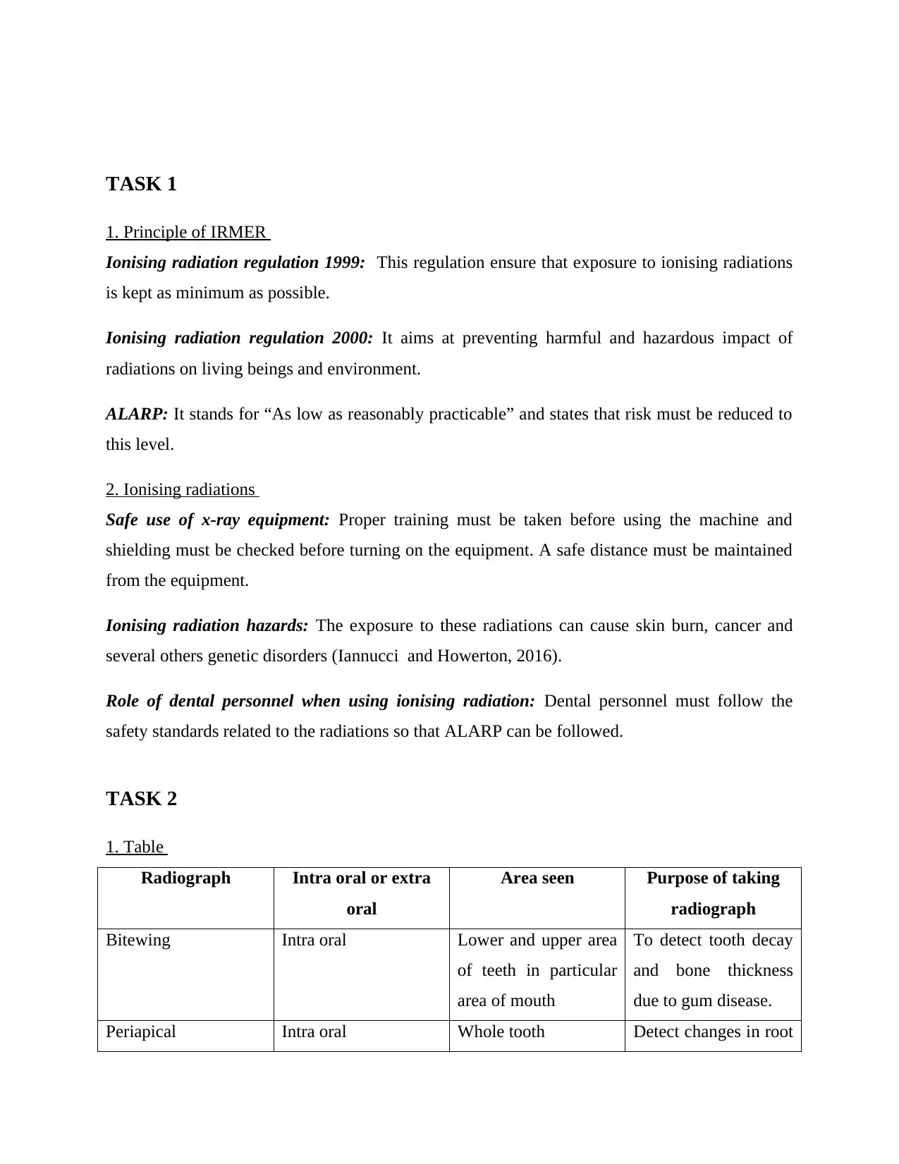
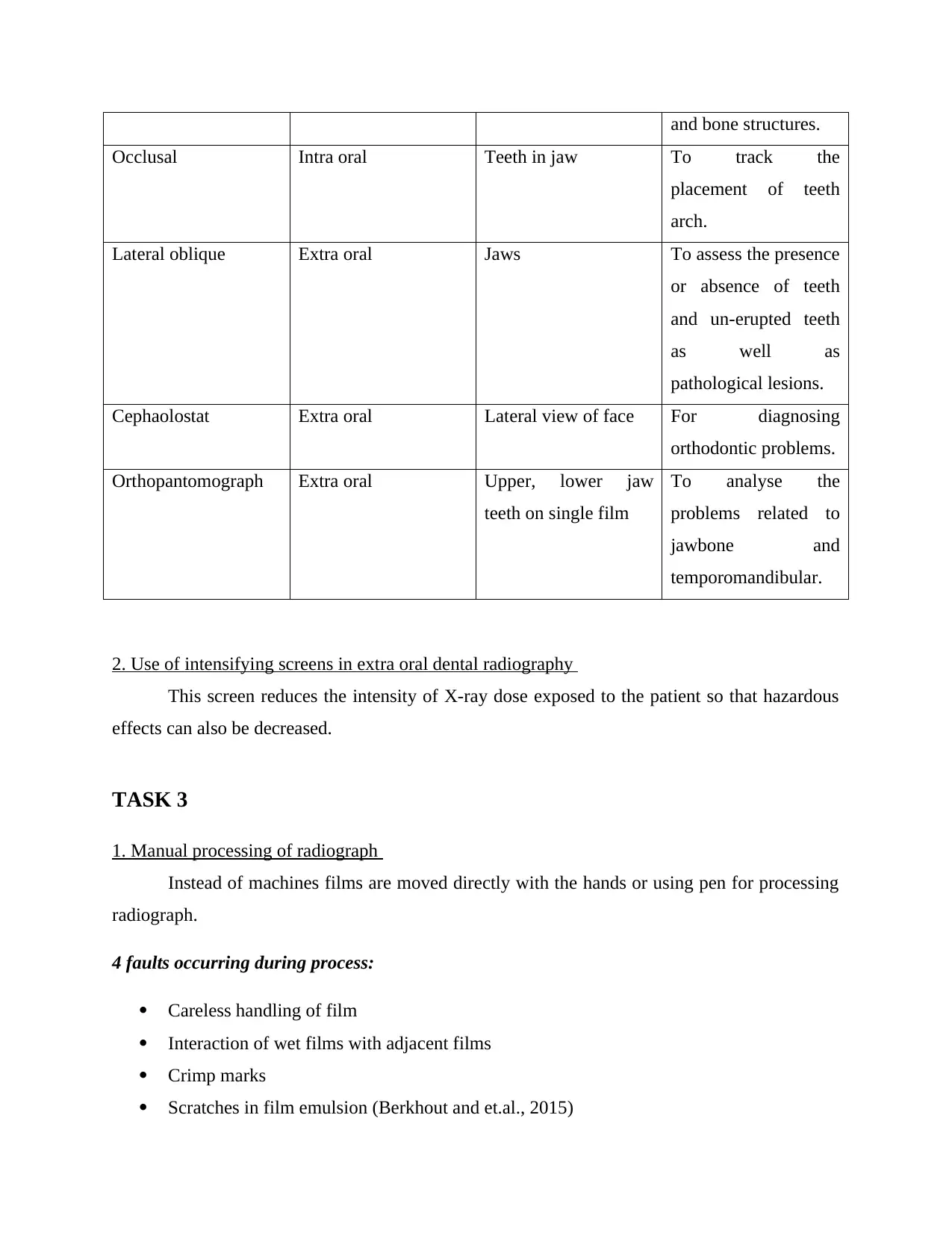
This report provides a comprehensive overview of the principles and techniques of dental radiography. It begins with an introduction to the Ionising Radiation (Medical Exposure) Regulations (IRMER) and the concept of 'As Low As Reasonably Practicable' (ALARP), emphasizing radiation safety and the hazards of ionizing radiation. The report then details various radiographic techniques, differentiating between intraoral (bitewing, periapical, occlusal) and extraoral (lateral oblique, cephalostat, orthopantomograph) methods, and explains the use of intensifying screens. The report also covers the manual and automatic processing of radiographs, including chemicals used, precautions, and potential faults. Furthermore, it addresses stock rotation, film storage, and the importance of using quality films. Finally, the report discusses methods of mounting radiographs, the quality assurance control system, and grading of radiographs, ensuring consistent and high-quality images with minimal radiation exposure. The report is well-structured with references to support the information provided.
Document Page
PRINCIPLES AND
TECHNIQUE OF DENTAL
RADIOGRAPHY
tabler-icon-diamond-filled.svg

Paraphrase This Document

Need a fresh take? Get an instant paraphrase of this document with our AI Paraphraser
Document Page
Table of Contents
TASK 1............................................................................................................................................3
1. Principle of IRMER.................................................................................................................3
2. Ionising radiations...................................................................................................................3
TASK 2............................................................................................................................................3
1. Table........................................................................................................................................3
2. Use of intensifying screens in extra oral dental radiography..................................................4
TASK 3............................................................................................................................................4
1. Manual processing of radiograph............................................................................................4
2. Chemicals used in developing radiograph...............................................................................5
3. Precautions...............................................................................................................................5
4. Developing radiograph using automatic processor..................................................................5
5. Digital radiograph....................................................................................................................5
TASK 4............................................................................................................................................6
1. Rationale to rotate stock..........................................................................................................6
2. Storage of radiographic films..................................................................................................6
3. Reasons to avoid use of deteriorated films..............................................................................6
TASK 5............................................................................................................................................6
1. Methods of mounting radiograph............................................................................................6
2. Quality assurance control system............................................................................................7
3. Grading of radiographs............................................................................................................7
REFERENCES................................................................................................................................8
Document Page
TASK 1
1. Principle of IRMER
Ionising radiation regulation 1999: This regulation ensure that exposure to ionising radiations
is kept as minimum as possible.
Ionising radiation regulation 2000: It aims at preventing harmful and hazardous impact of
radiations on living beings and environment.
ALARP: It stands for “As low as reasonably practicable” and states that risk must be reduced to
this level.
2. Ionising radiations
Safe use of x-ray equipment: Proper training must be taken before using the machine and
shielding must be checked before turning on the equipment. A safe distance must be maintained
from the equipment.
Ionising radiation hazards: The exposure to these radiations can cause skin burn, cancer and
several others genetic disorders (Iannucci and Howerton, 2016).
Role of dental personnel when using ionising radiation: Dental personnel must follow the
safety standards related to the radiations so that ALARP can be followed.
TASK 2
1. Table
Radiograph Intra oral or extra
oral
Area seen Purpose of taking
radiograph
Bitewing Intra oral Lower and upper area
of teeth in particular
area of mouth
To detect tooth decay
and bone thickness
due to gum disease.
Periapical Intra oral Whole tooth Detect changes in root
Document Page
and bone structures.
Occlusal Intra oral Teeth in jaw To track the
placement of teeth
arch.
Lateral oblique Extra oral Jaws To assess the presence
or absence of teeth
and un-erupted teeth
as well as
pathological lesions.
Cephaolostat Extra oral Lateral view of face For diagnosing
orthodontic problems.
Orthopantomograph Extra oral Upper, lower jaw
teeth on single film
To analyse the
problems related to
jawbone and
temporomandibular.
2. Use of intensifying screens in extra oral dental radiography
This screen reduces the intensity of X-ray dose exposed to the patient so that hazardous
effects can also be decreased.
TASK 3
1. Manual processing of radiograph
Instead of machines films are moved directly with the hands or using pen for processing
radiograph.
4 faults occurring during process:
Careless handling of film
Interaction of wet films with adjacent films
Crimp marks
Scratches in film emulsion (Berkhout and et.al., 2015)
tabler-icon-diamond-filled.svg

Paraphrase This Document

Need a fresh take? Get an instant paraphrase of this document with our AI Paraphraser
Document Page
Storage, disposal and handling of chemicals:
Chemicals must be handled with care. Safety data sheet must be prepared so that
chemicals are handled effectively. Durable and non reactive chemical storage tank and covers
must be used.
Managing chemical spillage:
The spilled or splashed chemicals must be cleaned immediately and clothes must be
rinsed so that its traces are washed away.
Accidental intrusion:
The undesired exposure of radiation into surrounding must be avoided so that radiations
do not cause harm to other individuals.
Quality management:
Through proper handling of film the quality of radiograph can be controlled. It is
necessary for assuring the accurate diagnosis.
2. Chemicals used in developing radiograph
Developers and fixer chemicals such as alkaline and reducing agents are used for
developing radiograph.
3. Precautions
The direct contact with chemicals must be avoided and gloves should be used so that
chemicals are handled carefully.
4. Developing radiograph using automatic processor
An automatic device consisting tray and bin is used for moving films so that radiograph
can be developed (Çalışkan and Sumer, 2017).
5. Digital radiograph
Faults:
Geometric distortion due to improper centring
Document Page
Poor radiographic contrast
Fogging
Actions to be taken during equipment failure:
In the event of equipment failure the reason of failure must be analysed so that it can be
repaired. It must be ensure that accidental intrusion is not caused by the failure.
TASK 4
1. Rationale to rotate stock
Stock is rotated so that older films are used first and deterioration can be reduced.
2. Storage of radiographic films
Radiographic film must be stored in a clean, dry place which is not away from heat,
humidity and chemical fumes (Iannucci and Howerton, 2016).
3. Reasons to avoid use of deteriorated films
The deteriorated film gives inaccurate and unclear interpretation to the practitioner.
TASK 5
1. Methods of mounting radiograph
Following methods can be used for mounting film:
Labial method: Labial perspective is used to view the radiograph and convex side of
identification point must face the viewer.
Lingual method: Lingual perspective is used and instead of convex, concave side is used.
Results of incorrect mounting:
The incorrect mounting cause confusion and make it hard to interpret the accurate results.
Document Page
2. Quality assurance control system
It regularly monitors the elements in x-ray imaging system. It is necessary to ensure that
consistent and high quality images are produced with minimum radiation exposure to personnel
and patient.
3. Grading of radiographs
Grade 1 No errors in exposure, processing or positioning so excellent quality
radiograph.
Grade 2 Some errors but radiograph is diagnostically acceptable.
Grade 3 Full of errors which make radiograph unacceptable.
tabler-icon-diamond-filled.svg

Paraphrase This Document

Need a fresh take? Get an instant paraphrase of this document with our AI Paraphraser
Document Page
REFERENCES
Berkhout, W.E.R., Suomalainen, A., and et.al., 2015. Justification and good practice in using
handheld portable dental X-ray equipment: a position paper prepared by the European
Academy of DentoMaxilloFacial Radiology (EADMFR). Dentomaxillofacial
Radiology. 44(6). p.20140343.
Çalışkan, A. and Sumer, A.P., 2017. Definition, classification and retrospective analysis of
photostimulable phosphor image artefacts and errors in intraoral dental
radiography. Dentomaxillofacial Radiology.46(3). p.20160188.
Iannucci, J. and Howerton, L.J., 2016. Dental Radiography-E-Book: Principles and Techniques.
Elsevier Health Sciences.
chevron_up_icon
1 out of 8
circle_padding
hide_on_mobile
zoom_out_icon
[object Object]