Prostate MRI: PROPELLER Technique in Patients with Hip Implants
VerifiedAdded on 2022/11/18
|14
|3285
|359
Report
AI Summary
This report examines the application of the PROPELLER technique in prostate MRI, focusing on its impact on image quality and artifact reduction, particularly in patients with hip replacements. The study explores the benefits of PROPELLER-DWI-FS and PROPELLER-DWI-NFS sequences, highlighting their ability to improve lesion assessment and reduce artifacts caused by metal implants. The report discusses the limitations of EPI sequences and the advantages of PROPELLER's radial k-space sampling. It presents findings from a study involving patients with hip replacements, demonstrating significant improvements in image quality and reduced distortion compared to standard EP-DWI. The analysis also considers the use of PROPELLER in different patient populations and the potential for further advancements in prostate MRI techniques.

Running head:PROSTATE MRI
Prostate MRI
Name of the student
Name of the university
Author’s name
Prostate MRI
Name of the student
Name of the university
Author’s name
Paraphrase This Document
Need a fresh take? Get an instant paraphrase of this document with our AI Paraphraser
1PROSTATE MRI
Table of Contents
INTRODUCTION...........................................................................................................................2
DISCUSSION..................................................................................................................................3
FIGURE 1 shows an improved lesion assessment of a 72-year-old patient, PSA 14.1 ng/ml
with PROPELLER-DWI-FS (Czarniecki et al., 2018)............................................................9
FIGURE 2 shows a reduction in artefact at non-fat saturated PROPELLER DWI seen in a
72-year-old patient — active surveillance for Gleason 3+3 disease (Czarniecki et al., 2018).
.................................................................................................................................................9
CONCLUSION..............................................................................................................................10
REFERENCES......................................................................................................................11
2PROSTATE MRI
INTRODUCTION
Magnetic resonance imaging (MRI) systems offer very high-resolution images of the
body's tissue. The devices identify and manage the electromagnetic waves produced by the
abundance in the tissue of hydrogen atoms, are positioned in a strong magnetic field and
energized by a resonant magnetic excitation pulse. Magnetic resonance imaging is one of the
most used assessments in neurology and neurosurgery. MRI provides excellent anatomy details
of the brain, spinal cord and vascular, and has the advantage of visualizing anatomy in the three
planes: axial, sagittal and coronal. MRI has a benefit over CT, which lets it detect the flowing
blood and cryptic vascular malformations. It can identify the demyelinating disease and does not
have beam-hardening artefacts, which can be found in CT. Thus, MRI can easily visualize the
posterior fossa compared to CT. Imaging without ionizing radiation also takes place. This report
aims to examine the standard EPI and periodically rotated overlapping parallel lines with
enhanced reconstruction (PROPELLER) image quality, artefact, and distortion for prostate
magnetic resonance imaging DWI in patients with hip replacement. MRI utilizes powerful
magnetic field, radio waves and a computer to generate a full photo of the prostate structure
within a man's prostate gland. It is mainly used to assess the degree of prostate cancer and
determine whether it has extended. It is also used to analyze the infection, enlarged prostate or
congenital irregularities (radiologyinfo.org, 2019).
⊘ This is a preview!⊘
Do you want full access?
Subscribe today to unlock all pages.
Trusted by 1+ million students worldwide
3PROSTATE MRI
DISCUSSION
With the rise in demand of the prostate MRI by the clinicians, imaging branches are
being tested to set up an imaging practice which will fulfil the latest criteria. By utilizing the
suggested multi-parametric method, image is created into three different sequences, which are T2
weighted, diffusion-weighted, and dynamic contrast-enhanced imaging. All sequence type has a
unique subset of artefacts that must be recognized when recording and excluded from the
scanning process. Diffusion-weighted MR imaging (DW-MRI) is a capable method that can
deliver together qualitative and quantitative data concerning the motion of water particles inside
the tissue, and can be provided in the present imaging procedures without a considerable rise in
the total analysis time (60-300 seconds), and does not need the management of exogenous
contrast material. Prostate cancer is the most common non-cutaneous cancer (Burton et al.,
2018). After the introduction of prostate-specific antigen (PSA) testing in the 1980s, there has
been a descending drift in cancer stage during the analysis. It is often believed that prostate
imaging comprises the use of a 3.0T MRI unit and multi-parametric MRI methods. Integrating
DW-MRI, MR spectroscopic imaging, dynamic contrast-enhanced MRI and in addition to
conventional T2-weighted images has shown an increase in the specificity for prostate cancer
recognition and localization.
Multiparametric MRI (mpMRI) has been an approved method for prostate lesion
identification with the help of vital sequences (Ahmed et al., 2016). In particular, Diffusion-
weighted magnetic resonance imaging (DWI) has demonstrated a converse correspondence to
tumour Gleason grade with the capability to differentiate Low-, medium- and high-risk prostate
cancer. DWI generally uses magnetization preparation gradients to sensitize the Nuclear
Paraphrase This Document
Need a fresh take? Get an instant paraphrase of this document with our AI Paraphraser
4PROSTATE MRI
Magnetic Resonance (NMR) water motion signal, trailed by visual record of the quick echo-
planar imaging (EPI). Though, EPI has limitations, however, it can contribute too many artefacts.
PROPELLER integrates a fast-spin echo (FSE) process with a radial sampling of k-space,
creating a blade-like acquisition. PROPELLER-DWI reduces the movement artefact in the skull,
face and throat, and body MRI. In prostate MRI, the movement artefact is problematic owing to
peristalsis or rectal spasm but the research that goes up from metal hip implants parallels is not
probable to be reported. PROPELLER is not well researched and is the main objective of this
report in reducing metal artifacts. mpMRI is now progressively used in distinct age categories to
examine presumed prostate cancer and therefore to reduce image distortion and artefacts there is
an increasing need for efficient scanning techniques.
21 individuals with an average age of 70 and PSA 9,2ng / mL were chosen for either
recognized prostate cancer and hip replacement with a clinical concept. They were screened
using an array head coil with 1.5 teslas. DWI was gained using one-shot EPI and PROPELLER
practices using fat saturation PROPELLER-DWI-FS and without PROPELLER-DWI-NFS. The
image performance (complete test performance impressions) was compared by the five-point
Likert scale with the T2-weighted (T2WI) imagery with diffusion patterns further marked by the
artifacts and four-point distance distortion with artifacts identified as the total of the impacted
prostate and distortion as the grades of organ tightness. The dimensions of T2W and DW images
were compared to quantitative distortion charts. A Wilcoxon two-sample trial contrasted quality
values with a variation of the interreader calculated using Cohen's kappa (Czarniecki et al.,
2018).
In the 21 patients who were selected had hip replacement metalwork, which was present
bilaterally in 3 patients, left-sided in 9, and right-sided in 9. PROPELLER-DWI-FS
5PROSTATE MRI
ominouslyenhanced image quality and decreased distortion when equated to standard EP-DWI.
The artefactdid not improve as much as the expectations. The last few patients in the trails were
furthermore imaged with PROPELLER-DWI-NFS, which caused in a noteworthydecrease in
artefact compared to EP-DWI. Quantitative distortion was significantly lower compared to EP-
DWI for both PROPELLER with fat saturation and without fat saturation.
PROPELLER mapping allows FSE to radially take k-space to reduce artefacts and
distortions and is hypothetically useful for the restriction of vulnerable metalworking sensitive
artefacts. Research had revealed that there is a significant improvement of the DWI image
quality with the help of PROPELLER and decreased the distortion level from metallic embeds in
individuals with hip metalwork, it was likened to standard EP-DWI with a significant reduction
in the artefact achieved by usage of PROPELLER-DWI-NFS.
Different researches revealed that using the PROPELLER imaging during the assessment
of pelvis were restricted; however, it has been beneficial decreasing movement adjustment,
including masculine and woman pelvis, in anatomic areas outside of the prostate. Deng et al.
performed another research. In the year 2006 in the abdomen which showed that utilizing the
PROPELLER-DWI improved picture quality, reducing the artifact quality and geometrical
distortion considerably; however, the study was done on nine healthy volunteers and three
patients (Deng et al., 2006). PROPELLER has been identified as a feasible method in prostate
MRI for T2 weighted imaging in patients, but not for DWI, the major asset for tumour detection
in the peripheral zone (Rosenkrantz et al., 2015). Another study was conducted on five male
patients by Robert et al. which underlined the possible advantages of PROPELLER DWI for
prostate testing by leaving artifacts connected with inhomogeneity of the magnetic field (Roberts
&Haider, 2004). None of the researches evaluated artefact lessening from metalwork, which is
⊘ This is a preview!⊘
Do you want full access?
Subscribe today to unlock all pages.
Trusted by 1+ million students worldwide
6PROSTATE MRI
the primary goal of this report. The study further assessed the earlier outcomes utilizing a group
of hip replacement patients, which was not evaluated earlier. This research resulted in
considerable improvement of the image distortion and provided better image quality with
reduced artefact. This qualitative result is verified by the deformity quantitative approach and
provides back up to the earlier researches in this region (Gill et al., 2017). These outcomes can
be ascribed to PROPELLER’s individual radial k-space sequencing achievement that lessens the
artefact in the path of phase encoding. The DW-PROPELLER is a licensed technology for the
implementation of a diffusion-weighted series based on FSE that comes in the metal sensitivity
artefact in comparison to the EPI-based series. Different producers provide the same; TSE (DW-
TSE or DW-HASTE) and the cartesian diffusion-weighted results are anticipated to relate to
Cartesian and radial acquisitions.
The little gathering of patients undertaking PROPELLER-DWI-NFS picture securing had
a huge factual advancement in the scores for every one of the three groups of picture quality,
antiquity, and contortion. Nonetheless, the example size was little, which recommended that the
obtainment of PROPELLER and denying the fat immersion may prompt further improvement of
results. It is accepted that the additional fat-immersion heartbeat brings about a new significant
drop in districts of off-reverberation that might be shaped in parts adjacent to the hip embed.
The between peruser changeability stayed great or generally excellent in classes of
surveying picture quality and ancient rarity, with the one avoidance being the translation of
picture contortion. This might be clarified by trouble in recognizing antiquity from depravity,
just as for picture quality; the parameters are not unrelated, possibly making the qualification
increasingly abstract and experience-subordinate. Consequently, the utilization of a quantifiable
technique for estimating twisting as delineated in this report and by Gill et al., along these lines,
Paraphrase This Document
Need a fresh take? Get an instant paraphrase of this document with our AI Paraphraser
7PROSTATE MRI
might be valuable in future investigations to gauge picture bending and to check quality
improvement (Gill et al., 2017). Thus with the utilization of this strategy, there is an
improvement in mutilation when contrasting EP-DWI with PROPELLER-DWI pictures.
There is a certain number of impediments including its review nature and constrained example
size, specifically for the correlation of PROPELLER pictures with and without fat immersion,
which was performed in just five cases. The patient people were assorted as far as a hip
substitution; in any case, the lion's share 18 of 21 patients were one-sided. Albeit all patients had
a hip substitution, the model and kind of metalwork were likewise not associated, and the
outcomes are not appropriate to patients with other pelvic metalwork, for example, hip
reemerging, dynamic hip screws or obsession plates. Also, the different appearance of the DWI
groupings implied blinding of these were impractical, which may have affected scoring. Separate
perusing sessions maintained a strategic distance from any inclination identifying with scoring
the DWI groupings in direct resistance. The quantitative Apparent Diffusion Coefficient (ADC)
values were likewise not surveyed or compared between the distinctive arrangement. It is likely,
in any case, that these would give various outcomes because of the picture bending. Even though
the rational utilization of PROPELLER isn't right now suggested for general prostate DWI, its
application in the subpopulation of patients following all-out hip substitution might be justified.
Pelvic imaging in these patients is regularly non-indicative in current clinical work on utilizing
EP-DWI, while the utilization of PROPELLER-DWI has been appeared to build picture quality
and decline antique and mutilation. As observed in the past inquires about by Rosenkrantz et al.
this system might be fundamental for tumour finding; notwithstanding, the past investigations
have demonstrated a considerable reduction conversely.
8PROSTATE MRI
FIGURE 1
FIGURE 2
FIGURE 1
FIGURE 2
⊘ This is a preview!⊘
Do you want full access?
Subscribe today to unlock all pages.
Trusted by 1+ million students worldwide
9PROSTATE MRI
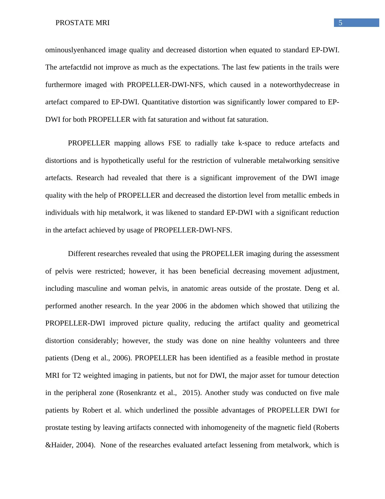
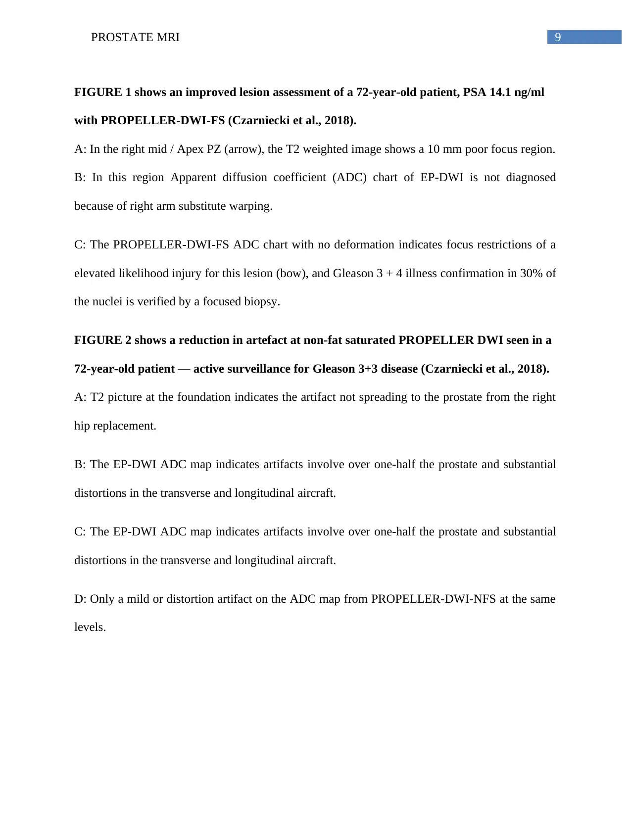
FIGURE 1 shows an improved lesion assessment of a 72-year-old patient, PSA 14.1 ng/ml
with PROPELLER-DWI-FS (Czarniecki et al., 2018).
A: In the right mid / Apex PZ (arrow), the T2 weighted image shows a 10 mm poor focus region.
B: In this region Apparent diffusion coefficient (ADC) chart of EP-DWI is not diagnosed
because of right arm substitute warping.
C: The PROPELLER-DWI-FS ADC chart with no deformation indicates focus restrictions of a
elevated likelihood injury for this lesion (bow), and Gleason 3 + 4 illness confirmation in 30% of
the nuclei is verified by a focused biopsy.
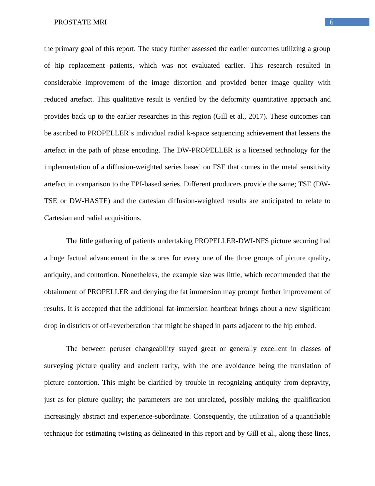
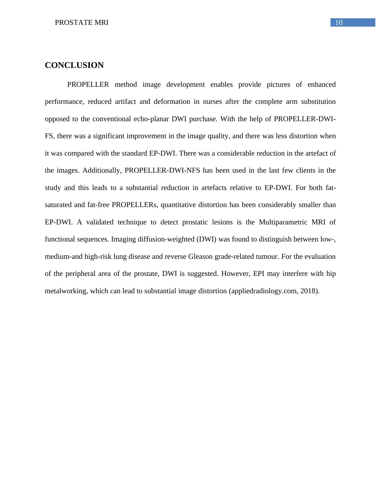
FIGURE 2 shows a reduction in artefact at non-fat saturated PROPELLER DWI seen in a
72-year-old patient — active surveillance for Gleason 3+3 disease (Czarniecki et al., 2018).
A: T2 picture at the foundation indicates the artifact not spreading to the prostate from the right
hip replacement.
B: The EP-DWI ADC map indicates artifacts involve over one-half the prostate and substantial
distortions in the transverse and longitudinal aircraft.
C: The EP-DWI ADC map indicates artifacts involve over one-half the prostate and substantial
distortions in the transverse and longitudinal aircraft.
D: Only a mild or distortion artifact on the ADC map from PROPELLER-DWI-NFS at the same
levels.
FIGURE 1 shows an improved lesion assessment of a 72-year-old patient, PSA 14.1 ng/ml
with PROPELLER-DWI-FS (Czarniecki et al., 2018).
A: In the right mid / Apex PZ (arrow), the T2 weighted image shows a 10 mm poor focus region.
B: In this region Apparent diffusion coefficient (ADC) chart of EP-DWI is not diagnosed
because of right arm substitute warping.
C: The PROPELLER-DWI-FS ADC chart with no deformation indicates focus restrictions of a
elevated likelihood injury for this lesion (bow), and Gleason 3 + 4 illness confirmation in 30% of
the nuclei is verified by a focused biopsy.
FIGURE 2 shows a reduction in artefact at non-fat saturated PROPELLER DWI seen in a
72-year-old patient — active surveillance for Gleason 3+3 disease (Czarniecki et al., 2018).
A: T2 picture at the foundation indicates the artifact not spreading to the prostate from the right
hip replacement.
B: The EP-DWI ADC map indicates artifacts involve over one-half the prostate and substantial
distortions in the transverse and longitudinal aircraft.
C: The EP-DWI ADC map indicates artifacts involve over one-half the prostate and substantial
distortions in the transverse and longitudinal aircraft.
D: Only a mild or distortion artifact on the ADC map from PROPELLER-DWI-NFS at the same
levels.
Paraphrase This Document
Need a fresh take? Get an instant paraphrase of this document with our AI Paraphraser
10PROSTATE MRI
CONCLUSION
PROPELLER method image development enables provide pictures of enhanced
performance, reduced artifact and deformation in nurses after the complete arm substitution
opposed to the conventional echo-planar DWI purchase. With the help of PROPELLER-DWI-
FS, there was a significant improvement in the image quality, and there was less distortion when
it was compared with the standard EP-DWI. There was a considerable reduction in the artefact of
the images. Additionally, PROPELLER-DWI-NFS has been used in the last few clients in the
study and this leads to a substantial reduction in artefacts relative to EP-DWI. For both fat-
saturated and fat-free PROPELLERs, quantitative distortion has been considerably smaller than
EP-DWI. A validated technique to detect prostatic lesions is the Multiparametric MRI of
functional sequences. Imaging diffusion-weighted (DWI) was found to distinguish between low-,
medium-and high-risk lung disease and reverse Gleason grade-related tumour. For the evaluation
of the peripheral area of the prostate, DWI is suggested. However, EPI may interfere with hip
metalworking, which can lead to substantial image distortion (appliedradiology.com, 2018).
11PROSTATE MRI
REFERENCES
Ahmed, H. U., El-ShaterBosaily, A., Brown, L. C., Kaplan, R. S., Colaco-Moraes, Y., Ward,
K., ... & Gabe, R. (2016). The PROMIS study: A paired-cohort, blinded confirmatory
study evaluating the accuracy of multi-parametric MRI and TRUS biopsy in men with an
elevated PSA.
Barrett, T., Priest, A. N., Lawrence, E. M., Goldman, D. A., Warren, A. Y., Gnanapragasam, V.
J., ... & Gallagher, F. A. (2015). Ratio of tumor to normal prostate tissue apparent
diffusion coefficient as a method for quantifying DWI of the prostate. American Journal
of Roentgenology, 205(6), W585-W593.
Barrett, T., Turkbey, B., &Choyke, P. L. (2015). PI-RADS version 2: what you need to
know. Clinical radiology, 70(11), 1165-1176.
Burton, L. J., Hawsawi, O., Loyd, Q., Henderson, V., Howard, S., Harlemon, M., ...&Odero-
Marah, V. (2018). Association of Epithelial Mesenchymal Transition with prostate and
breast health disparities. PloS one, 13(9), e0203855.
Chen, X., Xian, J., Wang, X., Wang, Y., Zhang, Z., Guo, J., & Li, J. (2014). Role of periodically
rotated overlapping parallel lines with enhanced reconstruction diffusion-weighted
imaging in correcting distortion and evaluating head and neck masses using 3 T
MRI. Clinical radiology, 69(4), 403-409.
Czarniecki, M., Caglic, I., Grist, J. T., Gill, A. B., Lorenc, K., Slough, R. A., ...& Barrett, T.
(2018). Role of PROPELLER-DWI of the prostate in reducing distortion and artefact
from total hip replacement metalwork. European journal of radiology, 102, 213-219.
⊘ This is a preview!⊘
Do you want full access?
Subscribe today to unlock all pages.
Trusted by 1+ million students worldwide
1 out of 14

Your All-in-One AI-Powered Toolkit for Academic Success.
+13062052269
info@desklib.com
Available 24*7 on WhatsApp / Email
![[object Object]](/_next/static/media/star-bottom.7253800d.svg)
Unlock your academic potential
Copyright © 2020–2026 A2Z Services. All Rights Reserved. Developed and managed by ZUCOL.